Adductor Muscles: The Psychology of Physical Alignment
- The Core Definition of Adductor Muscles
- Anatomical Overview and Groupings
- Detailed Anatomy of Individual Adductor Muscles
- Physiological Function and Biomechanics of Adduction
- Historical Understanding and Anatomical Discovery
- Practical Applications and Role in Movement
- Clinical Significance: Injuries and Conditions
- Diagnosis, Treatment, and Rehabilitation
- Connections to Other Musculoskeletal Systems
- Conclusion and Future Perspectives
The Core Definition of Adductor Muscles
The term adductor, within the realm of human and animal anatomy, precisely identifies a specific group of muscles whose primary physiological role is adduction. This fundamental anatomical action involves the movement of a limb or body part closer to the body’s central sagittal plane, or in the case of digits, towards the midline of the hand or foot. Situated predominantly within the medial compartment of the thigh, these powerful muscles are instrumental in a broad spectrum of functions, ranging from stabilizing the pelvis during locomotion to enabling precise lower body movements essential for daily activities and specialized athletic performance. Their intricate coordination with antagonist muscle groups is vital for maintaining musculoskeletal balance and ensuring efficient, injury-free movement patterns.
The underlying mechanism of adduction is initiated by muscle contraction, wherein the muscle fibers shorten, drawing their points of insertion towards their respective origins. For the thigh adductors, this translates into a medial pull on the femur, effectively bringing the thigh inward. This action is far from simplistic; it represents a complex symphony involving multiple muscles, each contributing uniquely and often with secondary roles beyond mere inward movement. Understanding this core principle of adduction is foundational for comprehending the intricate biomechanics of human gait, dynamic balance, and the generation of power in various physical exertions, highlighting the indispensable nature of these muscle groups.
Anatomical Overview and Groupings
The adductor muscle group of the thigh is traditionally comprised of the adductor longus, adductor brevis, and adductor magnus. Additionally, the pectineus and the gracilis are often included due to their significant contributions to adduction, despite having other primary functions. These five muscles collectively constitute the medial compartment of the thigh. Their origins are primarily located on the pubic bone and ischium, with insertions distributing along various points of the femur and, for the gracilis, extending to the tibia. This strategic anatomical placement allows them to exert considerable leverage on the thigh bone, facilitating robust inward movements and contributing to hip stability.
The distinct anatomical arrangement of each muscle within this group dictates its specific functional contributions. The adductor longus is the most superficial, lying anterior to the adductor brevis, which is shorter and deeper. The adductor magnus is the largest and most posterior, often recognized for its dual innervation and functional resemblance to both adductor and hamstring muscles. The pectineus, a quadrilateral muscle, forms the anterior-most part of the group, while the gracilis is the most medial and superficial muscle, uniquely crossing both the hip and knee joints. These individual characteristics are crucial for understanding their integrated roles in complex motor tasks and for targeted clinical interventions.
Detailed Anatomy of Individual Adductor Muscles
The adductor longus muscle originates from the anterior superior aspect of the pubic bone and inserts onto the middle third of the linea aspera of the femur. Its primary roles are adduction and assistance in hip flexion. The adductor brevis lies deep to the longus, originating from the inferior ramus of the pubic bone and inserting onto the upper linea aspera. It is a powerful adductor and also assists in hip flexion. The largest and most complex, the adductor magnus, has an extensive origin from the pubic ramus and ischial tuberosity, with a broad insertion along the linea aspera and adductor tubercle of the femur. This muscle is functionally distinct, with an adductor portion and a hamstring-like portion acting as a hip extensor, showcasing its versatility in hip kinematics.
Completing the adductor complex, the pectineus originates from the pectineal line of the pubic bone and inserts onto the pectineal line of the femur. It is the most anterior adductor, contributing significantly to both adduction and hip flexion. The gracilis is a long, slender muscle originating from the pubic symphysis and arch, extending to insert onto the medial surface of the tibia as part of the pes anserinus. Uniquely crossing both the hip and knee joints, the gracilis performs adduction of the thigh, flexion of the knee, and internal rotation of the flexed leg, making it a crucial component in coordinated lower limb movements.
Physiological Function and Biomechanics of Adduction
The core physiological function of the adductor muscle group is the powerful adduction of the thigh, a movement indispensable for maintaining dynamic balance and generating force across a wide array of activities. During unilateral stance, such as when one leg is lifted during walking, the adductors of the standing leg engage to stabilize the pelvis, preventing lateral sway and ensuring the body’s center of gravity remains appropriately aligned over the supporting limb. This stabilization mechanism is fundamental for efficient locomotion, postural control, and the prevention of falls.
Beyond their primary adductor role, the biomechanics of these muscles are sophisticated, encompassing contributions to hip flexion, extension, and rotation. The anterior fibers of muscles like the adductor longus and brevis assist in hip flexion, particularly from an extended hip position. Conversely, the posterior fibers of the adductor magnus act as potent hip extensors, especially when the hip is flexed, collaborating with the hamstrings and gluteus maximus. This multifaceted functionality underscores their importance as multi-joint movers, enabling fluid and powerful lower body kinematics crucial for both everyday movements and peak athletic performance.
Historical Understanding and Anatomical Discovery
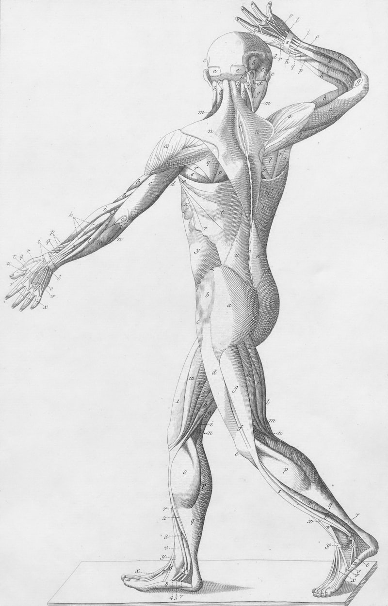
The journey to understanding the adductor muscle group, much like the broader field of human anatomy, spans centuries of dedicated observation and inquiry. Early anatomical insights from ancient civilizations, though rudimentary, laid foundational concepts of muscle function. However, it was during the Renaissance, spearheaded by pioneering figures like Andreas Vesalius, that modern anatomical precision began to emerge. Vesalius’s groundbreaking work, “De humani corporis fabrica libri septem” (1543), based on direct human dissection, provided the first highly accurate and detailed depictions of individual muscles, including the precise delineation of the adductor longus, brevis, and magnus within the medial thigh compartment.
The subsequent centuries witnessed continuous advancements in anatomical mapping and physiological understanding. The 19th and 20th centuries, in particular, brought forth sophisticated techniques such as histology, electromyography, and advanced imaging, which allowed researchers to delve deeper into the microscopic structure, innervation patterns, and precise biomechanical contributions of each adductor muscle. This cumulative body of knowledge, from macroscopic observation to intricate functional analysis, has forged our current comprehensive understanding of the adductor group’s vital structure and its multifaceted role in human movement and health.
Practical Applications and Role in Movement
The adductor muscles are critical for a vast array of daily activities and specialized athletic movements, serving both stabilizing and dynamic roles. In everyday activities like walking, the adductors of the stance leg are essential for stabilizing the pelvis and preventing excessive lateral sway as the opposite leg swings forward. They contribute to the powerful inward drive required for propulsion, ensuring an efficient and balanced gait. Without robust adductor function, maintaining upright posture and smooth ambulation would be significantly impaired, often resulting in an unstable, wide-based walking pattern.
In sports, the adductor muscles are indispensable for actions demanding lateral movement, rapid changes in direction, and powerful kicking. For instance, in soccer, the adductors are heavily recruited for the powerful leg swing during shooting, precise passing, and agile dribbling. Similarly, in sports such as ice skating, hockey, and skiing, these muscles are crucial for generating lateral force to propel forward, accelerate, and maintain balance during sharp turns. Their strength, endurance, and rapid responsiveness are paramount for athletes to execute explosive movements and sustain high-intensity performance, simultaneously mitigating the risk of injury. Furthermore, activities like horse riding rely heavily on adductor strength for gripping and control, demonstrating their broad impact on human kinetics.
Clinical Significance: Injuries and Conditions
Due to their extensive engagement in daily life and high-demand physical activities, the adductor muscles are highly susceptible to injury, leading to considerable pain and functional limitations. The most prevalent condition is an adductor strain, commonly known as a “groin pull.” This acute injury typically occurs during sudden, forceful, or eccentric movements such as sprinting, kicking, or abrupt changes in direction, which can result in overstretching or tearing of muscle fibers. Athletes in sports characterized by explosive lateral movements and rapid acceleration, like soccer, hockey, and track and field, face a particularly elevated risk of sustaining adductor strains.
Symptoms vary depending on the injury’s severity, ranging from mild tenderness and stiffness (Grade I) to significant pain, swelling, and partial loss of strength (Grade II), or even complete muscle rupture with intense pain and inability to move the limb (Grade III). Accurate diagnosis, often aided by physical examination and sometimes imaging such as MRI, is critical for guiding appropriate treatment and rehabilitation. Beyond acute strains, chronic conditions like adductor tendinopathy, characterized by tendon degeneration from repetitive overuse, or osteitis pubis, an inflammatory condition affecting the pubic symphysis and adductor attachments, can cause persistent debilitating groin pain, necessitating comprehensive and often prolonged therapeutic interventions.
Diagnosis, Treatment, and Rehabilitation
Diagnosing adductor muscle injuries typically commences with a comprehensive clinical evaluation. A healthcare professional assesses the patient’s symptoms, medical history, and the specific mechanism of injury. Physical examination involves palpation of the adductor muscles and their insertions, alongside specialized strength and flexibility tests, to accurately localize the injury and determine its severity. For instance, pain elicited during resisted adduction or passive abduction of the hip strongly indicates an adductor strain. In certain situations, imaging modalities like ultrasound or magnetic resonance imaging (MRI) may be employed to confirm the diagnosis, exclude other pathologies, and precisely evaluate the extent of muscle or tendon damage, especially in more severe or chronic presentations.
Treatment for acute adductor strains generally adheres to the RICE protocol: immediate Rest from aggravating activities, application of Ice to mitigate swelling and pain, targeted Compression with a bandage, and Elevation of the affected leg. Non-steroidal anti-inflammatory drugs (NSAIDs) may be prescribed for pain and inflammation management. Once acute symptoms subside, a structured, progressive rehabilitation program, supervised by a physical therapist, is crucial. This program typically advances from gentle stretching to restore flexibility, through a graded strengthening regimen that targets the adductors and surrounding musculature, aiming to restore balanced function and prevent recurrence. For chronic conditions like tendinopathies, a more specialized approach involving eccentric loading, manual therapy, and addressing biomechanical imbalances is often necessary, with surgery being a rare consideration for intractable cases.
Connections to Other Musculoskeletal Systems
The adductor muscles function not in isolation but as an integral component of the vast and interconnected musculoskeletal system. They maintain a crucial antagonistic relationship with the hip abductors, primarily the gluteus medius and minimus, which move the leg away from the midline. A synergistic balance in strength and flexibility between these opposing muscle groups is paramount for optimal pelvic stability, efficient gait mechanics, and the prevention of diverse musculoskeletal issues, including knee pain, hip impingement, and lower back discomfort. Imbalances, such as adductor tightness coupled with abductor weakness, can significantly disrupt normal biomechanics.
Furthermore, the adductors exhibit significant functional interplay with hip flexors and extensors. The adductor magnus, for example, serves as a potent hip extensor, especially when the hip is flexed, thereby synergistically supporting the hamstrings and gluteus maximus in powerful actions like running and jumping. The gracilis, as a bi-articular muscle, bridges the hip and knee joints, influencing not only hip adduction but also knee flexion and internal rotation. This demonstrates its pivotal role in coordinating complex movements across multiple joints, affecting the entire kinetic chain of the lower limb and highlighting the necessity of considering the adductors within a comprehensive biomechanical framework.
Conclusion and Future Perspectives
In summary, the adductor muscles constitute a vital and complex group within the human musculoskeletal system, fundamentally responsible for adduction—the critical movement of bringing the leg towards the body’s midline. Encompassing muscles such as the adductor longus, brevis, magnus, pectineus, and gracilis, they are indispensable for maintaining balance, facilitating efficient locomotion, and enabling a broad spectrum of daily and athletic movements. Their intricate anatomical structure and multifaceted physiological roles, extending beyond mere adduction to include contributions to hip flexion, extension, and rotation, underscore their profound significance in human biomechanics.
The historical evolution of our understanding of these muscles, from early anatomical observations to contemporary detailed analyses, has continuously refined our knowledge of their form and function. Their constant engagement across various physical activities makes them susceptible to common injuries like strains and tendinopathies, which can profoundly impact mobility and quality of life. Effective diagnosis, conservative treatment strategies, and diligent, progressive rehabilitation are paramount for managing these conditions and facilitating a full return to optimal function.
Looking forward, ongoing research continues to enhance our comprehension of adductor muscle physiology, particularly in areas concerning injury prevention, athletic performance optimization, and the development of personalized rehabilitation protocols. Advances in non-invasive imaging, sophisticated biomechanical analyses, and genetic research hold promise for new insights into muscle pathology and recovery mechanisms. The undeniable interconnectedness of the adductor group with other components of the musculoskeletal system reinforces the importance of a holistic approach to physical health, acknowledging these muscles as integral elements of the body’s dynamic and adaptive framework, crucial for advancing human health and maximizing athletic potential.