Amorphagnosia: When the Brain Cannot See Shape
Definition and Etymology of Amorphagnosia
Amorphagnosia is a specialized neurological deficit characterized by the profound inability to consciously recognize or comprehend the three-dimensional form or shape of objects, despite having intact primary sensory input. The term itself is derived from Greek roots, offering immediate insight into the nature of the disorder: the prefix a- signifies “without” or “absence,” morphē translates to “form” or “shape,” and gnōsis denotes “knowledge” or “consciousness.” Therefore, amorphagnosia fundamentally represents an absence of consciousness concerning shape. It is essential to delineate this condition from simple blindness or peripheral sensory loss; the patient retains the capacity to detect touch, pressure, and temperature, and often possesses normal visual acuity, yet the higher-order cognitive processing required to synthesize these inputs into a meaningful perception of form is compromised.
This specific form of agnosia often manifests most clearly within the tactile domain, where it is frequently discussed as a component or precursor to astereognosia, the broader inability to identify an object by touch alone. However, amorphagnosia isolates the failure specifically to the geometric properties of the object—the contour, dimensions, and structural relationships—as opposed to characteristics like texture, weight, or density. A patient suffering from this condition, when blindfolded, might correctly report that an object feels smooth or cold, but would be utterly incapable of determining if that object is a sphere, a cube, or a triangular prism. The integration of spatial information necessary for form recognition has been disrupted due to damage in specific cortical association areas, leading to a breakdown in the perceptual synthesis pathway.
The distinction between primary sensory deficits and agnosia is paramount in understanding amorphagnosia. If a patient cannot feel the object due to peripheral neuropathy or spinal cord injury, the problem lies in sensation (anesthesia). In contrast, the patient with amorphagnosia possesses fully functional tactile receptors and afferent pathways; the information successfully reaches the brain, but the cerebral cortex fails to interpret the data stream into a recognizable geometric concept. This failure occurs at the level of association cortex function, highlighting the complexity of human perception where raw sensory data must be compared, categorized, and spatialized against learned templates stored within memory.
The clinical significance of amorphagnosia extends beyond mere academic classification. Its presence immediately flags a highly specific type of focal neurological damage, offering crucial localizing information to the examining physician. Furthermore, the functional impairment it causes profoundly impacts daily living, as the manipulation of tools, recognition of common household items, and execution of basic self-care tasks often rely heavily on the unconscious, automatic recognition of object shapes and sizes, particularly when vision is unavailable or diverted. Understanding this precise deficit is the foundation upon which effective diagnostic and rehabilitative strategies are built.
Clinical Presentation and Symptomatology
The clinical manifestation of amorphagnosia revolves around profound difficulties in discriminating and identifying the shape and size of objects, frequently tested through the tactile modality. A classic presentation involves the patient being asked to identify common geometric shapes or household items placed in their hand while their vision is occluded. While they can articulate the object’s material properties—for instance, noting that a key is metallic, cold, and has ridges—they cannot articulate its defining shape, such as recognizing the contour of the key’s head or the serrated edge. This inability makes activities requiring fine motor control and object manipulation extremely challenging.
A critical aspect of the symptomatology involves the dissociation between the perception of fundamental attributes and the perception of form. Patients may accurately gauge the size of an object relative to another (e.g., “This apple is bigger than that marble”), but they struggle severely when asked to describe or replicate the specific shape of the object (e.g., drawing the apple’s round contour or matching the marble to a circular cutout). This suggests that the brain’s ability to process simple magnitude comparison remains somewhat preserved, while the integrative function necessary for complex geometric processing is selectively impaired. The difficulty is not merely a lack of naming (anomia); the deficit lies in the failure to achieve the internal, conceptual recognition of the form itself.
The impairment in amorphagnosia is often compounded when the patient is required to perform bimanual tasks or coordinate object manipulation based on tactile feedback. For example, fitting a key into a lock, buttoning a shirt, or selecting the correct tool from a pouch all require instantaneous, unconscious recognition of shape and orientation. The amorphagnosic patient may resort to excessive visual reliance, constantly looking at their hands and the object to compensate for the missing tactile form recognition. This compensation, however, slows down processes and fails completely in darkness or when the hands are obscured. The severity of the agnosia determines the level of functional disability, ranging from minor inconvenience to complete dependency in complex manipulative tasks.
In some cases, amorphagnosia can present with a degree of laterality, meaning the deficits are more pronounced in one hand, corresponding to damage in the contralateral hemisphere. When the lesion is highly focal, for example, affecting only the sensory association cortex of the dominant parietal lobe, the patient might exhibit profound amorphagnosia in the non-dominant hand while retaining comparatively better form recognition in the dominant hand. This lateralized presentation is a powerful diagnostic indicator, helping to precisely pinpoint the neurological site of the damage and guiding the scope of subsequent neuroimaging studies.
Neurological Basis and Localization
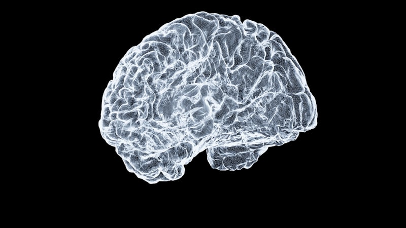
Amorphagnosia, like most forms of agnosia, results from focal damage to the brain’s association cortices, specifically those areas responsible for synthesizing and interpreting sensory data into complex perceptions. The condition is classically associated with lesions in the parietal lobe, particularly involving the superior parietal lobule and the post-central gyrus (primary somatosensory cortex) and its adjacent association areas. The parietal lobe is the critical hub for processing spatial awareness, body orientation, and integrating tactile information into a coherent three-dimensional map of the environment and the objects within it.
The underlying mechanism involves a disruption in the pathway connecting the primary sensory reception area (S1) to the secondary and tertiary somatosensory processing areas (S2 and beyond). While S1 registers basic sensation (touch, pressure), it is the higher-order processing regions, often located in the posterior parietal cortex, that are responsible for the complex task of geometric analysis—determining the object’s contour, volume, and spatial orientation. Damage to these areas prevents the raw tactile data from being transformed into a coherent mental model of the object’s shape, leading directly to the symptoms of amorphagnosia. This region is sometimes referred to as the tactile recognition pathway, analogous to the visual streams (ventral and dorsal) that process visual object recognition.
Specific neural pathways that are implicated include those involved in the integration of proprioceptive (sense of self-movement and position) and tactile information. Recognizing a shape requires constantly updating the mental map of the object as the fingers explore its contours. This dynamic integration relies heavily on the integrity of the white matter tracts connecting the somatosensory cortex with the posterior parietal cortex. Lesions, such as infarcts or traumatic injury, that sever these critical communication lines, even if they spare the gray matter of the primary sensory cortices, can result in a pure form of amorphagnosia, underscoring the role of connectivity in complex perceptual tasks.
Furthermore, amorphagnosia may sometimes be seen in conjunction with damage extending into the temporo-occipital region, particularly if the lesion is large or complex. While classic amorphagnosia is tactile, if the condition involves a failure to recognize shape visually (though this is typically classified as visual form agnosia), the lesion tends to involve the inferior temporal cortex or areas along the ventral visual stream—the “what” pathway. However, when the deficit is purely tactile and focused on shape recognition, the pathology is overwhelmingly localized to the somatosensory association areas of the dominant or non-dominant posterior parietal lobe.
Differentiation from Related Conditions
Accurate diagnosis requires careful differentiation of amorphagnosia from several related neurological and sensory conditions, particularly other types of agnosia and primary sensory deficits. The most important distinction is made between amorphagnosia and astereognosia (also known as tactile agnosia). Astereognosia is the general term for the inability to identify an object by touch, encompassing failures in recognizing texture, weight, density, and material, alongside shape. Amorphagnosia is often considered a specific, isolated component of astereognosia, where the failure is restricted primarily to the perception of the object’s geometric form, while other tactile qualities (e.g., thermal or textural discrimination) may remain relatively intact.
Another crucial differentiation is made against primary sensory loss, or anesthesia. In patients with anesthesia, the peripheral nerves or spinal tracts conveying touch sensation are damaged, meaning the information never reaches the cortex effectively. A simple clinical test—such as two-point discrimination or light touch testing—will reveal the sensory loss. Conversely, the amorphagnosic patient reports normal sensation upon testing, but fails the interpretation test (e.g., identifying a key). This confirms that the deficit is associative and cortical, not peripheral.
Amorphagnosia must also be distinguished from visual agnosias, such as apperceptive or associative visual agnosia. While visual agnosia impairs the recognition of objects via sight, amorphagnosia specifically targets the tactile domain. It is entirely possible for a patient to have severe amorphagnosia (cannot recognize a sphere by touch) yet immediately recognize and name the sphere when they look at it. This sensory modality specificity is the defining characteristic that separates tactile agnosias from their visual counterparts, although concurrent damage can lead to multi-modal deficits.
Finally, it is necessary to rule out cognitive deficits that might masquerade as agnosia, such as severe memory impairment or language deficits (aphasia). If a patient fails to name an object, the reason might be a difficulty retrieving the name (anomia) rather than a failure to recognize the shape. The diagnostic process often includes asking the patient to non-verbally demonstrate recognition, such as drawing the perceived shape or matching it to a template. If they can mentally recognize the shape but cannot retrieve the linguistic label, the diagnosis leans towards anomia; if they cannot internally represent the shape regardless of naming attempts, amorphagnosia is confirmed.
Etiology and Causes
The underlying causes of amorphagnosia are invariably linked to conditions that result in focal damage to the somatosensory association areas of the parietal lobe, particularly those involving the non-dominant hemisphere (often the right hemisphere) for spatial processing, or bilateral damage. The most common etiology is cerebrovascular accident (CVA), or stroke. Ischemic strokes affecting the territory supplied by the Middle Cerebral Artery (MCA), especially its parietal branches, frequently lead to this type of deficit. The sudden interruption of blood flow causes necrosis in the highly sensitive association cortex, leading to the rapid onset of shape recognition failure.
Another significant cause is traumatic brain injury (TBI). Severe head trauma, particularly deceleration injuries, can result in contusions, hemorrhages, or diffuse axonal injury (DAI) concentrated in the posterior regions of the brain. The impact of the brain against the skull’s bony ridges often damages the cortical surfaces and underlying white matter tracts in the parietal and occipital lobes, disrupting the critical communication pathways necessary for complex sensory synthesis and resulting in amorphagnosia, often in conjunction with other parietal lobe syndromes.
Progressive neurological disorders, such as brain tumors (neoplasms) and neurodegenerative conditions, also contribute to the incidence of amorphagnosia. Slow-growing tumors, such as meningiomas or gliomas, that compress or infiltrate the superior parietal lobule can gradually erode the function of the shape recognition pathways. Similarly, certain focal presentations of Alzheimer’s disease or Posterior Cortical Atrophy (PCA) may initially present with isolated agnosias, including amorphagnosia, before the generalized cognitive decline becomes apparent. In these cases, the onset is insidious and the progression is typically chronic.
Less common, but clinically relevant, etiologies include infectious diseases resulting in encephalitis, demyelinating diseases (e.g., Multiple Sclerosis) where plaques form in critical white matter tracts, and certain metabolic or toxic encephalopathies that selectively target vulnerable neuronal populations in the association cortices. Regardless of the primary cause, the common neuropathological endpoint is the selective damage or functional impairment of the neural substrate responsible for converting raw tactile data into a meaningful, conscious perception of geometric form.
Diagnostic Procedures
The diagnosis of amorphagnosia is achieved through a combination of detailed clinical examination, standardized neuropsychological testing, and advanced neuroimaging. The initial step involves a thorough neurological assessment to confirm the integrity of primary sensory pathways. This includes testing for light touch, pain, temperature, vibration, and proprioception. If these primary sensations are normal, the examiner proceeds to specialized tests for tactile recognition.
Neuropsychological testing for amorphagnosia employs specific tasks designed to isolate the failure of shape recognition. The most common method involves the use of standardized stereognosis test kits, which contain objects of varying shapes (spheres, cubes, pyramids) and materials. The patient is asked to identify the shape of the object placed in their hand without visual input. Critical tasks include:
- Matching the object held in the hand to a visual representation of the shape.
- Drawing the outline of the object after palpation.
- Discriminating between two similar shapes (e.g., a slightly oblong sphere and a perfect sphere).
If the patient fails these shape-specific tests while successfully recognizing texture or weight, amorphagnosia is strongly indicated.
To confirm the underlying neurological damage, neuroimaging studies are essential. Magnetic Resonance Imaging (MRI) is the gold standard, providing high-resolution images of the brain structure. MRI scans, particularly T2-weighted and FLAIR sequences, can clearly visualize ischemic lesions, hemorrhage, tumors, or areas of atrophy in the parietal lobe that correspond precisely to the somatosensory association cortex. In acute settings, Computed Tomography (CT) scans may be used initially to rule out hemorrhagic stroke. Functional imaging techniques, such as functional MRI (fMRI) or Positron Emission Tomography (PET), may occasionally be utilized in a research context to observe reduced activity in the parietal cortex during attempted shape recognition tasks, further corroborating the diagnosis.
Finally, the comprehensive diagnostic workup must include tests to exclude related conditions, such as ruling out severe memory or naming deficits via standardized cognitive batteries. The physician must ensure that the patient understands the instructions and possesses the necessary motor skills (apraxia must be ruled out) to manipulate the object, confirming that the failure truly lies in the perceptual interpretation of form.
Management and Therapeutic Approaches
The management of amorphagnosia primarily focuses on rehabilitation, centered on maximizing functional recovery and developing compensatory strategies, as pharmacological interventions generally do not directly address the cortical processing deficit. Rehabilitation is highly individualized and typically involves a multidisciplinary team, including neurologists, occupational therapists (OTs), and neuropsychologists. The success of rehabilitation is heavily dependent on the extent of the underlying brain damage and the patient’s capacity for neuroplasticity.
Occupational therapy (OT) plays the most crucial role in managing amorphagnosia. Therapists use structured exercises designed to retrain the brain to integrate sensory input. These techniques often involve repetitive, focused practice aimed at improving shape discrimination. Techniques may include:
- Haptic Training: Using increasingly complex shapes for tactile exploration, initially paired with visual confirmation, gradually fading the visual component.
- Sensory Substitution: Training the patient to rely more heavily on intact sensory modalities, such as visual scanning or auditory cues, to compensate for tactile deficits during daily activities.
- Errorless Learning: Providing immediate feedback to prevent the repetition of errors, helping to rebuild the correct association between tactile input and geometric concept.
Compensatory strategies are vital, especially in chronic or severe cases where full recovery is unlikely. Patients are taught to consciously use vision for tasks that were previously performed automatically by touch. For instance, instead of reaching blindly into a drawer for a tool, the patient is instructed to look at the tool before grasping it, thus utilizing the intact visual recognition pathway to guide manipulation. This shift in sensory reliance helps maintain independence in activities of daily living (ADLs) and reduces frustration associated with object interaction.
Furthermore, cognitive rehabilitation focuses on enhancing the patient’s awareness of their deficit (anosognosia must be ruled out) and teaching them adaptive problem-solving skills. Understanding the precise nature of the failure—that the shape is not recognizable by touch—allows the patient to implement proactive compensatory measures. Psychosocial support is also necessary, as the frustration and functional limitations imposed by amorphagnosia can lead to anxiety and depression.
Prognosis and Quality of Life Implications
The prognosis for recovery from amorphagnosia is highly variable and depends largely on the etiology, size, and location of the lesion, and the patient’s age and overall health. If the condition is caused by an acute event such as a small, isolated stroke, significant recovery, particularly in the first six months post-injury, can occur due to spontaneous resolution of swelling (edema) and neuroplastic reorganization. However, if the damage is extensive or caused by a progressive neurodegenerative disease, the prognosis for complete recovery of function is guarded.
Amorphagnosia has significant implications for the quality of life, primarily by impacting the performance of routine activities that require automatic tactile-spatial processing. Simple tasks become laborious and require conscious visual attention, leading to reduced efficiency and increased mental fatigue. These implications include:
- Difficulties with fine motor skills, such as dressing, cooking, and handling small implements.
- Impairment in vocational activities, especially those requiring manual dexterity or operating machinery.
- Safety risks, as tactile cues about object shape and orientation (e.g., handling a hot utensil or sharp tool) are misinterpreted or absent.
Long-term adaptation often involves the masterful use of compensatory strategies and environmental modifications. Patients may need specialized tools or adaptive equipment that provide clearer visual or auditory feedback regarding shape and orientation. The goal of long-term care is not necessarily to restore the original neurological function, but rather to ensure the patient achieves maximum functional independence through learned visual-manual substitutions. Ongoing occupational therapy and periodic assessments are necessary to adjust these strategies as the patient’s environment or needs change.
Ultimately, while amorphagnosia presents a challenging and specific deficit in higher-order sensory processing, robust rehabilitation and psychological support can significantly mitigate the negative effects on daily living. The success hinges upon the patient’s ability to leverage remaining cognitive and sensory strengths to bypass the damaged shape recognition pathway, thereby maintaining engagement with their environment.