Developmental Psychology: Understanding Testicular Descent
Definition and Nomenclature
The term arrested testis refers specifically to a condition where one or both testes fail to complete their normal descent into the scrotum, becoming lodged or fixed along the typical path of descent, most commonly within the inguinal canal. This specific diagnosis implies a mechanical obstruction or a structural limitation preventing the final migration into the scrotal sac, distinguishing it slightly from the broader medical term cryptorchidism (meaning ‘hidden testis’), which encompasses any undescended testis, regardless of the cause or location, including those that may be ectopic or truly absent. A testis classified as arrested is typically palpable within the canal but cannot be manipulated into the scrotum, confirming the physical barrier to movement. The descriptive synonym frequently utilized in clinical practice is undescended testicle, although the specificity of ‘arrested’ emphasizes the physical containment due to a blockage of the passage, rather than solely hormonal or neurogenic failure. Understanding this nuance is crucial, as the therapeutic management for a mechanically arrested testis is almost invariably surgical, necessitating the removal of the obstruction to ensure proper placement and mitigate long-term health risks.
The normal physiological process of testicular descent is a complex, biphasic event that is highly sensitive to hormonal signals and physical guidance mechanisms. When this process halts due to fibrous bands, an inadequate processus vaginalis, or a short spermatic cord, the condition is defined as an arrested testis. Crucially, the location of the arrest—most often high in the superficial inguinal pouch or lower in the canal—dictates the complexity of the surgical repair and influences the degree of risk associated with long-term complications, such as impaired spermatogenesis. Because the scrotum provides a cooler environment essential for the viability of germ cells, any prolonged retention of the testis at the higher, warmer body temperature of the inguinal canal or abdomen compromises its functionality, making timely intervention paramount. Therefore, the diagnosis of an arrested testis immediately signals the need for definitive therapeutic action, focusing on correcting the underlying anatomical impediment.
While cryptorchidism affects a significant percentage of premature male infants, the incidence of true arrested testis—a fixed, non-retractile organ stuck in the inguinal canal—is lower but represents a more definitive pathological entity requiring intervention. The classification of the arrested testis must be meticulous, differentiating it from a retractile testis, which can be easily moved into the scrotum because the cremasteric reflex is overactive but the anatomical passage is clear. In contrast, the arrested testis resists all attempts at manual manipulation, confirming the presence of the obstructive pathology. This rigid distinction guides the clinical pathway, ensuring that surgical resources are appropriately utilized for the mechanical blockage identified in the arrested testis, while surveillance may be adequate for the temporary retractile variety.
Etiology and Developmental Pathways
The etiology of the arrested testis is fundamentally linked to a failure during the second, or inguinoscrotal, phase of testicular descent, which typically occurs between the seventh month of gestation and birth. This complex migration is orchestrated by a delicate interplay between hormonal cues, specifically Insulin-like factor 3 (INSL3) produced by the Leydig cells, and physical guidance provided by the gubernaculum testis, a mesenchymal cord that links the caudal pole of the testis to the developing scrotum. INSL3 is primarily responsible for the transabdominal phase, guiding the testis to the internal inguinal ring. The subsequent passage through the inguinal canal and into the scrotum, however, is heavily dependent on the maturation and regression of the gubernaculum, alongside the influence of androgens, which promote the widening of the inguinal canal. Arrested descent occurs when anatomical factors physically obstruct this final pathway, even if the hormonal signals were initially correct.
Structural impediments that lead to an arrested testis include several possible defects. One common cause is the presence of inadequate development or fibrosis of the gubernaculum, leading to a tethering effect that prevents the full excursion into the scrotum. Alternatively, the obstruction may arise from a narrow or malformed external inguinal ring, effectively creating a bottleneck that the testis cannot pass through. Furthermore, an unduly short spermatic cord or associated vascular structures can physically restrict the downward movement of the gonad, fixing its position high within the canal. These anatomical failures are often compounded by anomalies of the processus vaginalis, the peritoneal outpouching that normally precedes the descent of the testis; if the processus vaginalis fails to obliterate properly or forms restrictive adhesions, it can mechanically bind the testis to the canal wall. Therefore, while hormonal deficiencies (such as low testosterone or lack of androgen receptor sensitivity) might cause a testicle to halt entirely in the abdomen, the specific diagnosis of an arrested testis suggests the failure is predominantly mechanical or structural at the level of the inguinal passage itself.
Genetic predisposition also plays a role in the development of arrested testis. Although most cases are sporadic, studies have demonstrated an increased familial incidence, suggesting polygenic inheritance patterns that influence the development of the supporting structures, such as the gubernaculum or the inguinal ring architecture. Environmental factors, including exposure to certain endocrine-disrupting chemicals (EDCs) during critical windows of gestation, have also been implicated, potentially interfering with the delicate hormonal balance required for normal descent. However, regardless of whether the initial impetus is genetic or environmental, the final common pathway leading to the arrested testis remains the physical failure to navigate the inguinal canal. This highlights why surgical correction, which physically clears the passage and repositions the testis, is the established standard of care, addressing the anatomical consequence of these underlying etiological factors.
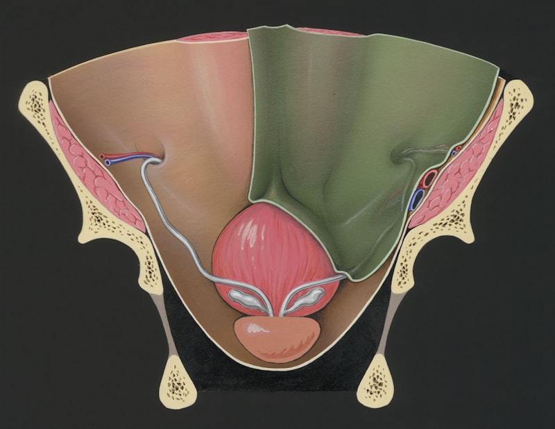
Classification and Location
The classification of the arrested testis is primarily based upon its physical location, which is crucial for determining the surgical approach and assessing potential risks. Arrested testes are typically categorized as being located along the path of normal descent, distinguishing them from ectopic testes, which have strayed significantly from the standard route (e.g., in the femoral triangle or perineum). For the arrested testis, the location is usually described relative to the inguinal canal: high intracanalicular, low intracanalicular, or suprascrotal (just outside the external ring but unable to fully descend). The majority of arrested testes are palpable upon physical examination, lying within the inguinal region, which makes diagnosis relatively straightforward compared to the non-palpable abdominal testis, which requires advanced imaging.
Specific classifications help surgeons plan the procedure. A high intracanalicular arrest implies the testis is deep within the inguinal canal, often near the internal ring, requiring a more extensive dissection to mobilize the spermatic cord sufficiently for tension-free placement in the scrotum. Conversely, a low intracanalicular arrest or suprascrotal position involves the testis being closer to the external ring, making mobilization simpler, but still requiring the correction of the mechanical tethering—often a short cord or fibrous bands—that prevents final descent. It is vital to differentiate the arrested testis from a ‘gliding’ or retractile testis during classification. The arrested testis is fixed and cannot be manually brought down, whereas the retractile testis, despite its proximity to the scrotum, is mobile and returns to the inguinal region spontaneously due to an overactive cremasteric muscle but does not suffer from a true anatomical obstruction. Misclassification can lead to unnecessary surgery for a retractile testis or delayed surgery for a truly arrested one.
Further anatomical classification involves whether the condition is unilateral (affecting one testis) or bilateral (affecting both). Unilateral arrest is far more common and usually indicates a localized anatomical problem. Bilateral arrest, while rarer, often suggests a greater systemic or hormonal etiology affecting both sides, although mechanical obstruction may still be present. When a testis is definitively classified as arrested, it signifies that the gonad is viable but physically entrapped. This contrasts sharply with cases of testicular agenesis (absence) or testicular atrophy (vanishing testis syndrome), which are also non-palpable but require different management strategies. Accurate location and classification through physical examination and, occasionally, supplementary imaging (such as high-resolution ultrasonography) ensures that the subsequent orchiopexy procedure is tailored precisely to the anatomical constraints presented by the arrested gonad.
Clinical Presentation and Diagnosis
The clinical presentation of an arrested testis is typically straightforward: the absence of a testis in the scrotal sac, coupled with the presence of a palpable mass in the inguinal region. The diagnosis is primarily established through a thorough physical examination, ideally conducted by an experienced pediatrician or urologist. This examination requires the infant or young boy to be relaxed, often achieved in a warm room, to minimize the activation of the cremasteric reflex, which can cause a normally descended testis to temporarily retract. The examiner systematically palpates the scrotum and the entire inguinal path, starting from the anterior superior iliac spine down to the scrotum. If a firm, ovoid mass is felt in the inguinal canal, attempts are made to gently manipulate the testis into the scrotal sac. If the testis is fixed, firm, and resists all attempts at repositioning, the diagnosis of an arrested testis due to mechanical obstruction is highly probable.
Differentiating a truly arrested testis from a retractile testis is the most critical diagnostic step, as it determines the need for immediate surgical intervention. A retractile testis can be manipulated into the scrotum and remains there momentarily before snapping back, confirming the viability of the passage. An arrested testis, however, is tethered and cannot be brought past the neck of the scrotum. If the testis is non-palpable, the clinical approach shifts to ruling out an abdominal testis or agenesis, which requires further investigation. While the physical exam is generally sufficient for a palpable, arrested testis, imaging may be utilized, especially in cases where the clinical findings are ambiguous or to rule out associated pathologies like an inguinal hernia, which frequently coexists with cryptorchidism. High-resolution ultrasonography is the primary imaging modality used to confirm the presence and size of the arrested testis and, importantly, to verify the presence of testes on the contralateral side.
The timing of diagnosis is crucial for optimizing prognosis. The American Academy of Pediatrics recommends that all male infants be examined shortly after birth and again at the six-month well-child visit. If the testis has not descended spontaneously by six months of age, spontaneous descent thereafter is rare, and the risk of permanent damage to the germ cells begins to increase significantly due to prolonged exposure to supra-scrotal temperatures. Therefore, the definitive diagnosis of an arrested testis often triggers surgical planning to occur between six and eighteen months of age. Furthermore, in rare cases of bilateral arrested testis, or when there are signs of associated genital anomalies, hormonal testing, such as the HCG stimulation test, may be employed to assess the functional capacity of the Leydig cells and distinguish between anatomical failure and primary hormonal insufficiency, although for a simple mechanical arrest, this is usually unnecessary.
Associated Risks and Complications
The failure of the testis to descend fully into the scrotum, as seen in the arrested testis, carries significant long-term health risks, primarily related to the function and malignant potential of the gonad. The foremost concern is infertility or subfertility. Normal sperm production (spermatogenesis) requires a temperature approximately 2 to 3 degrees Celsius lower than core body temperature, which the scrotum naturally provides. When the testis is arrested in the warmer inguinal canal, the prolonged exposure to elevated temperatures causes irreversible damage to the delicate germ cells, particularly the spermatogonia. Even if only one testis is arrested, the overall sperm count and quality can be significantly diminished, and if the condition is bilateral, the risk of azoospermia and absolute infertility approaches certainty, underscoring the urgency of surgical correction during infancy.
A second, and perhaps the most serious, complication is the dramatically increased risk of developing testicular germ cell tumor (TGCT). Men with a history of undescended testis have a risk of developing testicular cancer that is approximately three to eight times higher than the general population. While the precise mechanism linking maldescent and malignancy is complex, it is hypothesized that the thermal stress and dysgenesis caused by the supra-scrotal location contribute to the malignant transformation of the germ cells. Importantly, while surgical relocation (orchiopexy) significantly improves the long-term prognosis for fertility and may reduce the risk of malignancy compared to leaving the testis arrested, it does not completely normalize the cancer risk, especially if the surgery is performed after puberty. This persistent, albeit reduced, risk necessitates lifelong self-examination and clinical surveillance for these individuals.
Furthermore, an arrested testis is highly susceptible to mechanical injury and complications, most notably testicular torsion. When the testis is mobile within the inguinal canal, it may undergo torsion, where the spermatic cord twists, cutting off the blood supply. This constitutes a surgical emergency, as prolonged ischemia (typically exceeding six hours) leads to irreversible tissue death and loss of the gonad. Because the fixation of the arrested testis is often incomplete or abnormal, it may be more prone to this catastrophic twisting event. Finally, the presence of an arrested testis is frequently associated with an accompanying patent processus vaginalis, creating a communicating defect that often results in an indirect inguinal hernia. Therefore, the surgical correction of the arrested testis (orchiopexy) is often performed simultaneously with the repair of the coexisting hernia, addressing both anatomical defects in a single intervention.
Therapeutic Management: Surgical Intervention
The definitive treatment for an arrested testis is surgical relocation, a procedure known as orchiopexy (or orchidopexy). The primary goal of this intervention is two-fold: to securely place the testis within the scrotum without tension on the spermatic cord and to fix it in that position to prevent retraction or torsion. The universally accepted optimal timing for orchiopexy is between 6 and 18 months of age. Performing the surgery during this window maximizes the potential for normal germ cell development, as the critical proliferative phase of spermatogonia occurs primarily in early infancy, and the damage caused by supra-scrotal temperatures accelerates after the first year of life. Delaying surgery beyond 18 months significantly increases the risk of permanent infertility.
The standard approach for a palpable arrested testis in the inguinal canal involves an open inguinal incision. The surgeon carefully identifies the testis, dissects the spermatic cord, and releases all anatomical tethers, including the fibrous attachments of the gubernaculum and any restrictive adhesions surrounding the cord. A crucial step involves the high ligation and closure of the patent processus vaginalis (hernia sac), which almost always accompanies the arrested testis. The cord must be fully mobilized to achieve a tension-free placement. Once sufficient length is achieved, a small counter-incision is made in the scrotum, and the testis is secured within a surgically created scrotal pouch (Dartos pouch) to prevent re-ascent. The success rate of this standard orchiopexy for an arrested inguinal testis is extremely high, typically exceeding 95%.
In cases where the testis is high-lying or the spermatic cord is exceptionally short, achieving adequate length for tension-free scrotal placement may be challenging. Advanced techniques may be required, particularly the Fowler-Stephens procedure, which is usually reserved for high abdominal testes but can be adapted if necessary. This technique involves deliberately dividing the main testicular blood vessels (the internal spermatic artery and vein) while relying on collateral blood flow from the vessel of the vas deferens to sustain the testis. This procedure allows for greater mobilization but carries a higher risk of testicular atrophy and is usually performed in either a one-stage or two-stage approach. For the typical arrested testis, however, meticulous dissection and release of all surrounding fascia are usually sufficient to achieve the necessary length, making the less risky standard inguinal orchiopexy the preferred surgical method.
Non-Surgical and Hormonal Treatments
While surgery is the gold standard for the mechanically arrested testis, non-surgical approaches, primarily involving hormonal manipulation, have been explored historically and are still used selectively, particularly for high-lying or borderline cases of cryptorchidism that lack a clear anatomical obstruction. The most common hormonal treatment involves the administration of Human Chorionic Gonadotropin (hCG). hCG mimics Luteinizing Hormone (LH), stimulating the Leydig cells to produce testosterone. Theoretically, this surge in androgen production promotes the final maturation and descent of the testis, mirroring the normal hormonal environment in late gestation.
However, the efficacy of hCG therapy is highly dependent on the underlying etiology of the undescended testis. For a truly arrested testis, where descent is blocked by a fixed anatomical obstruction (such as a short cord, narrow ring, or fibrous bands), hormonal therapy is generally ineffective and is not recommended as a primary treatment. Studies suggest that hormonal therapy achieves successful descent in only a small percentage of cases, primarily those where the descent failure was due to a minor hormonal insufficiency or when the testis was retractile or already very low in the canal. Furthermore, hormonal treatment can cause temporary side effects, including pubertal signs like increased penile size and pubic hair growth, which rapidly regress upon discontinuation.
Another hormonal agent occasionally utilized is Gonadotropin-Releasing Hormone (GnRH) agonists, which also aim to stimulate the hypothalamic-pituitary-gonadal axis to induce descent. Like hCG, GnRH is most successful when used for high-lying testes or those with suspected hormonal etiology, not for the mechanically fixed arrested testis. Current consensus guidelines generally prioritize timely surgical repair (orchiopexy) over prolonged hormonal trials for the arrested testis. If a hormonal trial is considered, it must be initiated early (ideally before one year of age) and limited in duration, ensuring that it does not delay the necessary surgical intervention past the critical 18-month window, which would compromise long-term germ cell viability. Ultimately, for the mechanical obstruction characterizing the arrested testis, surgery remains the only reliable and definitive cure.
Long-Term Prognosis and Follow-Up Care
The long-term prognosis for a patient who has undergone successful orchiopexy for an arrested testis is generally positive, provided the surgery was performed within the optimal timeframe (6 to 18 months). The immediate post-operative success is measured by the stable, tension-free placement of the testis in the scrotum. Long-term follow-up, however, focuses on two key outcomes: the functional integrity of the testis (measured by growth and fertility potential) and the surveillance for malignancy risk. Post-operative monitoring is essential to ensure the testis remains securely descended and does not suffer from secondary testicular atrophy or re-ascent, which can occur due to scar tissue formation or insufficient cord length.
Regarding fertility, studies indicate that men who had unilateral arrested testis corrected early exhibit overall fertility rates comparable to the general population, although they may have slightly reduced sperm quality compared to those who never had the condition. For individuals with bilateral arrested testes, even with early correction, the risk of subfertility or infertility remains significantly higher, necessitating fertility assessments later in life. Post-operative catch-up growth of the testis is an important prognostic indicator; testes that grow to a normal size post-orchiopexy tend to have better functional outcomes. Urological follow-up typically continues until puberty to monitor testicular size and consistency.
The most critical aspect of long-term care is the ongoing surveillance for testicular germ cell tumors. Even after successful orchiopexy, the underlying cellular predisposition to malignancy remains higher than average. Patients and their families must be educated on the importance of regular testicular self-examination (TSE) starting in adolescence. Clinical follow-up protocols, often involving yearly checks by a primary care physician or urologist, are recommended throughout adulthood. While surgery reduces the risk relative to an uncorrected testis, the lifelong commitment to surveillance is necessary to ensure that any developing malignancy is detected at its earliest, most curable stage. Thus, the successful treatment of an arrested testis is not just the surgical relocation, but the implementation of a comprehensive, lifelong surveillance plan.