Autocrine Signaling: How Cells Master Self-Regulation
- The Core Definition of Autocrine Communication
- Molecular Mechanism and Cellular Components
- Historical Discovery and Context
- Autocrine Function in the Central Nervous System
- Practical Example: Neural Homeostasis
- Significance and Impact
- Connections to Other Signaling Modalities
- Therapeutic and Research Implications
The Core Definition of Autocrine Communication
The concept of Autocrine signaling represents one of the fundamental modes of chemical communication employed by cells within complex organisms. Fundamentally, it is a mechanism of self-regulation where a cell produces and secretes an extracellular mediator, such as a hormone, growth factor, or neurotransmitter, which then binds to specific Receptors located on the surface of that very same secreting cell. This creates a highly localized and instantaneous feedback loop, allowing the cell to monitor and adjust its own activity based on the output it is generating. Unlike the broad, systemic communication employed by the endocrine system, or the local neighborhood chatter characteristic of paracrine signaling, the autocrine loop is intensely personal, enabling fine-tuned control over cellular processes necessary for survival and function.
This definition highlights the core principle of autocrine action: the cell acts upon itself. When the signaling molecule is released, it diffuses into the immediate extracellular space, but instead of exclusively traveling to neighboring or distant cells, a significant portion re-engages with the machinery of its origin. This self-stimulation or self-inhibition is crucial for processes that demand immediate feedback, such as maintaining specific cell populations, coordinating rapid immune responses, or ensuring the stable firing rate of a neuron within the central nervous system. The efficiency of this system relies heavily on the proximity between the release site and the receptor site, minimizing latency and maximizing the impact of the secreted molecule on its source.
Furthermore, autocrine regulation often operates in conjunction with other signaling pathways, creating complex regulatory networks. While sometimes viewed as a simple loop, the strength and duration of the autocrine signal are modulated by numerous intracellular factors, including the density of the receptors, the rate of signal molecule synthesis, and the speed of signal degradation. This complexity ensures that the self-regulatory signal is appropriate for the current physiological context, preventing runaway positive feedback loops that could lead to pathology, such as uncontrolled cell proliferation or excitotoxicity in the nervous system. Understanding this localized control is paramount for deciphering how specialized cells, including certain neurons and glial cells, maintain their unique functional identities within dense tissue environments.
Molecular Mechanism and Cellular Components
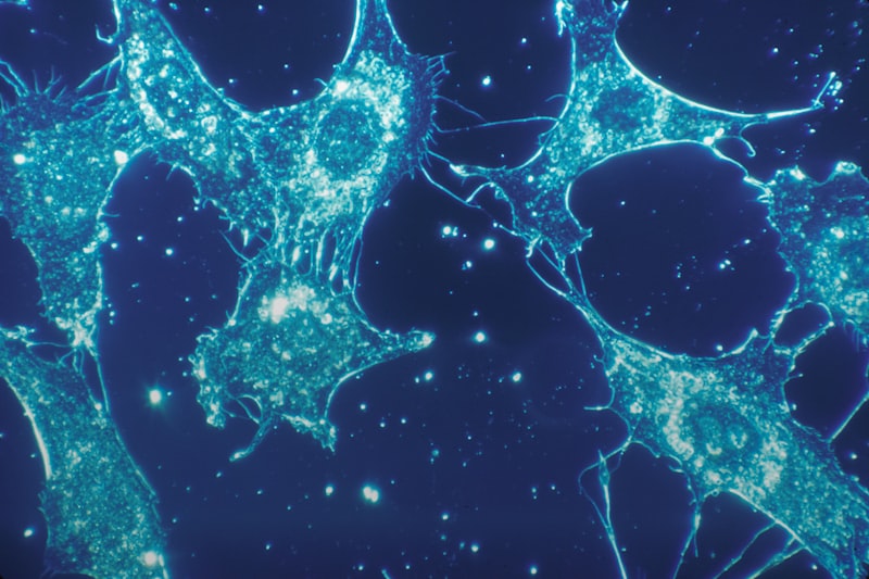
The molecular mechanism underpinning autocrine function is highly conserved across various cell types and involves a precise sequence of events. The process begins with the intracellular synthesis of the signaling molecule—which could be a peptide, a small lipid, or a monoamine—often stored in vesicles near the cell membrane. Upon receiving an appropriate stimulus (e.g., electrical activity, chemical input, or mechanical stress), the cell executes exocytosis, releasing the molecule into the surrounding milieu. This release is the critical first step that defines the subsequent autocrine action. Once released, the molecule must navigate the immediate extracellular space to find the appropriate receptor on the surface of the originating cell.
Receptors are the key transducers of the autocrine signal. These are specialized protein structures embedded within the cell membrane (or sometimes located intracellularly) that possess high affinity for the specific signaling molecule. The binding of the ligand to the Receptor initiates a cascade of intracellular events, known as signal transduction. This cascade often involves the activation of secondary messengers (like cAMP, calcium ions, or specific kinases) that ultimately alter the cell’s behavior. For instance, the signal might lead to changes in gene expression, modification of ion channel permeability, or alteration of the cell’s metabolic rate. The diversity of receptors ensures that a single type of secreted molecule can elicit a wide array of responses, depending on the specific receptor subtypes present on the cell surface.
A particularly fascinating aspect of autocrine signaling involves the concept of positive and negative feedback loops. In a positive autocrine loop, the secreted factor stimulates the cell to produce more of that same factor, leading to amplification of the initial signal. This is frequently observed in developmental biology and, unfortunately, in many forms of cancer where uncontrolled proliferation is driven by self-sustaining growth factor loops. Conversely, negative autocrine loops, which are more common in Homeostasis and mature neural systems, involve the secreted factor inhibiting its own synthesis or release, acting as a brake on excessive activity. This inhibitory mechanism is vital for maintaining physiological stability and preventing cellular exhaustion or damage caused by overstimulation.
Historical Discovery and Context
While the functional concept of self-regulating cellular output has likely existed since the evolution of multicellularity, the formal identification and naming of Autocrine signaling as a distinct mode of communication occurred relatively late in the history of cell biology. The term gained prominence primarily in the 1970s and 1980s, driven largely by advancements in endocrinology and oncology research. Early investigations into how cancer cells manage to achieve uncontrolled, sustained growth hinted at mechanisms where these cells were providing their own necessary growth signals, effectively becoming independent of external regulatory control. Researchers observed that many tumor cell lines secreted growth factors that also bound to receptors on their own surface, thus fueling their own proliferation in a self-perpetuating manner.
Key researchers focused heavily on peptide growth factors and their receptors, particularly in the context of fibroblast and epithelial cell lines. The discovery that cells could produce, secrete, and respond to the same signaling molecule revolutionized the understanding of cellular autonomy and tissue maintenance. Simultaneously, research into the immune system provided strong evidence for autocrine function. For instance, T-lymphocytes, when activated, secrete Cytokines, such as Interleukin-2 (IL-2), which act back on the same T-cells to promote clonal expansion and sustain the immune response. These findings established autocrine communication not merely as a pathological curiosity but as a fundamental mechanism essential for normal physiological responses, especially those requiring rapid amplification.
The integration of autocrine concepts into neuroscience came later, but proved equally crucial. Early studies on presynaptic regulation of Neurotransmitter release revealed that the releasing neuron often possessed autoreceptors that sensed the concentration of the released chemical. This mechanism, though sometimes categorized under the broader umbrella of paracrine signaling due to the involvement of the synaptic cleft, is fundamentally autocrine because the signal is initiated by the cell and acts back upon the machinery of that same cell. This historical progression—from oncology to immunology and finally to neuroscience—demonstrates how this core biological principle underlies critical regulatory processes across diverse physiological systems.
Autocrine Function in the Central Nervous System
In the realm of psychology and neuroscience, autocrine signaling plays a vital, albeit often subtle, role in regulating the minute-to-minute operations of the brain. Neurons, the primary signaling units, utilize autocrine loops to control their own excitability and the efficiency of synaptic transmission. For example, many neurons possess autoreceptors located on the presynaptic terminal, which are designed to detect the concentration of the neurotransmitter that the neuron has just released. If the concentration in the synaptic cleft becomes excessively high, the autoreceptor is activated, triggering an internal signaling cascade that typically inhibits further release of the neurotransmitter. This negative feedback mechanism is essential for preventing depletion of synaptic vesicles and managing the temporal precision of neural communication.
Beyond traditional neurotransmission, autocrine mechanisms are deeply involved in neuroplasticity and neural development. During the critical periods of brain development, growth factors and neurotrophins secreted by developing neurons often act in an autocrine manner to promote their own survival, guide axonal pathfinding, and regulate the formation of new synapses. This self-sustaining support system ensures that neurons facing high environmental demands or developmental stress can maintain viability. Furthermore, the ability of neurons to self-regulate the expression of certain genes via autocrine loops allows for long-term adaptation to changes in activity, providing a mechanism for experience-dependent remodeling of neural circuits, which is the biological basis for learning and memory.
Crucially, glial cells—including astrocytes, oligodendrocytes, and microglia—also rely heavily on autocrine communication. Microglia, the resident immune cells of the CNS, are particularly reliant on autocrine loops involving Cytokines and chemokines to respond to injury or inflammation. When activated by a pathogen or tissue damage, a microglial cell will secrete pro-inflammatory cytokines, some of which bind back to the microglial cell itself, promoting its sustained activation and migration to the injury site. While necessary for clearing debris and initiating repair, dysregulated autocrine loops in microglia are implicated in chronic neuroinflammation, a contributing factor to various neurodegenerative disorders, demonstrating the fine line between beneficial self-regulation and pathological self-perpetuation.
Practical Example: Neural Homeostasis
To illustrate the immediate and functional importance of autocrine signaling, consider the practical example of maintaining stable electrical activity, or neural Homeostasis, within a specific circuit in the hippocampus, a brain region critical for memory formation. Imagine a principal pyramidal neuron that is receiving a high volume of excitatory input, perhaps during an intense learning episode or a stressful event. This intense stimulation causes the neuron to fire rapidly and release a large quantity of its primary Neurotransmitter, Glutamate, into the synapse.
The application of the autocrine principle follows a clear, step-by-step regulatory sequence.
- High Activity and Release: The prolonged, intense firing of the presynaptic pyramidal neuron leads to the massive release of Glutamate.
- Autoreceptor Engagement: In addition to binding to receptors on the postsynaptic neuron, a portion of the released Glutamate diffuses back and engages specific autoreceptors (often inhibitory metabotropic receptors) located on the presynaptic terminal of the releasing neuron.
- Internal Signal Transduction: The binding of Glutamate to these autoreceptors initiates a negative feedback signal within the presynaptic terminal. This internal signal typically involves the activation of G-proteins which ultimately reduce the influx of calcium ions necessary for vesicle fusion and neurotransmitter release.
- Modulation of Future Release: The resulting drop in calcium influx immediately reduces the probability that subsequent action potentials will trigger the release of more Glutamate. The neuron effectively applies its own brake.
- Achievement of Homeostasis: By dampening its own output, the neuron prevents potentially damaging excitotoxicity (overstimulation) while conserving its metabolic resources and maintaining the fidelity of the circuit, ensuring the system remains in a state of controlled Homeostasis.
This example demonstrates a crucial feature: the autocrine loop provides an immediate, localized, and highly adaptive mechanism for preserving the integrity of individual cellular function against excessive environmental demands. Without this self-regulatory capacity, neural circuits would be far more susceptible to fatigue, damage, and instability, severely compromising psychological functions dependent on reliable signal processing.
Significance and Impact
The significance of Autocrine signaling to the broader field of psychology and medicine cannot be overstated, primarily because of its fundamental role in both normal development and pathological states. In developmental psychology, autocrine mechanisms are essential for establishing the precise circuitry of the brain. The self-support provided by neurotrophins ensures that only properly connected and healthy neurons survive the pruning process, shaping the final structure of the cortex and subcortical regions that govern complex behavior, emotion, and cognition. Disruptions in these early autocrine loops can lead to developmental disorders with profound psychological and behavioral consequences.
In clinical application, the pathological impact of dysregulated autocrine loops is most dramatically evident in oncology, where the self-sustaining growth loops are the target of many modern therapeutic strategies. However, its relevance extends deeply into mental health and neurological illness. For instance, chronic stress and associated mood disorders are often linked to altered immune profiles and chronic, low-grade inflammation. The autocrine signaling of Cytokines among immune cells and glial cells contributes significantly to the maintenance of this inflammatory state, which can directly affect neural function, synaptic integrity, and ultimately, mood regulation and stress resilience. Targeting these self-perpetuating inflammatory cycles is an emerging strategy in treating depression resistant to standard pharmacotherapy.
Furthermore, understanding autocrine regulation is critical in pharmacodynamics. Many psychotropic medications, particularly those affecting monoamine Neurotransmitters (like serotonin or dopamine), exert their effects partly by interacting with autoreceptors. For example, some antidepressant mechanisms involve modifying the sensitivity or availability of presynaptic autoreceptors to enhance the overall release of the neurotransmitter. Thus, the autocrine mechanism is not just a biological curiosity; it is a fundamental leverage point for pharmacological intervention aimed at restoring mental balance. The field’s impact lies in its ability to explain how individual cells maintain their identity and function, and how a breakdown in this self-control leads to systemic illness.
Connections to Other Signaling Modalities
Autocrine signaling is one of four primary communication modalities used by cells, and its unique function is best understood when contrasted with its related counterparts: paracrine, endocrine, and juxtacrine signaling. The key differentiating factor is the distance the signaling molecule must travel and the identity of the target cell. In paracrine signaling, the secreted factor targets neighboring cells within the immediate vicinity, influencing local tissue function (e.g., wound healing). In endocrine signaling, the factor (a hormone) enters the bloodstream and travels long distances to affect remote target organs, providing systemic control (e.g., cortisol release). Autocrine signaling, however, closes the loop, with the cell acting solely upon itself, representing the shortest distance and most self-focused communication path.
Despite these clear structural distinctions, there is significant functional overlap. A single signaling molecule, such as a prostaglandin or a specific growth factor, may simultaneously function in all three modes. For instance, a neuron might release a neuropeptide that binds to its own autoreceptors (autocrine), diffuses across the synapse to affect an adjacent neuron (paracrine), and, if released in sufficient quantities into the cerebrospinal fluid, might travel to a distant neural population (acting like a local endocrine signal). This ability for molecules to multitask allows for immense flexibility and complexity in tissue organization and response.
The concept of autocrine signaling is broadly situated within the subfield of Cellular and Molecular Psychology, a branch of neuroscience that seeks to explain psychological phenomena through the lens of cellular events and chemical communication. It also has strong ties to Neuroimmunology, particularly in understanding how the brain communicates with and responds to the immune system, often mediated by self-regulating cytokine loops in glial cells. The study of autocrine loops provides the molecular foundation for understanding higher-level concepts like neural plasticity, addiction mechanisms, and stress response, all of which rely on the dynamic, self-adjusting capacity of individual cells to maintain functional stability.
Therapeutic and Research Implications
The profound understanding of autocrine loops has opened critical avenues for therapeutic intervention, particularly in diseases where cellular self-perpetuation is a dominant feature. In cancer research, the goal is often to interrupt the positive autocrine feedback loops that drive uncontrolled cell division. This can involve developing drugs that specifically block the growth factor Receptors on the cancer cell surface, thus starving the cell of its self-generated growth signal and forcing it back into a regulated state, or triggering apoptosis. These targeted therapies represent a significant advance over broad-spectrum chemotherapy, focusing precisely on the cellular pathology.
In neuroscience and psychiatry, future research is increasingly focused on modulating autocrine mechanisms to restore healthy neural function. For example, researchers are investigating whether boosting the function of inhibitory autoreceptors could be a viable strategy for calming hyperactive neural circuits implicated in anxiety disorders or epilepsy. By enhancing the natural “brake” on Neurotransmitter release, clinicians could achieve more nuanced control over neural activity than is possible with current generalized receptor agonists or antagonists. Similarly, manipulating autocrine cytokine pathways in microglia offers a potential route for mitigating chronic neuroinflammation that underlies many persistent mood and cognitive disorders.
Finally, autocrine signaling provides a powerful model for understanding adaptive resilience. By studying how cells successfully maintain Homeostasis under stress, researchers can glean insights into developing treatments that enhance natural cellular protective mechanisms. The ability of a cell to initiate a self-repair or self-protective cascade, mediated by its own released factors, suggests that therapeutic strategies might move beyond simply replacing deficient substances to empowering the cell itself to manage its environment. This shift represents a move toward personalized cellular self-management strategies in medicine and psychology.