Axon Hillock: The Gatekeeper of Neural Communication
- Introduction and Definition of the Axon Hillock
- Anatomical Location and Structural Specialization
- The Role of Membrane Potential Integration
- The Concept of Threshold Potential
- Voltage-Gated Channels and Action Potential Initiation
- Functional Importance in Neural Communication
- Clinical Relevance and Pathophysiology
- Summary of Key Features
Introduction and Definition of the Axon Hillock
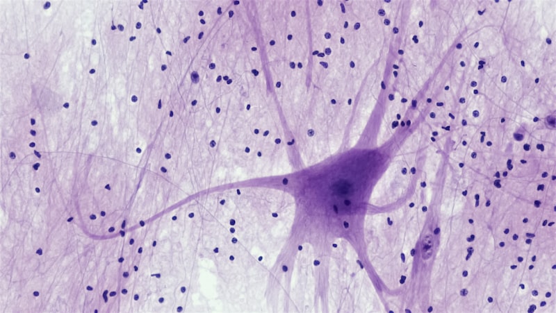
The axon hillock represents a fundamental anatomical and physiological structure within the intricate network of the nervous system. Serving as the critical junction between the neuronal cell body, or soma, and the filamentous extension known as the axon, this specialized region dictates whether an electrical signal will be successfully transmitted to subsequent neurons, muscles, or glands. Its primary defining characteristic is its unique shape: a distinct, cone-shaped protrusion emerging directly from the soma, marking the exact point where the integration of thousands of incoming synaptic signals culminates in a decisive electrical event. This location is not merely a physical nexus; it is the computational center responsible for determining the output of the entire neuron, effectively acting as the neuron’s decision-making gatekeeper regarding the initiation of an action potential, often referred to as a nerve impulse. The integrity and precise function of the axon hillock are absolutely essential for coherent neural communication and the swift processing of information throughout the central and peripheral nervous systems.
Historically, the axon hillock was primarily viewed through a morphological lens, identified simply as the tapering boundary of the soma. However, modern neurophysiology has revealed that this area possesses a dense concentration of specialized components that distinguish it functionally from the rest of the neuronal membrane. Unlike the dendritic tree, which primarily receives signals, or the soma, which houses the metabolic machinery, the axon hillock is dedicated almost exclusively to signal processing and initiation. This initiation process requires that the cumulative electrical input—derived from excitatory and inhibitory postsynaptic potentials (EPSPs and IPSPs)—reaches a specific, critical level of depolarization. Failure to achieve this threshold means the signal rapidly decays, whereas successful attainment triggers a cascade of events that propagate the signal faithfully down the axon, ensuring long-distance electrical fidelity.
Understanding the axon hillock necessitates appreciating its role as the final summation point. All incoming electrical changes, regardless of their origin on the dendrites or soma, converge here. This constant integration process is complex, involving temporal summation (signals arriving in rapid succession) and spatial summation (signals arriving at different points simultaneously). The consequence of this integration is a fluctuating membrane potential at the hillock. It is the ability of this region to precisely measure and respond to these fluctuations that underscores its immense physiological importance. Therefore, the simple definition of a cone-shaped origin belies its complex role as the master control switch for neuronal firing, profoundly influencing the patterns and rhythms of neural activity necessary for perception, cognition, and motor control.
Anatomical Location and Structural Specialization
The anatomical positioning of the axon hillock is strategically optimized for its functional role as the integration center. It is situated immediately adjacent to the soma, forming a seamless transition zone that differentiates the signal-receiving apparatus from the signal-transmitting pathway. While the cell body contains the nucleus and major organelles responsible for protein synthesis and cellular maintenance, the axon hillock itself is notable for what it lacks and what it possesses in abundance. It typically lacks the rough endoplasmic reticulum (Nissl bodies) that characterizes the rest of the soma, indicating a shift away from protein production and toward electrical machinery.
Structural analysis reveals that the membrane of the axon hillock, particularly the segment immediately following it known as the initial segment (AIS), is highly specialized. This membrane segment harbors the highest density of specific ion channels found anywhere in the neuron. These channels are anchored in place by a dense, underlying cytoskeletal matrix composed primarily of specialized proteins like ankyrin G and spectrin. This structural scaffolding is crucial because it ensures that the voltage-gated ion channels—which are the machinery necessary for action potential generation—are precisely positioned and maintained in the appropriate location to execute the rapid depolarization and repolarization cycle required for signal firing. The unique cytoskeletal framework helps define the geometrical constraints and electrical properties of this critical zone.
The morphology of the axon hillock is crucial for directing the flow of current. Its tapering shape concentrates the electrical current generated by the summed postsynaptic potentials. Because the membrane capacitance and resistance change dramatically between the soma and the axon, this funneling structure helps ensure that the current density is sufficiently high when it reaches the initial segment. The initial segment, which often includes the very distal portion of the hillock, is typically the site where the action potential physically originates due to its exceptionally high density of voltage-gated sodium channels (Nav channels), often Type 1.6 and 1.2 isoforms, which possess distinct kinetic properties optimized for low-threshold activation. This precise structural arrangement highlights evolutionary optimization for electrical efficiency.
The Role of Membrane Potential Integration
The primary function of the axon hillock is the integration of diverse synaptic inputs. A typical neuron in the central nervous system may receive thousands of synaptic contacts, some of which are excitatory (tending to depolarize the membrane) and others inhibitory (tending to hyperpolarize or stabilize the membrane). The electrical changes induced by these inputs—the EPSPs and IPSPs—travel across the dendritic tree and the soma, gradually decaying in magnitude as they move toward the hillock. The hillock must continuously weigh the strength and timing of these competing signals to decide the appropriate output.
This integration occurs through two main mechanisms: spatial summation and temporal summation. Spatial summation involves the simultaneous arrival of signals from different synaptic locations. For instance, multiple weak excitatory signals arriving concurrently at various points on the dendrites might individually be insufficient to cause firing, but their combined effect at the axon hillock can push the membrane potential past the critical threshold. Conversely, temporal summation occurs when a single synapse fires repeatedly and rapidly. If the subsequent inputs arrive before the preceding postsynaptic potential has completely decayed, the effects stack up over time, allowing the potential to rise incrementally until the firing threshold is met. The axon hillock is the physical site where both forms of summation are mathematically and electrically computed in real-time.
The result of this complex integration process is the continuous fluctuation of the membrane potential at the axon hillock. The membrane potential is a dynamic measure of the electrical difference across the cell membrane, typically resting around -70 mV. If the balance of inputs favors excitation, the potential becomes more positive (depolarization). If inhibition dominates, the potential becomes more negative (hyperpolarization). The integration mechanism ensures that only inputs that are sufficiently strong, synchronized, and sustained—overcoming the constant inhibitory influence—will succeed in driving the membrane potential high enough to initiate the rapid, all-or-nothing event known as the action potential. This intricate balance underscores the subtlety required for sophisticated neural processing.
The Concept of Threshold Potential
Central to the function of the axon hillock is the concept of the threshold potential. This is the specific membrane voltage level that, when reached, irrevocably triggers the opening of the voltage-gated sodium channels necessary for action potential generation. For most mammalian neurons, this threshold potential typically lies between -55 mV and -40 mV, significantly higher (less negative) than the resting membrane potential of approximately -70 mV. The establishment of this fixed threshold is essential for maintaining the fidelity and integrity of neural signaling; it acts as a noise filter, preventing small, random electrical fluctuations from resulting in unwanted or spurious firing.
The critical nature of the threshold potential stems from the biophysical properties of the voltage-gated sodium channels concentrated in the initial segment. These channels are exquisitely sensitive to changes in voltage. Once the membrane potential depolarizes to the threshold level, these channels undergo a rapid conformational change, snapping open instantaneously. This sudden influx of positively charged sodium ions (Na+) causes a massive, self-reinforcing depolarization—the rising phase of the action potential—that quickly drives the membrane potential far past zero, toward the equilibrium potential for sodium (around +30 mV to +50 mV). Because the density of these channels is highest at the hillock and initial segment, this region has the lowest activation threshold compared to the rest of the soma or dendrites.
If the depolarization reaching the axon hillock falls even marginally short of the threshold, the voltage-gated channels remain largely closed, and the incoming excitatory current dissipates harmlessly. This adherence to the threshold potential embodies the all-or-nothing principle of action potential generation: either the threshold is met, resulting in a full-sized action potential, or it is not met, resulting in no action potential whatsoever. The magnitude of the input signal, once above threshold, does not increase the size or speed of the action potential; rather, it increases the frequency (rate) at which subsequent action potentials are generated. Thus, the threshold potential is the binary switch that converts graded synaptic potentials into the stereotyped, propagating digital signal of the nervous system.
Voltage-Gated Channels and Action Potential Initiation
The unique physiological properties of the axon hillock and the initial segment are dictated by the specific complement of voltage-gated ion channels embedded in their membranes. The initiation of the action potential is fundamentally reliant on the high concentration and precise distribution of voltage-gated sodium channels (Nav) and voltage-gated potassium channels (Kv), which together orchestrate the rapid depolarization, peak, and repolarization phases of the impulse.
The initiation process begins with the Nav channels. As discussed, once the hillock reaches threshold, these channels activate extremely quickly, allowing a massive surge of sodium ions into the cell. This rapid influx constitutes the depolarization phase, serving as a powerful positive feedback loop: depolarization opens more Nav channels, leading to more sodium influx, causing further depolarization. This rapid reversal of membrane polarity ensures the signal is strong enough to travel long distances without decrement. Following their brief activation, the Nav channels rapidly inactivate, effectively stopping the influx of sodium and setting the stage for repolarization, which prevents the immediate re-firing of the signal and ensures unidirectional propagation.
The repolarization phase is primarily managed by the delayed activation of the Kv channels. These potassium channels open slightly slower than the sodium channels, peaking their activity just as the sodium channels are inactivating. The outward flow of positively charged potassium ions (K+) rapidly restores the negative potential inside the cell. In some neurons, this outward current briefly overshoots the resting potential, leading to a period of hyperpolarization (the refractory period), which further stabilizes the neuron and controls the maximum firing rate. The precise kinetic interplay between the rapid Nav channel activation and the slower Kv channel activation is what defines the characteristic shape and speed of the neuronal action potential originating at the axon hillock.
Furthermore, the axon hillock also contains specialized types of potassium channels, such as M-channels, which are crucial for regulating the excitability of the neuron over longer timescales. By modulating the resting potential and the ease with which the threshold is reached, these channels help determine the intrinsic firing patterns—such as bursting or tonic firing—that characterize different types of neurons. The entire complement of channels works synergistically, ensuring that the integration decision made at the hillock is not only accurate but also appropriately tailored to the specific functional requirements of that particular neuron type.
Functional Importance in Neural Communication
The axon hillock is arguably the single most important functional component governing the output of a neuron. Its role as the integration site transforms the analog, graded inputs received across the soma and dendrites into the digital, all-or-nothing output signal transmitted along the axon. This conversion from graded potential to action potential is critical for long-distance communication because graded potentials decay quickly over distance, making them unsuitable for transmitting information across the extensive tracts of the nervous system. The action potential, once initiated at the hillock, regenerates itself at periodic intervals along the axon (at the Nodes of Ranvier in myelinated axons), ensuring signal integrity.
This functional specialization allows for sophisticated information processing. For example, the phenomenon of adaptation, where a neuron fires rapidly upon stimulus onset but then slows its firing rate despite continuous stimulation, is often regulated by channels concentrated at the hillock that modify the threshold potential over time. Similarly, the ability of certain neurons to fire in distinct rhythmic bursts—essential for generating patterns like breathing or walking—is heavily dependent on the intrinsic excitability properties established by the channel configuration at the axon hillock and initial segment. The hillock thus determines not only if a neuron fires, but how often and in what pattern.
Moreover, the axon hillock is crucial for establishing the directionality of neural signaling. Because the concentration of Nav channels is highest and the threshold is lowest at this point, the action potential preferentially initiates here and propagates unidirectionally down the axon, away from the soma. While the impulse can theoretically propagate backward into the soma (back-propagation), the structural and electrical characteristics of the hillock ensure that the primary, output signal moves efficiently toward the synaptic terminals. This directional control is fundamental to maintaining the organized, sequential flow of information required for complex cognitive and motor functions.
Clinical Relevance and Pathophysiology
Given its central role in initiating all neuronal output, the axon hillock is a vulnerable target in various neurological and psychiatric disorders. Any pathological process that compromises the structural integrity of the initial segment or alters the function or distribution of its key ion channels can severely impair neural communication, leading to widespread system dysfunction. Conditions affecting the myelin sheath, such as Multiple Sclerosis (MS), often have downstream effects on the excitability of the axon hillock, forcing the neuron to rely on exposed membrane segments, which may lack the necessary channel density to fire effectively.
Specific channelopathies—diseases caused by defects in ion channel function—frequently manifest through altered axon hillock excitability. For instance, mutations affecting the precise isoforms of voltage-gated sodium channels found in the initial segment have been linked to various forms of epilepsy. If the channels become hypersensitive, the threshold potential drops too low, leading to neuronal hyperexcitability and uncontrolled, synchronous firing characteristic of seizures. Conversely, if the channels are hypoactive, transmission may fail entirely, contributing to symptoms seen in some motor neuron diseases or peripheral neuropathies. The study of these localized channel defects provides critical insight into the mechanisms underlying these complex disorders.
Furthermore, the physical structure of the axon hillock and initial segment is sensitive to chronic stress, trauma, and neurodegenerative processes. In conditions like Alzheimer’s disease or traumatic brain injury, the structural proteins that anchor the critical ion channels (like ankyrin G) can become damaged or mislocalized. This dislocation disrupts the precise high-density arrangement necessary for low-threshold firing, increasing the effort required for the neuron to reach threshold. Research is increasingly focusing on the axon hillock as a potential therapeutic target, aiming to restore proper channel function or structural organization to mitigate the debilitating effects of various central nervous system pathologies, thereby highlighting its immense clinical importance beyond basic physiology.
Summary of Key Features
The functionality of the axon hillock can be summarized by a set of defining characteristics that underscore its indispensable role in neurophysiology. It serves as the obligatory bridge between the input structure (dendrites/soma) and the output structure (axon), ensuring that only meaningful, sufficiently strong signals are translated into propagating action potentials. This specialized region is the final checkpoint where all electrical activity converges and is critically evaluated before transmission.
- Integration Center: It sums all incoming excitatory and inhibitory postsynaptic potentials (EPSPs and IPSPs) via spatial and temporal summation.
- Threshold Determination: It possesses the lowest firing threshold in the neuron, typically due to the high concentration of voltage-gated sodium channels in the adjacent initial segment.
- All-or-Nothing Decision: It enforces the binary decision rule for action potential generation, acting as the neuron’s principal output switch.
- Structural Specialization: Its membrane lacks Nissl bodies but is richly endowed with cytoskeletal anchor proteins (like ankyrin G) that maintain the precise location of necessary ion channels.
- Signal Conversion: It transforms graded, analog electrical signals into uniform, digital action potentials suitable for long-distance, high-fidelity transmission.
In conclusion, the axon hillock is far more than a simple anatomical transition point; it is the physiological epicenter of neuronal computation. Its ability to flawlessly integrate complex inputs and instantaneously determine the firing status of the neuron is foundational to the operational capacity of the entire nervous system, making it a pivotal area of study in neuroscience and related clinical fields.