Electrocorticogram: Brain Mapping at the Source
- The Core Definition of Electrocorticogram (ECoG)
- Historical Development and Key Pioneers
- Mechanism of Action: Data Acquisition and Processing
- ECoG vs. Other Neurophysiological Techniques
- Practical Applications and Real-World Scenarios
- Clinical Significance and Therapeutic Impact
- Connections to Related Neuroscientific Concepts
- Advantages and Limitations of the Technique
The Core Definition of Electrocorticogram (ECoG)
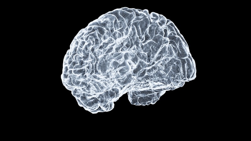
The Electrocorticogram (ECoG) is a highly specialized neurophysiological technique used to record the electrical activity generated by the brain, specifically by placing electrodes directly onto the surface of the cerebral cortex. Unlike the more commonly known Electroencephalogram (EEG), where electrodes are situated on the scalp, ECoG requires an intracranial procedure, meaning the electrodes are placed surgically beneath the skull, resting either directly on the pial surface or beneath the dura mater. This direct contact provides a recording resolution far superior to non-invasive methods, capturing neural signals with high fidelity, particularly those originating from localized cortical columns or specific functional areas of interest.
The fundamental mechanism behind ECoG relies on capturing the summed postsynaptic potentials generated by large populations of cortical neurons. Because the physical distance between the recording electrode and the active neuronal tissue is minimized—often mere millimeters—the resulting signal experiences significantly less attenuation and distortion compared to signals filtered through the skull, cerebrospinal fluid, and scalp tissues. This proximity allows ECoG to detect high-frequency oscillations, such as gamma band activity (typically 30–150 Hz), which are often obscured in standard EEG recordings but are crucial indicators of localized cognitive processing, motor planning, and pathological states like seizure onset.
The application of ECoG is almost exclusively limited to clinical environments, particularly in the context of neurosurgical planning and evaluation. It serves as an invaluable tool for precise localization, providing neurosurgeons and neurologists with a real-time, high-resolution map of cortical function and dysfunction. The resulting data provides detailed insights into both the normal functioning of the brain regions—such as language and motor control areas—and the precise origins of abnormal electrical discharges associated with neurological disorders, requiring a deep understanding of neuroanatomy and electrophysiology for accurate interpretation.
Historical Development and Key Pioneers
The development of ECoG is intrinsically linked to the advancement of neurosurgery in the early to mid-20th century, particularly efforts focused on treating intractable epilepsy. While the concept of recording brain electricity dates back to Richard Caton in the late 19th century, the technique of applying electrodes directly to the exposed human cortex was pioneered primarily by Dr. Wilder Penfield and his colleague Dr. Herbert Jasper at the Montreal Neurological Institute starting in the 1930s. Their groundbreaking work centered on mapping the functions of the human brain during awake surgery, utilizing both electrical stimulation and direct electrical recording to understand the relationship between specific cortical areas and various sensory, motor, and cognitive functions.
Penfield and Jasper’s methodology was driven by the clinical necessity of identifying and safely removing the epileptogenic focus—the exact region of the brain where seizures originate—without damaging adjacent areas critical for speech, movement, or memory. By performing ECoG recordings intraoperatively, they could visualize the abnormal electrical spikes and rhythmic discharges characteristic of seizure activity, thereby guiding the surgical resection with unparalleled precision. This historical context cemented ECoG’s role as a critical diagnostic technique, moving beyond mere research into a standard procedure for managing severe neurological conditions that were otherwise untreatable by medication alone.
Following their initial successes, the technique evolved significantly. Early methods involved placing temporary electrodes during the surgical procedure itself (intraoperative ECoG). Later advancements involved the development of subdural electrode grids and strips, which could be left in place for several days or weeks post-craniotomy while the patient recovered in the hospital (extraoperative ECoG). This allowed for continuous, long-term monitoring of spontaneous seizure activity, significantly improving the chances of accurately localizing the epileptogenic zone and defining functional boundaries, thereby optimizing the outcomes of subsequent curative surgeries.
Mechanism of Action: Data Acquisition and Processing
The mechanism of ECoG data acquisition begins with the surgical placement of the electrode arrays. These arrays typically consist of flexible silicone sheets embedded with multiple platinum or stainless steel contacts, often arranged in grids (two-dimensional arrays) or strips (linear arrays). The choice of array type and placement is highly individualized, determined by pre-surgical imaging (MRI, PET) and EEG data, specifically targeting the suspected area of pathology. These electrodes are placed directly on the dura or the surface of the cortex, and leads are tunneled through the skin and connected to external recording equipment, allowing the patient mobility within a specialized monitoring unit.
The recording equipment amplifies and filters the minute electrical potentials generated by the brain, which are typically in the microvolt range. A major characteristic differentiating ECoG from scalp EEG is its ability to capture higher frequency components of brain activity, notably the aforementioned high-gamma band (70–150 Hz). These high-gamma signals are often correlated with local neural computation and neuronal firing rates, suggesting they are a robust marker for active cortical processing. Analyzing these distinct frequency bands requires sophisticated signal processing techniques, including Fourier transforms and wavelet analyses, to decompose the raw signal into its constituent frequencies for clinical and research interpretation.
Furthermore, ECoG provides exceptional spatial resolution, often distinguishing activity across electrodes spaced as little as 5–10 millimeters apart. This precision allows researchers to perform detailed functional mapping, identifying subtle differences in electrical activity when a patient performs specific tasks, such as speaking or moving a limb. This high-resolution mapping is critical not just for identifying pathological tissue but also for demarcating the boundaries of eloquent cortex—areas vital for function—that must be preserved during surgical resection, minimizing the risk of post-operative neurological deficit.
ECoG vs. Other Neurophysiological Techniques
ECoG stands apart from other common neurophysiological techniques primarily due to its invasive nature, which simultaneously represents its greatest limitation and its most significant advantage. Compared to the standard EEG, ECoG offers significantly enhanced spatial localization and temporal resolution. EEG signals must pass through multiple layers (meninges, skull, scalp), resulting in a blurred or attenuated signal, particularly for high-frequency activity. Conversely, ECoG bypasses these impediments, providing a clean, direct signal that accurately reflects underlying cortical activity, making it vastly superior for pinpointing the exact origin of pathological discharges.
When compared to Magnetoencephalography (MEG), which measures the magnetic fields generated by neural currents, ECoG retains the advantage of direct contact. While MEG offers excellent temporal resolution and is non-invasive, its spatial resolution is limited, particularly deep within sulci (the grooves of the cortex), and it is highly susceptible to external magnetic noise. ECoG provides a more direct measure of the electrical voltage changes at the cortical surface, unaffected by external magnetic interference and offering a closer proximity to the neural source, which is indispensable for detailed functional mapping within the surgical context.
The primary trade-off, however, lies in the invasiveness. Techniques like EEG and MEG are safe, repeatable, and easily scalable for widespread use. ECoG, being a surgical procedure, carries inherent risks including infection, hemorrhage, and the general complications associated with craniotomy. Therefore, ECoG is reserved exclusively for patients for whom non-invasive techniques have failed to provide the necessary clarity, typically those with medication-resistant epilepsy or those undergoing focused tumor removal where precise functional mapping is essential to preserve neurological integrity.
Practical Applications and Real-World Scenarios
The most prominent real-world application of ECoG is in the pre-surgical evaluation and mapping of patients suffering from intractable focal epilepsy. Consider a patient whose seizures cannot be controlled by medication and whose pre-operative imaging (MRI) suggests a potentially resectable lesion. Traditional scalp EEG might indicate the general hemisphere or lobe where the seizure originates, but it lacks the precision needed for a surgeon to confidently remove the tissue. ECoG provides this crucial localization by allowing continuous monitoring over several days.
- Step 1: Surgical Implantation. The patient undergoes a craniotomy where subdural grid electrodes are meticulously placed over the suspected seizure onset zone and adjacent functionally critical areas (like the primary motor or language cortex).
- Step 2: Continuous Monitoring. Over the next week, the patient is monitored in an Epilepsy Monitoring Unit. ECoG continuously records the brain’s electrical activity, waiting for spontaneous seizures to occur. This allows neurologists to precisely identify the “seizure onset zone” by observing where the pathological, high-amplitude discharges first emerge.
- Step 3: Functional Mapping. While the electrodes are in place, the medical team performs cortical stimulation mapping. A small, low-intensity electrical current is applied sequentially through the ECoG electrodes. If stimulation of a specific electrode causes the patient to temporarily cease speaking or twitch a finger, that area is identified as eloquent cortex (language or motor area, respectively) and must be avoided during resection.
- Step 4: Surgical Planning and Resection. Combining the data from seizure onset localization (pathological tissue) and functional mapping (eloquent cortex), the neurosurgeon develops a precise plan to resect the epileptogenic tissue while sparing all essential functional areas, maximizing the chance of seizure freedom while minimizing neurological deficits.
This step-by-step process illustrates how ECoG transforms imprecise, low-resolution data into an actionable, high-precision surgical map. Without the high spatial and temporal resolution afforded by ECoG, many complex epilepsy surgeries would be too risky to attempt, potentially resulting in severe, life-altering deficits.
Clinical Significance and Therapeutic Impact
The clinical significance of ECoG extends far beyond its diagnostic capabilities; it fundamentally impacts therapeutic outcomes in complex neurological cases. For patients with refractory epilepsy, ECoG offers the best chance for curative treatment. By precisely delimiting the pathological tissue, surgeons can perform highly targeted resections, leading to seizure freedom in a significant proportion of carefully selected patients who would otherwise face a lifetime of debilitating seizures and heavy medication regimens.
In oncology, ECoG plays a vital role in removing brain tumors located near critical functional areas. When a tumor infiltrates or presses against the motor or language cortex, the surgeon must operate within very tight margins. Intraoperative ECoG, often combined with stimulation mapping, ensures that healthy, functional tissue is not inadvertently removed along with the cancerous mass. This meticulous mapping preserves the patient’s quality of life by safeguarding essential cognitive and motor capabilities, demonstrating ECoG’s powerful impact on both survival and functional prognosis.
Furthermore, ECoG is becoming increasingly significant in the planning stages for the placement of Deep Brain Stimulation (DBS) electrodes. While typically used for movement disorders like Parkinson’s disease, ECoG can assist in refining the target selection for DBS in certain forms of epilepsy or psychiatric disorders, providing real-time feedback on the electrical environment surrounding the target area before permanent implantation. This high-resolution data allows clinicians to optimize the placement and programming of neurostimulators, enhancing the therapeutic effect while minimizing side effects.
Connections to Related Neuroscientific Concepts
ECoG is deeply interconnected with several fundamental neuroscientific concepts, forming a bridge between basic research and clinical application. One major connection is its central role in the advancement of Brain-Computer Interfaces (BCI). Because ECoG provides a stable, high-bandwidth signal directly from the cortex, it has emerged as one of the most promising modalities for invasive BCI research. Researchers leverage ECoG signals, particularly the high-gamma activity associated with motor planning, to decode intended movements, allowing paralyzed individuals to control robotic prosthetics, cursors, or communication devices with high accuracy and speed.
Another key relationship exists with the concept of functional connectivity. By simultaneously recording activity from numerous discrete points on the cortex, ECoG data allows scientists to analyze how different brain regions communicate with each other during rest or during specific tasks. Analyzing coherence and phase-locking between widely separated electrodes helps map functional networks, revealing how connectivity might be altered in disease states like schizophrenia or Alzheimer’s disease, thus contributing profoundly to cognitive neuroscience.
ECoG also relates closely to the study of event-related potentials (ERPs). While ERPs are typically measured non-invasively using EEG, ECoG allows for the measurement of “cortical” ERPs with exceptional clarity. These signals are time-locked to sensory, motor, or cognitive events, and ECoG’s resolution enables the identification of specific cortical layers or microcircuits responsible for generating these potentials, offering crucial insights into the precise timing and localization of cognitive processing stages, such as attention, decision-making, and memory encoding.
Advantages and Limitations of the Technique
The primary advantage of ECoG is its unparalleled combination of spatial and temporal resolution compared to non-invasive methods. The direct placement on the cerebral cortex eliminates the blurring effect of the skull and scalp, enabling the detection of subtle, high-frequency oscillations that are critical for understanding both normal function and highly localized pathology. This high-fidelity data is crucial for precise clinical decision-making, especially in complex neurosurgical cases where margins of error are minute. Furthermore, the ability to perform long-term extraoperative monitoring provides a window into spontaneous brain events, such as rare seizures, that might be missed during a short, acute recording session.
Despite these powerful advantages, ECoG is severely limited by its invasive nature. The requirement for a craniotomy means the procedure carries all the risks associated with major brain surgery, including infection, hemorrhage, and general anesthesia complications. This inherently restricts its use to individuals with severe, refractory conditions. Moreover, the recording window is temporary; typically, electrodes remain implanted only for the duration of the hospital stay (often one to three weeks), meaning researchers and clinicians cannot track neural activity long-term outside of the immediate clinical context.
Another limitation pertains to the coverage area. ECoG grids and strips can only cover a localized region of the brain, typically the area suspected to be involved in the pathology. Therefore, ECoG provides a high-resolution view of a small area, but it cannot offer a comprehensive, whole-brain perspective simultaneously, unlike techniques such as functional MRI (fMRI) or generalized EEG. The interpretation of ECoG data also requires highly specialized expertise in both neurosurgery and clinical neurophysiology, making it a resource-intensive procedure available only at specialized medical centers.