Psychosomatic Heart: The Mind-Body Connection to Inflammation
Definition and Etiology of Endocarditis
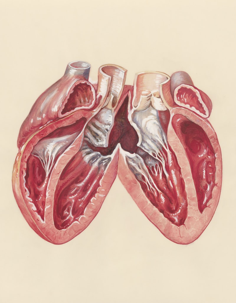
Endocarditis is defined as a serious inflammatory condition affecting the endocardium, which constitutes the thin, delicate inner lining of the heart chambers and the surfaces of the cardiac valves. This crucial anatomical structure, when compromised, initiates a complex pathological process that can severely impair cardiac function. Historically, endocarditis was often rapidly fatal, but modern medical interventions have significantly improved outcomes, though it remains a condition associated with high morbidity and mortality. The essence of the disease lies in the formation of vegetations—amalgamations of fibrin, platelets, inflammatory cells, and the causative microorganisms—primarily on the heart valves. These vegetations disrupt normal laminar blood flow and can lead to valvular destruction or systemic embolization, demanding prompt diagnosis and aggressive therapeutic management.
The primary cause of endocarditis is almost universally an infectious agent, typically either bacteria or fungi, which gain access to the bloodstream, a phenomenon known as bacteremia or fungemia. Once these pathogens circulate, they tend to adhere preferentially to sites of pre-existing endothelial damage or turbulent blood flow, most commonly involving the mitral and aortic valves, although the tricuspid valve is frequently affected in cases related to intravenous drug use. The causative microorganisms vary widely, but Staphylococcus aureus and Streptococcus species are the most frequent culprits in acute and subacute presentations, respectively. Fungal endocarditis, though less common, is particularly aggressive and often affects immunocompromised individuals or those with prosthetic valves, requiring intensive and prolonged antifungal therapy often combined with surgical debridement due to the dense nature of fungal vegetations.
The transition from transient bacteremia to established endocarditis involves several complex steps. Initially, damage to the endocardium creates a nidus for platelet and fibrin deposition, forming a sterile thrombus, known as non-bacterial thrombotic endocarditis (NBTE). When circulating microbes encounter this NBTE lesion, they adhere, proliferate, and become embedded within the protective matrix of the vegetation. This embedding shields them effectively from host immune defenses and makes antibiotic penetration challenging, necessitating prolonged, high-dose intravenous antimicrobial regimens. Understanding this etiology is crucial, as the severity of the illness often correlates directly with the virulence of the invading organism and the extent of subsequent valvular damage, which dictates the urgency of surgical consultation.
Anatomy and Pathophysiology
The endocardium is critical for maintaining the structural integrity and functional efficiency of the heart. It provides a smooth, non-thrombogenic surface that minimizes friction and prevents clot formation under normal physiological conditions. In endocarditis, the inflammation and infectious process primarily targets the cardiac valves, which are crucial for directional blood flow. The turbulent flow created by regurgitation or stenosis due to damaged valves further exacerbates the inflammatory cycle, leading to rapid destruction of valvular leaflets or cusps. This destruction can result in acute valvular insufficiency (e.g., severe aortic regurgitation), which constitutes a hemodynamic emergency requiring urgent surgical intervention to prevent acute decompensated heart failure and cardiogenic shock.
Pathophysiologically, the formation of infectious vegetations is the hallmark of the disease. These masses are dynamic entities that grow over time and are inherently friable. Their growth poses twin dangers: first, they mechanically impede valve function, leading directly to heart failure; and second, fragments of the vegetation can break off and enter the systemic circulation, causing embolic events. Embolization is a major source of morbidity, potentially leading to stroke (cerebral emboli), pulmonary embolism (right-sided endocarditis), splenic infarcts, or renal complications. The location of the infection dictates the pattern of embolization; left-sided vegetations (mitral and aortic) cause systemic emboli, while right-sided vegetations (tricuspid and pulmonic) usually cause septic pulmonary emboli, often resulting in pulmonary infarcts or lung abscesses.
Furthermore, the persistent presence of infection activates a systemic inflammatory response, often leading to the formation of circulating immune complexes that deposit in various distant organs, a phenomenon termed immunologically mediated damage. A classic example is the development of glomerulonephritis, vasculitis, or the characteristic peripheral signs of endocarditis such as Osler’s nodes. The systemic symptoms, including fever, malaise, and elevated acute-phase reactants, are manifestations of this immune response. The relentless nature of the infection, coupled with continuous hemodynamic stress from damaged valves, often culminates in congestive heart failure, which remains the most common cause of death in patients diagnosed with active infective endocarditis.
Classification and Distinct Clinical Types
Endocarditis is traditionally classified based on the clinical tempo of the illness and the state of the affected valve. The duration and severity of symptoms allow for categorization into acute endocarditis and subacute endocarditis. Acute endocarditis, often caused by highly virulent organisms like Staphylococcus aureus, develops rapidly over days, presenting with high spiking fever, profound toxicity, and swift valvular destruction, demanding immediate and aggressive treatment. Conversely, subacute endocarditis, frequently linked to less virulent organisms such as Viridans streptococci, progresses insidiously over weeks or months, characterized by low-grade fever, fatigue, and nonspecific symptoms, often leading to significant diagnostic delay until major complications arise.
A more functional classification differentiates between native valve endocarditis (NVE) and prosthetic valve endocarditis (PVE). NVE occurs on previously healthy or damaged native cardiac valves. PVE is a particularly challenging variant that occurs on artificial valves (mechanical or bioprosthetic) and carries a higher risk of complications like ring abscesses and valve dehiscence. PVE is further subdivided into early PVE (occurring within 60 days of surgery) and late PVE (occurring after 60 days). Early PVE is usually associated with perioperative contamination and often involves highly resistant, hospital-acquired organisms, such as coagulase-negative staphylococci or fungi, carrying a very high mortality rate. Late PVE often mimics NVE and is generally caused by community-acquired pathogens, often streptococci.
A third important classification considers the patient population, specifically distinguishing between non-intravenous drug user (IVDU) endocarditis and IVDU-associated endocarditis. The latter group, due to repeated skin piercing and non-sterile injection practices, typically develops infection in the right side of the heart, predominantly the tricuspid valve. This presentation often involves highly resistant Staphylococci and carries a high risk of recurrent infection if drug use continues. In contrast, non-IVDU endocarditis typically affects the left-sided valves (aortic and mitral). Recognizing these distinct clinical phenotypes is essential for guiding empirical antimicrobial selection, anticipating common complications, and determining the urgency of surgical intervention.
Clinical Presentation and Systemic Manifestations
The clinical presentation of endocarditis is notoriously variable, leading to its description as a “disease of a thousand faces.” The most common initial symptom is a persistent or intermittent fever, frequently accompanied by chills, night sweats, and profound malaise. However, many of the classical signs are related to the systemic sequelae of embolization and immune complex deposition. Classic peripheral signs, while less common today due to earlier diagnosis, include petechiae (small, non-blanching spots), splinter hemorrhages under the fingernails, and pathognomonic findings such as Osler’s nodes and Janeway lesions, all signaling microvascular compromise.
Osler’s nodes are painful, tender, reddish-purple nodules typically found on the pads of the fingers and toes, resulting from localized immune complex deposition and small vessel vasculitis. In contrast, Janeway lesions are non-tender, erythematous macules found on the palms and soles, which are highly indicative of septic microemboli. Ophthalmic examination may reveal Roth spots—retinal hemorrhages with pale centers—another manifestation of immune vasculitis. Crucially, the presence of a new or changing heart murmur is a highly suggestive finding, indicating acute valvular damage or insufficiency, and demands immediate investigation, as this finding signifies structural cardiac compromise.
The most devastating clinical manifestations are usually neurological. Approximately 20% to 40% of patients experience neurological complications, ranging from subtle changes in cognition to large embolic strokes, transient ischemic attacks, development of mycotic aneurysms (infected arterial wall dilatations), or intracranial hemorrhage. The sudden onset of focal neurological deficits in a febrile patient should immediately raise suspicion for endocarditis, necessitating urgent brain imaging. Other major systemic complications include septic arthritis, splenic abscesses, and acute kidney injury due to immune complex glomerulonephritis or renal artery embolization, illustrating the pervasive, multi-organ nature of this systemic infection.
Diagnostic Procedures and Criteria
Diagnosis of endocarditis requires integrating clinical findings, microbiological evidence, and imaging results. The cornerstone of diagnosis relies on the modified Duke Criteria, a standardized set of guidelines that categorizes findings into major and minor criteria to establish a definite, possible, or rejected diagnosis. The major criteria typically include positive blood cultures demonstrating typical organisms consistent with endocarditis and objective evidence of endocardial involvement, usually confirmed by echocardiography, which visualizes the structural damage caused by the vegetations.
Echocardiography, specifically transthoracic echocardiography (TTE) and transesophageal echocardiography (TEE), is the primary imaging modality. TTE is non-invasive and provides a good initial assessment, especially for large vegetations or significant valvular dysfunction, and is typically the first test performed. However, TEE offers superior resolution, particularly for visualizing small vegetations (less than 5 mm), identifying abscesses, confirming prosthetic valve involvement, and evaluating the extent of perivalvular extension into surrounding cardiac tissue. Definitive echocardiographic evidence includes the visualization of an oscillating intracardiac mass (vegetation) on a valve or supporting structure, the formation of an abscess, or new partial dehiscence of a prosthetic valve.
Microbiological confirmation requires obtaining at least three sets of blood cultures drawn from separate venipuncture sites over a specified period before initiating antibiotic therapy. Proper culturing is critical, as identification of the specific pathogen dictates targeted antimicrobial treatment, moving away from broad empirical coverage. In cases where routine cultures remain negative, specialized serological testing or polymerase chain reaction (PCR) assays may be necessary to identify fastidious organisms like Coxiella burnetii (Q fever), Bartonella species, or organisms previously suppressed by partial antibiotic treatment. Laboratory markers such as elevated C-reactive protein (CRP) and erythrocyte sedimentation rate (ESR) support the presence of systemic inflammation but are non-specific for definitive diagnosis.
Risk Factors and Vulnerable Populations
While endocarditis can technically affect anyone suffering from bacteremia, certain pre-existing cardiac conditions and behaviors significantly increase susceptibility. The presence of abnormal turbulent blood flow is the most potent risk factor for adherence of microbes. Patients with pre-existing valvular heart disease, such as bicuspid aortic valve, mitral valve prolapse with significant regurgitation, or degenerative valvular calcification, are highly vulnerable. Similarly, individuals who have undergone previous cardiac surgery, particularly those with prosthetic heart valves or implanted cardiac devices (e.g., pacemakers or cardiac defibrillators), face a lifetime risk of developing PVE due to the foreign material acting as a colonization site.
Another critical risk group encompasses individuals with conditions that frequently introduce bacteria into the bloodstream. This includes those undergoing frequent invasive medical or dental procedures, patients with central venous catheters (CVCs) or hemodialysis access lines, and, significantly, intravenous drug users (IVDUs). IVDU-associated endocarditis is characterized by unique microbiological profiles (often highly resistant Staphylococci) and anatomical location (right-sided valves), often complicating management due to challenges in adherence and high rates of recurrence. Poor dental hygiene is also a major, often overlooked, risk factor, as the transient bacteremia resulting from dental infections or procedures can seed the endocardium, particularly in those with underlying valvular defects.
Immunocompromised status further increases susceptibility to infection and often leads to more aggressive forms of endocarditis, frequently involving opportunistic fungal pathogens or unusual bacteria. Conditions such as HIV/AIDS, malignancy, chronic liver or renal failure, and those receiving immunosuppressive therapies (e.g., post-transplantation) fall into this category. The confluence of structural heart disease and systemic infection risk factors necessitates meticulous patient education and, in specific high-risk scenarios, mandates prophylactic antibiotic administration prior to certain high-risk invasive procedures to mitigate the risk of infectious seeding.
Treatment Modalities and Management Strategy
The management of endocarditis is complex and requires prompt initiation of treatment, typically involving a combination of prolonged, high-dose intravenous antibiotics and, frequently, cardiac surgery. The initial antibiotic regimen is empirical, based on the clinical setting (e.g., community-acquired vs. healthcare-associated, native vs. prosthetic valve), but must be adjusted immediately once blood culture results and antibiotic sensitivity profiles are available to ensure optimal efficacy against the specific pathogen. Treatment duration is extensive, typically ranging from four to six weeks, necessary to eradicate the pathogens deeply embedded within the fibrin-platelet matrix of the vegetations; failure to complete the full course significantly increases the risk of relapse and treatment failure.
Surgical intervention is required in approximately half of all endocarditis cases and is often life-saving, serving both to remove the infectious burden and to repair the resulting structural damage. Indications for surgery are based on preventing systemic complications and treating refractory heart failure. The primary indications include the presence of severe or refractory heart failure due to acute valvular insufficiency, uncontrolled infection despite optimal antibiotic therapy (e.g., persistent bacteremia or fungal endocarditis), large vegetations (typically greater than 10 mm) that carry a high risk of embolization, and the development of perivalvular complications such as abscess formation, pseudoaneurysms, or fistulas. The procedure involves debridement of infected tissue and vegetation removal, often necessitating valve repair or, more commonly, valve replacement with a prosthetic valve.
Perioperative management is crucial, requiring multidisciplinary input from cardiologists, infectious disease specialists, and cardiac surgeons. Following surgery, patients must continue their antimicrobial therapy to ensure that any remaining microscopic infectious foci are eradicated. Furthermore, careful monitoring for complications, particularly new embolic events, neurological deterioration, or cardiac conduction abnormalities secondary to abscess extension, is mandatory during the entire course of treatment. The decision to pursue surgical intervention is highly nuanced, balancing the significant risks of open-heart surgery in a critically ill patient against the almost certain morbidity and mortality associated with conservative management in the face of major complications.
Prevention and Prognosis
Prevention of endocarditis focuses primarily on reducing the incidence of bacteremia in high-risk individuals. This is achieved through meticulous attention to personal hygiene, especially oral and dental health, and careful management of indwelling catheters or central lines, ensuring minimal opportunity for bacterial ingress. The controversial practice of antibiotic prophylaxis before invasive procedures is now restricted to a very small, well-defined group of patients who face the highest risk of adverse outcomes from endocarditis. These high-risk patients include those with prosthetic cardiac valves or prosthetic material used for cardiac valve repair, those with a history of previous infective endocarditis, and patients with specific forms of unrepaired congenital heart disease.
Prophylaxis is specifically recommended only for these highest-risk patients undergoing certain dental procedures that involve manipulation of the gingival tissue or the periapical region of the teeth, where bacteremia is highly anticipated. Routine prophylaxis is generally not recommended for gastrointestinal or genitourinary procedures unless there is an active localized infection present at the site of manipulation. The emphasis shifted away from broad prophylactic use because the risk of endocarditis from routine daily activities (like chewing or brushing teeth) far outweighs the risk from most medical procedures, and the widespread overuse of antibiotics contributes significantly to the global challenge of antimicrobial resistance.
Despite advancements in diagnosis and treatment, the prognosis for infective endocarditis remains guarded, with in-hospital mortality rates typically ranging from 15% to 30%, depending on the cohort studied. Prognostic factors are heavily dependent on the causative microorganism (fungal and staphylococcal infections carry a poorer prognosis), the presence of underlying comorbidities, the development of heart failure, and whether urgent surgery is required. Long-term management involves diligent follow-up, ensuring adherence to preventative strategies, and monitoring for signs of recurrent infection, as survivors remain at an elevated lifetime risk for subsequent episodes. Early diagnosis, rapid initiation of appropriate, pathogen-specific antibiotics, and timely surgical intervention remain the most crucial determinants of a favorable clinical outcome and improved long-term survival.