Cellular Biology: The Architecture of Your Mind
- Introduction: Definition and Overview
- Structural Organization and Morphology
- The Rough Endoplasmic Reticulum (RER): Function in Protein Synthesis
- The Smooth Endoplasmic Reticulum (SER): Lipid Metabolism and Detoxification
- Role in Calcium Homeostasis
- Post-Translational Modification and Quality Control
- ER Stress and the Unfolded Protein Response (UPR)
- Clinical Relevance and Disease States
Introduction: Definition and Overview
The endoplasmic reticulum (ER) is a vast, highly dynamic network of interconnected membranous tubules, sacs, and flattened sheets, collectively known as cisternae, that permeates the cytoplasm of eukaryotic cells, extending continuously from the nuclear envelope to the plasma membrane vicinity. This complex system represents a significant proportion of the total membrane surface area within the cell, acting as a crucial central processing and manufacturing hub. Initially described by Keith Porter and colleagues in the mid-20th century using electron microscopy, the ER’s fundamental role is to manage the flow of materials destined for secretion, incorporation into cellular membranes, or delivery to other organelles, such as lysosomes and the Golgi apparatus. Its structural integrity and extensive reach allow it to coordinate numerous cellular functions, effectively compartmentalizing the cytosol and creating a distinct internal environment essential for protein folding, lipid biosynthesis, and calcium storage.
Functionally, the ER is indispensable for the life of the cell, serving as the primary site for the synthesis and modification of proteins and lipids that are destined for the secretory pathway or for insertion into cellular membranes. The original definition highlights its dual role: structural extension from the nucleus and functional processing of macromolecules. Proteins synthesized on ribosomes tethered to the ER surface—the Rough ER—are threaded into the ER lumen, where they undergo complex folding, glycosylation, and quality control checks before being transported further. Simultaneously, the specialized regions of the ER—the Smooth ER—are critical for the production of various lipids, including phospholipids and steroids, and play a vital role in the detoxification of metabolic byproducts and exogenous toxins. The integration of these diverse functions underscores the ER’s status as a master regulator of cellular physiology and membrane biogenesis.
The lumen, or the internal aqueous space enclosed by the ER membrane, is distinct from the surrounding cytosol and maintains a unique biochemical environment crucial for its operations. This environment is highly oxidative, facilitating disulfide bond formation in proteins, and contains a specialized suite of chaperone proteins, such as BiP (Binding Immunoglobulin Protein) and calnexin, that assist in proper protein folding. The ER membrane itself is a highly permeable barrier that selectively regulates the transport of molecules, especially calcium ions, maintaining steep concentration gradients necessary for signaling cascades. The close physical association of the ER with other organelles, particularly the mitochondria (forming Mitochondria-Associated Membranes, or MAMs) and the Golgi complex, ensures efficient communication and transfer of metabolic intermediates, highlighting its role not merely as an isolated factory but as an integrated component of the endomembrane system.
Structural Organization and Morphology
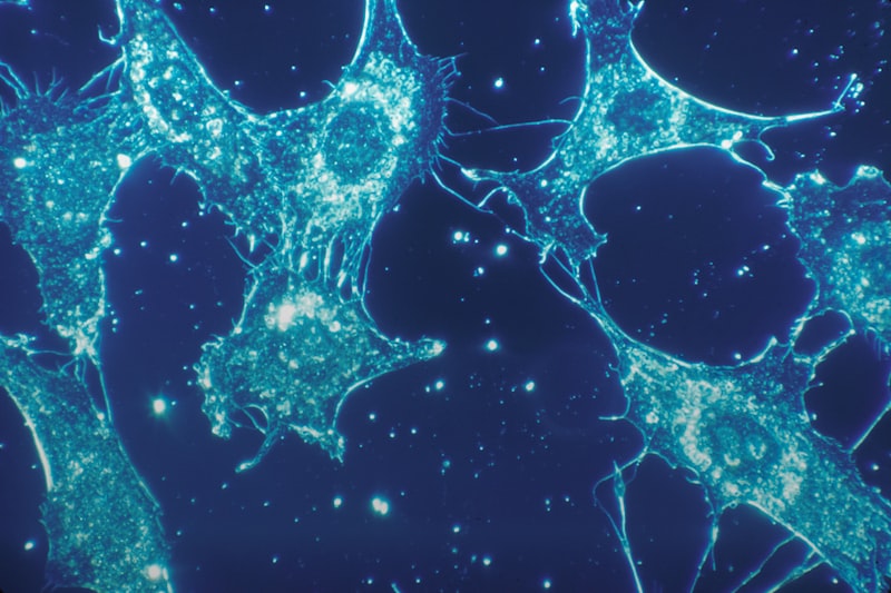
The morphology of the endoplasmic reticulum is characterized by a high degree of plasticity, allowing it to adapt its structure based on the specific metabolic demands of the cell. Structurally, the ER is classified into two distinct, yet continuous, domains: the Rough Endoplasmic Reticulum (RER) and the Smooth Endoplasmic Reticulum (SER). The RER is predominantly composed of flattened sacs, or cisternae, which are studded on their cytosolic surface with ribosomes, giving them their characteristic rough appearance under electron microscopy. These cisternae are often arranged in parallel stacks, particularly prominent in cells specialized for protein secretion, such as plasma cells or pancreatic acinar cells. The RER’s continuous association with the outer nuclear membrane ensures that the processes initiated at the nucleus, such as transcription and translation, are seamlessly linked to protein processing and modification within the ER lumen.
In contrast, the SER typically appears as a network of highly curved, branching tubules that lack associated ribosomes. The proportion and structural complexity of the SER vary dramatically across cell types; for example, hepatocytes (liver cells) and skeletal muscle cells (where it is referred to as the sarcoplasmic reticulum) possess extensive networks of SER due to their specialized roles in detoxification and calcium storage, respectively. The transition zone between the RER and SER is dynamic, often marked by the presence of transitional elements, which are crucial sites for the formation of transport vesicles that bud off the ER and travel to the Golgi apparatus. This structural division allows for functional specialization, wherein the RER focuses primarily on protein synthesis and translocation, while the SER concentrates on lipid metabolism and ion regulation.
The membrane of the ER, which defines the boundaries of the lumen, is critical for defining the function of the organelle. It houses numerous integral membrane proteins, including translocons (protein channels for translocation), protein-modifying enzymes, and specialized transporters. The thickness and lipid composition of the ER membrane differ subtly from those of the plasma membrane, reflecting its role as a biosynthetic hub rather than a signaling interface. Furthermore, the structural scaffolding required to maintain the complex geometry of the tubular SER network involves interactions with the cytoskeleton, ensuring that the ER is properly distributed throughout the cytoplasm and positioned adjacent to key sites of cellular activity, thereby facilitating rapid communication and substrate exchange.
The Rough Endoplasmic Reticulum (RER): Function in Protein Synthesis
The primary function of the Rough Endoplasmic Reticulum (RER) is the synthesis, folding, and initial modification of proteins destined for export from the cell, insertion into membranes, or delivery to other components of the endomembrane system. This critical process begins when a ribosome translating an mRNA molecule encounters a signal sequence on the nascent polypeptide chain. This signal sequence targets the ribosome and the growing chain to the RER membrane, where the ribosome docks onto a protein channel known as the translocon (Sec61 complex). The polypeptide chain is then either threaded through the translocon into the ER lumen, or, if it is destined to be a membrane protein, it is integrated laterally into the lipid bilayer.
Once inside the ER lumen, proteins immediately encounter a complex environment designed for quality control. They undergo significant post-translational modifications, the most prevalent of which is N-linked glycosylation, where a pre-formed oligosaccharide tree is attached to specific asparagine residues. This glycosylation serves multiple purposes, including protecting the protein from degradation, assisting in proper folding, and acting as a signal tag for the ER’s quality control system, often involving lectin chaperones like calnexin and calreticulin. These chaperones monitor the folding status of the newly synthesized protein, ensuring that it achieves its correct three-dimensional structure before being allowed to exit the ER. Defective or misfolded proteins are typically retained in the RER and eventually targeted for degradation via the ER-associated degradation (ERAD) pathway.
The RER’s commitment to meticulous quality control is paramount because many of the proteins processed here are critical for extracellular communication, enzymatic activity, and structural integrity. For proteins that are secretory, the RER ensures efficient disulfide bond formation, a reaction catalyzed by protein disulfide isomerase (PDI) within the oxidative environment of the lumen. Only correctly folded and assembled proteins are permitted to leave the RER, typically packaged into COPII-coated vesicles that bud from the transitional ER and proceed to the cis-face of the Golgi apparatus. This rigorous screening mechanism minimizes the release of potentially toxic or non-functional proteins into the cellular environment or circulatory system.
The Smooth Endoplasmic Reticulum (SER): Lipid Metabolism and Detoxification
The Smooth Endoplasmic Reticulum (SER) performs functions that are fundamentally distinct from those of the RER, centering primarily on lipid biosynthesis, detoxification, and carbohydrate metabolism. The SER is the main site within the cell for the synthesis of most membrane lipids, including phospholipids, cholesterol, and ceramides. Enzymes embedded in the SER membrane catalyze the synthesis of fatty acids and glycerol into phospholipids, which are then integrated into the ER membrane itself. Since phospholipids are synthesized exclusively on the cytosolic face of the ER membrane, specialized enzymes called scramblases and flippases are required to transfer these lipids to the luminal leaflet, ensuring symmetrical expansion of the bilayer necessary for membrane growth and vesicle formation.
A significant role of the SER, particularly in liver cells (hepatocytes), is the detoxification of both endogenous metabolic waste products and exogenous lipophilic drugs and toxins. This process is largely carried out by a family of enzymes known as the cytochrome P450 monooxygenases. These enzymes utilize molecular oxygen to catalyze hydroxylation reactions, which increase the solubility of lipid-soluble compounds, making them easier to excrete from the cell or body. Exposure to high levels of certain toxins or drugs, such as phenobarbital, can lead to a massive proliferation of the SER in hepatocytes, a classic example of cellular adaptation demonstrating the organelle’s functional plasticity in response to environmental demands.
Furthermore, the SER plays a crucial part in steroid hormone synthesis, particularly in endocrine cells of the adrenal cortex and gonads. These cells possess abundant SER networks that house the necessary enzymatic machinery to convert cholesterol into steroid hormones like testosterone and cortisol. Finally, in hepatocytes, the SER contains glucose-6-phosphatase, an enzyme vital for glucose homeostasis. This enzyme removes a phosphate group from glucose-6-phosphate, allowing the resulting free glucose to be transported out of the cell and into the bloodstream, a critical step in maintaining blood sugar levels during periods of fasting.
Role in Calcium Homeostasis
One of the most universally critical functions of the endoplasmic reticulum, especially the smooth domain, is its ability to act as the cell’s major intracellular storage depot for calcium ions (Ca2+). The concentration of free calcium within the ER lumen is maintained at levels significantly higher than that in the cytosol (often millimolar range in the ER versus nanomolar range in the cytosol). This steep concentration gradient is established and maintained by specialized calcium pumps, primarily the Sarco/Endoplasmic Reticulum Ca2+-ATPase (SERCA), which actively transports calcium from the cytosol into the ER lumen, utilizing ATP hydrolysis.
This high calcium concentration within the ER is not merely for storage; it is essential for the function of many ER-resident proteins, including chaperone proteins like calnexin and calreticulin, which are calcium-dependent. More importantly, the ER’s controlled release of stored calcium acts as a powerful and rapid intracellular signaling mechanism. When cells receive specific external signals, such as hormones or neurotransmitters, receptors on the plasma membrane activate phospholipase C, generating inositol 1,4,5-trisphosphate (IP3). IP3 then binds to IP3 receptor channels located on the ER membrane, triggering a rapid efflux of calcium into the cytosol.
The transient rise in cytosolic calcium concentration initiates numerous downstream cellular processes, including muscle contraction (where the SER is known as the sarcoplasmic reticulum, playing a specialized role in excitation-contraction coupling), neurotransmitter release, fertilization events, and gene expression regulation. The precise spatial and temporal control of calcium release and reuptake by the ER is thus fundamental to cellular communication and responsiveness. Dysfunction in ER calcium handling, such as leakiness or impaired SERCA activity, can lead to cellular pathology and is often implicated in conditions ranging from heart failure to neurodegenerative diseases.
Post-Translational Modification and Quality Control
The endoplasmic reticulum is the gatekeeper for the secretory pathway, employing rigorous mechanisms of post-translational modification and quality control (QC) to ensure that only correctly folded and assembled proteins proceed to the Golgi apparatus. The QC system relies heavily on a complex interplay between chaperones, modifying enzymes, and receptors that continuously monitor the folding status of nascent proteins. If a protein folds correctly within a prescribed timeframe, it is released from the QC cycle and enters the exit site of the ER. If folding is delayed or incorrect, the protein remains bound to chaperones for repeated attempts at achieving the native conformation.
A key modification monitored by the QC system is N-linked glycosylation. After the initial attachment of the oligosaccharide tree, the trimming and re-addition of glucose residues act as a folding sensor. Monoglucosylated proteins are recognized by the lectin chaperones (calnexin/calreticulin), which temporarily hold the protein. If the protein fails to fold properly after multiple cycles of glucose addition and removal, it is eventually recognized as terminally misfolded. This highly regulated process ensures that proteins with critical structural requirements, such as antibodies or cell surface receptors, achieve maximum functional integrity before deployment.
For proteins deemed irreversibly misfolded, the ER initiates the ER-Associated Degradation (ERAD) pathway. This process involves identifying the terminally misfolded protein, retrotranslocating it back across the ER membrane into the cytosol, and marking it for destruction. Once in the cytosol, the protein is ubiquitinated—tagged with ubiquitin molecules—and subsequently degraded by the 26S proteasome. ERAD is essential for maintaining proteostasis and preventing the accumulation of potentially toxic protein aggregates, thereby safeguarding cellular health and preventing the onset of numerous protein-misfolding disorders.
ER Stress and the Unfolded Protein Response (UPR)
When the demands placed on the ER exceed its capacity to correctly fold and process proteins—a condition known as ER stress—a critical adaptive signaling cascade called the Unfolded Protein Response (UPR) is activated. ER stress can be triggered by various factors, including nutrient deprivation, changes in calcium concentration, viral infection, mutations leading to misfolded proteins, or high secretory demand. The accumulation of unfolded or misfolded proteins in the ER lumen is the primary trigger for the UPR, which aims to restore ER homeostasis.
The UPR is mediated by three primary transmembrane sensor proteins embedded in the ER membrane: PERK (PKR-like ER kinase), IRE1 (Inositol-requiring enzyme 1), and ATF6 (Activating transcription factor 6). Under non-stress conditions, these sensors are kept inactive by binding to the chaperone BiP. When BiP dissociates to bind to accumulating unfolded proteins, the sensors become activated. PERK activation leads to the attenuation of global protein synthesis, reducing the load on the ER. IRE1 activation initiates the splicing of the mRNA encoding XBP1, producing a potent transcription factor that upregulates genes involved in folding, ERAD, and lipid biosynthesis. ATF6 is cleaved and migrates to the nucleus to induce the expression of other UPR target genes, including those encoding BiP and other chaperones.
The UPR is fundamentally an attempt to restore balance by expanding the ER folding capacity and degrading excess misfolded proteins. If the stress is transient, the UPR successfully resolves the problem. However, if the stress is severe or chronic, and the UPR fails to restore homeostasis, the signaling pathway shifts from adaptive protective mechanisms toward initiating programmed cell death, or apoptosis. This transition highlights the critical balance the cell maintains: attempting to survive stress, but ultimately sacrificing itself if the damage to the ER integrity is irreversible, preventing harm to the organism.
Clinical Relevance and Disease States
The central role of the endoplasmic reticulum in protein processing, quality control, and lipid metabolism means that ER dysfunction is implicated in a vast array of human diseases, often referred to as ER stress pathologies. Many neurodegenerative disorders, including Alzheimer’s disease, Parkinson’s disease, and amyotrophic lateral sclerosis (ALS), are linked to chronic ER stress resulting from the accumulation of misfolded proteins that overwhelm the ERAD pathway and induce sustained UPR activation, leading to neuronal apoptosis. Targeting the UPR signaling branches, particularly the PERK pathway, has become a major focus for therapeutic intervention in these conditions.
Furthermore, disruption of ER function is central to metabolic diseases. In Type 2 Diabetes Mellitus, chronic ER stress in pancreatic beta cells, often caused by high nutrient load and increased insulin demand, impairs insulin synthesis and secretion and ultimately leads to beta cell death. Similarly, ER dysfunction in the liver contributes to insulin resistance and non-alcoholic fatty liver disease (NAFLD) by disrupting lipid metabolism and signaling pathways. The ability of the ER to modulate inflammation and immune responses further links its integrity to autoimmune disorders and chronic inflammatory states.
Finally, specific genetic disorders directly involve components of the ER. Cystic fibrosis (CF), for instance, is often caused by a mutation (CFTR ΔF508) that leads to the misfolding of the CFTR chloride channel protein. Although the mutated protein might retain partial function, the ER quality control system recognizes it as misfolded and efficiently targets it for degradation via ERAD, preventing its insertion into the plasma membrane. This strict fidelity of the ER QC system, while normally protective, becomes pathological in CF. Understanding and potentially overriding the ERAD pathway in such cases represents a significant challenge and opportunity for developing novel pharmacological chaperones that can assist the mutated protein in escaping ER retention.