Epilepsy Surgery: Navigating the Path to Cognitive Relief
- Introduction and Core Definition
- Historical Context and Evolution of Techniques
- Indications for Surgical Intervention
- Primary Resective Surgical Techniques
- Neuromodulation and Advanced Methods
- Illustrative Practical Scenario
- Outcomes, Significance, and Quality of Life
- Risks, Complications, and Related Concepts
Introduction and Core Definition
Epilepsy is a chronic, non-communicable neurological disorder characterized by recurrent, unprovoked seizures, which are transient occurrences of signs and symptoms due to abnormal, excessive, or synchronous neuronal activity in the brain. While pharmacological treatments—specifically anti-epileptic drugs (AEDs)—are the primary and highly successful initial line of defense, a significant subset of patients, often estimated to be around one-third, will develop what is termed drug-resistant or refractory epilepsy. For these individuals, where medication fails to control seizure activity despite adequate trials of appropriate drugs, epilepsy surgery emerges as a potentially curative or significantly palliative treatment option. The core mechanism behind considering surgery rests on the principle that if the specific brain region responsible for initiating the seizures (the epileptogenic focus) can be accurately identified and safely removed or disrupted, the patient may achieve long-term seizure freedom or a substantial reduction in seizure frequency and severity.
The definition of epilepsy surgery is fundamentally an intervention aimed at addressing the neurological substrate of the disorder. It is not a uniform procedure but rather a collection of highly specialized neurosurgical techniques tailored to the unique topography and functional architecture of the patient’s brain. The goal extends beyond merely controlling the symptoms; it seeks to improve the patient’s overall quality of life, cognitive function, employment stability, and psychosocial well-being, which are often severely compromised by frequent, unpredictable seizures. Therefore, the decision to pursue surgical intervention is the culmination of a rigorous, multidisciplinary evaluation process typically conducted at specialized epilepsy centers, involving neurologists, neurosurgeons, neuropsychologists, and neuroradiologists, ensuring that the procedure is both necessary and highly likely to succeed without causing unacceptable functional deficits.
Historical Context and Evolution of Techniques
The concept of surgically treating epilepsy traces its origins back to ancient times, but modern epilepsy surgery began to take shape in the late 19th and early 20th centuries. Early pioneers, such as Sir Victor Horsley in London, performed some of the first systematic surgical resections for focal epilepsy, demonstrating that removal of scar tissue or visible lesions could sometimes halt seizure activity. However, the field was truly revolutionized by the work of neurosurgeons like Wilder Penfield and Herbert Jasper at the Montreal Neurological Institute starting in the 1930s. Their meticulous development of electrocorticography (ECoG), which involved mapping the brain’s electrical activity during surgery while the patient was awake under local anesthesia, allowed for the precise identification of the seizure focus while simultaneously mapping critical functional areas, such as language and motor control, thereby minimizing post-operative deficits.
This historical approach paved the way for the establishment of the modern standardized procedure for Temporal Lobe Epilepsy, known as the anterior temporal lobectomy, which remains the most common form of curative epilepsy surgery today. The work of Penfield and his colleagues provided the crucial framework for localizing the epileptic focus (the “where”) and understanding the necessary limits of resection (the “how much”). Following their foundational contributions, the mid-to-late 20th century saw refinement in imaging technology, particularly the advent of Magnetic Resonance Imaging (MRI), which dramatically improved the non-invasive identification of structural lesions that cause epilepsy, such as hippocampal sclerosis or focal cortical dysplasia. This shift from relying solely on invasive monitoring to integrating advanced imaging techniques marked a significant evolution in the pre-surgical evaluation process, greatly increasing the pool of suitable candidates for surgical intervention by making the localization of the epileptogenic zone far more accurate and efficient.
Indications for Surgical Intervention
The primary and most critical indication for considering epilepsy surgery is the diagnosis of medically refractory seizures, often defined by the International League Against Epilepsy (ILAE) as the failure of adequate trials of two tolerated and appropriately chosen anti-epileptic drug schedules, whether used as monotherapy or in combination, to achieve sustained seizure freedom. This resistance to pharmacological intervention signals that the underlying pathology is likely structural or localized in a manner that drugs cannot effectively modulate or suppress without causing severe systemic side effects. Beyond the sheer frequency of seizures, the decision is heavily influenced by the patient’s overall quality of life and safety profile. For instance, frequent falls or injuries directly attributable to convulsive seizure events provide a compelling argument for surgical intervention due to the immediate risk of severe physical harm, including fractures or head trauma.
Furthermore, indications extend into neuropsychological and social domains. Patients experiencing significant cognitive decline, memory loss, or pronounced emotional comorbidities, such as severe depression or anxiety linked directly to their seizure disorder, may benefit substantially from surgery, even if complete seizure freedom is not guaranteed. The persistent electrical discharges associated with uncontrolled epilepsy can cause progressive neuronal damage, a concept sometimes referred to as ‘kindling,’ which underscores the urgency of early intervention. The presence of a clearly defined epileptogenic lesion identified through advanced imaging strongly suggests a high probability of success with surgical resection, making these patients prime candidates. Ultimately, the indication for surgery is a meticulous risk-benefit calculation, weighing the low risks associated with the procedure against the profound long-term risks associated with uncontrolled epilepsy, including potential progressive neurological damage and the elevated mortality risk known as Sudden Unexpected Death in Epilepsy (SUDEP).
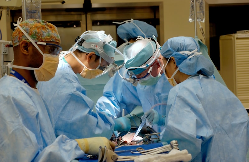
Primary Resective Surgical Techniques
The overarching goal of all curative epilepsy surgical techniques is the precise localization and targeted removal or disruption of the seizure-generating area, while meticulously preserving surrounding functionally critical brain tissue. Among the curative methods, Lesionectomy stands out as the most straightforward approach when a discrete structural abnormality, such as a cavernoma, vascular malformation, or low-grade tumor, is clearly visible on imaging and coincides exactly with the electrical focus of the seizures confirmed by EEG. This technique involves the selective removal of only the identified lesion, often resulting in excellent outcomes because the surrounding healthy brain tissue is left intact, minimizing the risk of widespread functional deficits.
When the epileptogenic focus is broader or involves tissue that appears structurally normal but is functionally abnormal (as is often the case in mesial temporal lobe epilepsy with hippocampal sclerosis), a more extensive procedure called Functional Cortical Resection is employed. The most common example of this is the anterior temporal lobectomy. This technique involves removing the cortical area deemed responsible for generating seizures, including adjacent structures like the amygdala and hippocampus. Crucially, this resection must be guided by pre-operative functional mapping (using fMRI or Wada testing) and often intra-operative monitoring to ensure that essential functions—like speech, comprehension, and motor control—are spared. This careful balance between maximal removal of pathological tissue and preservation of function dictates the success and safety of the procedure, demanding a high degree of neurosurgical skill and technical precision.
Other specialized resective or disconnective techniques include hemispherectomy or hemispherotomy (functional disconnection or removal of one cerebral hemisphere), typically reserved for severe, catastrophic epilepsies in children, such as Rasmussen’s encephalitis, where the entire hemisphere is pathologically involved. Another technique, multiple subpial transection (MST), is a palliative method used when the focus lies in functionally critical cortex that cannot be removed; it involves making shallow, parallel cuts in the cortex to interrupt the horizontal spread of seizure activity without damaging the vertical columns responsible for local function.
Neuromodulation and Advanced Methods
When the epileptogenic focus cannot be safely localized, is multifocal, or involves areas that are functionally critical and cannot be resected, non-ablative techniques known as Neuromodulation provide an alternative palliative solution. These techniques do not remove brain tissue but instead use electrical stimulation to interrupt or suppress abnormal seizure activity. The primary neuromodulatory devices currently utilized include Vagus Nerve Stimulation (VNS), which involves stimulating the vagus nerve in the neck to modulate brain activity; Responsive Neurostimulation (RNS); and Deep Brain Stimulation (DBS). DBS, specifically, involves the placement of electrodes deep within specific brain nuclei, such as the anterior nucleus of the thalamus, which are intricately connected to seizure generation and propagation circuits.
A significant advancement is the development of the Responsive Neurostimulation (RNS) system, which represents a major technological leap toward personalized, closed-loop treatment. RNS involves placing electrodes directly over or within the seizure focus, often requiring stereotactic placement with high precision. Unlike VNS or standard DBS, RNS continuously monitors the brain’s electrical activity and, upon detecting abnormal patterns indicative of an impending seizure, delivers targeted stimulation to abort the event before it clinically manifests or fully generalizes. These technologies are invaluable for patients deemed ineligible for traditional resective surgery because their seizure foci are diffuse or located in eloquent cortex. While generally associated with a lower rate of complete seizure freedom compared to resective surgery, neuromodulation techniques are highly effective at reducing seizure frequency (often 50% to 70% reduction) and severity, and are typically associated with fewer permanent cognitive side effects.
Illustrative Practical Scenario
Consider a patient, Mark, a 40-year-old accountant, who has suffered from focal seizures originating in his left temporal lobe since his early twenties. Despite trying three different combinations of high-dose anti-epileptic medications over a period of seven years, he continues to suffer from frequent complex partial seizures, typically occurring once every week. These episodes often begin with a sensation of fear and déjà vu (an aura) and progress to periods of non-responsiveness where he stares blankly and picks at his clothes, lasting several minutes. The unpredictability of these events has made him unable to drive and has severely curtailed his professional opportunities, leading to significant depression.
The application of epilepsy surgery principles in Mark’s case would follow a meticulous, multi-stage process. Step one: Pre-surgical evaluation. This involves high-resolution MRI showing clear evidence of hippocampal sclerosis in the left mesial temporal lobe. Video-EEG monitoring confirms that all his habitual seizures consistently originate from this localized area. Step two: Functional mapping and risk assessment. Since the left hemisphere is typically dominant for language, advanced testing, such as the intracarotid amobarbital test (Wada test) or functional MRI (fMRI), is performed to confirm the exact location of his language center and memory encoding areas. This assessment determines the acceptable extent of resection to avoid disabling aphasia or severe memory impairment. Step three: Surgical intervention. Mark undergoes an anterior temporal lobectomy. The “how-to” here involves the neurosurgeon meticulously removing the epileptogenic focus—the sclerotic hippocampus and associated cortex—guided by pre-operative imaging and intra-operative electrophysiological monitoring. Following successful surgery, Mark achieves complete seizure freedom and is eventually able to reduce his reliance on anti-epileptic drugs, demonstrating the transformation of a life previously dominated by chronic illness.
Outcomes, Significance, and Quality of Life
The significance of epilepsy surgery lies in its potential to offer a genuine cure or profound palliation for a condition that is otherwise debilitating and progressive. Outcomes data consistently demonstrate that surgical intervention provides superior long-term seizure control compared to continuing medical therapy indefinitely in patients with refractory epilepsy. Specifically, resective procedures like Lesionectomy and temporal lobectomy are associated with highly favorable seizure reduction rates, often resulting in complete seizure freedom for 60% to 90% of appropriately selected patients, depending on the underlying pathology and precision of localization. Even when complete freedom is not achieved, a significant reduction in seizure frequency (greater than 50%) is highly valuable, often being the difference between institutionalization and independent living.
Beyond seizure control, the true impact of epilepsy surgery is measured in the improvement of overall Quality of Life (QoL). Studies have repeatedly shown that successful surgery leads to substantial improvements across multiple domains: cognitive functioning often stabilizes or improves once continuous seizure activity and high doses of AEDs are reduced; psychological health improves dramatically, with reductions in rates of depression and anxiety, which are often severe comorbidities of chronic epilepsy; and social function is enhanced, evidenced by increased employment rates, better educational attainment, and greater independence. The ability to drive legally, hold a consistent job, and participate fully in social life signifies the profound clinical and societal importance of making epilepsy surgery available to suitable candidates. It is an intervention that changes the trajectory of a chronic disease, moving it from progressive disability toward stability and recovery, thereby justifying the intensive pre-surgical workup and the associated surgical risks.
Risks, Complications, and Related Concepts
While highly effective, epilepsy surgery is a major neurosurgical procedure associated with inherent risks, which must be thoroughly discussed during the consent process. The risk of serious immediate complications, such such as intracranial infection, hemorrhage, or stroke, is generally low (under 5%) but requires intensive post-operative monitoring. A more nuanced and common concern is the risk of postoperative cognitive decline, particularly involving memory and verbal fluency, especially following extensive resections in the dominant hemisphere. Patients must be carefully counseled regarding the trade-off: eliminating life-threatening seizures versus accepting a manageable, localized cognitive deficit. For instance, resection of the hippocampus, even in the non-dominant hemisphere, carries a risk of new-onset memory difficulties, though these must be weighed against the memory difficulties already caused by uncontrolled seizures. Additionally, patients who have undergone epilepsy surgery may experience transient neuropsychiatric disturbances, such as depression and anxiety, requiring psychological support and monitoring.
The interdisciplinary nature of surgical evaluation links this field directly to several related psychological and neurological concepts. The concept of Lateralization of Function is central, as the surgical approach depends heavily on identifying which hemisphere controls critical functions like language and executive function. Furthermore, the effectiveness of the procedure relies fundamentally on the accuracy of Seizure Focus Localization, connecting it to advanced neuroimaging (like PET, SPECT, and MEG) and invasive monitoring (like Stereo-EEG, or sEEG). This highlights the crucial role of the neuropsychologist in the pre-operative assessment (to establish baseline cognitive function) and post-operative rehabilitation.
Epilepsy surgery also relates closely to the broader field of Neuroplasticity, as the brain, especially in children, often exhibits remarkable capacity to reorganize function following removal of the epileptogenic zone. Finally, the development and application of Neuromodulation techniques connect this treatment modality directly to biomedical engineering and closed-loop control systems. These related concepts demonstrate that epilepsy surgery is not merely a surgical technique but a highly integrated discipline within clinical neuroscience, utilizing sophisticated diagnostic tools and drawing upon fundamental knowledge of brain function and pathology.