Auditory Perception: How Ear Health Shapes Your Mental State
- The Eustachian Tube: A Foundational Overview
- Anatomical Structure and Components
- Physiological Mechanisms of Function
- Historical Discovery and Early Understanding
- Everyday Manifestations and Psychological Implications
- The Broader Significance: Medical and Psychological Impact
- Related Concepts and Subfields of Psychology
The Eustachian Tube: A Foundational Overview
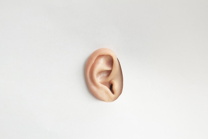
The Eustachian tube, also known as the auditory tube, represents a crucial anatomical structure within the human auditory system. It is a small, curved canal that establishes a vital connection between the middle ear cavity and the posterior region of the nasopharynx, which is the upper part of the throat behind the nose. This intricate passage is typically lined with specialized ciliated epithelium, a type of tissue characterized by hair-like projections that play an active role in its function. Fundamentally, the Eustachian tube serves as a dynamic pathway for air exchange, facilitating the movement of air between the sealed environment of the middle ear and the external atmosphere, thereby playing an indispensable role in maintaining optimal ear health and auditory perception.
The primary functions of the Eustachian tube are twofold, both critical for proper auditory function and preventing discomfort. Firstly, it is instrumental in equalizing air pressure between the middle ear and the surrounding environment. This pressure balance is essential for the eardrum to vibrate freely and efficiently, which is necessary for clear hearing. Without proper pressure equalization, the eardrum can become retracted or bulging, leading to muffled hearing, pain, or a sense of fullness. Secondly, the Eustachian tube is responsible for draining accumulated mucus and debris from the middle ear cavity into the nasopharynx. This mucociliary clearance mechanism helps to prevent the buildup of fluid, which could otherwise create a fertile breeding ground for bacteria and viruses, thereby precluding ear infections. Any dysfunction of this tube can precipitate a cascade of otologic issues, including but not limited to hearing loss, recurrent ear infections, and the distressing symptom of tinnitus, underscoring its profound importance in both physiological and psychological well-being.
Anatomical Structure and Components
The Eustachian tube is a relatively narrow conduit, measuring approximately 36 millimeters in length in adults, and is intricately structured to perform its specialized functions. It is generally divided into three distinct anatomical portions, each contributing to its overall integrity and mechanics. The most superior segment is the meatal portion, also referred to as the bony part, which originates directly from the anterior wall of the middle ear, situated just lateral to the tympanic membrane or eardrum. This initial section is rigid and relatively unchanging in its structure, providing a stable anchor for the entire tube. Following this, the tube transitions into the cartilaginous portion, which constitutes the middle and most substantial segment. This part is composed predominantly of hyaline cartilage, a flexible yet supportive tissue, which forms a significant part of the tube’s wall, allowing for its dynamic opening and closing.
The final and most inferior segment is the pharyngeal portion, which culminates in an opening into the lateral wall of the nasopharynx. This opening, known as the pharyngeal orifice, is particularly significant as it is the point where the tube interacts with the throat and where its ventilatory function is primarily regulated. The entire lumen of the Eustachian tube, particularly the cartilaginous and pharyngeal portions, is lined with pseudostratified ciliated columnar epithelium, interspersed with goblet cells. These ciliated cells work in a coordinated, wave-like motion, propelling mucus and any trapped particles from the middle ear cavity down towards the nasopharynx, where they can be swallowed or expelled. The dynamic nature of the cartilaginous portion, influenced by surrounding musculature, is pivotal for the intermittent opening and closing required for effective pressure regulation.
A notable anatomical difference exists between adults and children concerning the Eustachian tube. In pediatric populations, the tube is typically shorter, wider, and positioned more horizontally compared to its adult counterpart. This anatomical variance renders children particularly susceptible to Eustachian tube dysfunction, as the shorter length and more horizontal angle make it easier for pathogens from the nasopharynx to ascend into the middle ear, and for fluid to accumulate due to less efficient drainage. Understanding these developmental differences is critical for comprehending the higher incidence of ear infections, such as otitis media, in younger individuals and for devising appropriate diagnostic and therapeutic interventions.
Physiological Mechanisms of Function
The primary physiological role of the Eustachian tube revolves around the intricate process of pressure equalization. The middle ear is an air-filled cavity, and for optimal hearing, the air pressure within this cavity must closely match the atmospheric pressure outside. When there is a significant difference in pressure, the tympanic membrane’s ability to vibrate effectively is compromised, leading to muffled hearing, discomfort, and sometimes pain. The Eustachian tube acts as a pressure relief valve, intermittently opening to allow air to flow in or out of the middle ear. This opening mechanism is largely controlled by the coordinated action of specific muscles of the soft palate, primarily the tensor veli palatini and the levator veli palatini, which contract during activities such as swallowing, yawning, or chewing. These contractions exert traction on the cartilaginous portion of the tube, momentarily pulling its walls apart and creating a pathway for air exchange.
During a swallow or yawn, the muscular contraction creates a momentary negative pressure gradient within the nasopharynx relative to the middle ear, or physically pulls the tube open, facilitating the influx of air from the nasopharynx into the middle ear. This influx replenishes the air absorbed by the middle ear lining and helps to normalize the pressure. Conversely, if external pressure drops rapidly (e.g., during ascent in an airplane), the tube needs to open to release air from the middle ear. This dynamic process ensures that the pressure on both sides of the eardrum remains balanced, allowing it to vibrate optimally in response to sound waves. Without this crucial function, activities involving changes in ambient pressure, such as flying, diving, or even ascending in an elevator, would cause significant pain and temporary hearing impairment due to the inability to adapt to external pressure changes, a condition known as barotrauma.
Beyond pressure regulation, the Eustachian tube plays a vital role in the protective mechanism of mucus drainage from the middle ear. The ciliated epithelium lining the tube forms part of the mucociliary escalator system. These microscopic cilia beat rhythmically and unidirectionally, sweeping mucus, trapped pathogens, and cellular debris from the middle ear cavity down towards the nasopharynx. Once in the nasopharynx, this material can be swallowed and subsequently neutralized by stomach acid. This continuous cleansing action is paramount in preventing the accumulation of fluid and infectious agents within the middle ear, which would otherwise lead to recurrent infections and potential long-term damage to the auditory structures. A compromised mucociliary clearance due to inflammation, infection, or anatomical anomalies significantly increases the risk of developing conditions like chronic otitis media with effusion, highlighting the protective importance of the tube’s physiological integrity.
Historical Discovery and Early Understanding
The existence of the Eustachian tube has been recognized since antiquity, with early anatomical observations hinting at its presence. However, its most definitive and systematic description is attributed to the Italian anatomist Bartolomeo Eustachi (1510/1513–1574) during the 16th century. Eustachi, a pioneer in the field of anatomy, provided a detailed account of this structure in his seminal work, “Epistula de auditus organis” (Letter on the Organs of Hearing), published posthumously in 1707. His meticulous dissections and comprehensive anatomical studies in Rome allowed him to accurately depict the tube connecting the middle ear to the throat, a discovery that fundamentally advanced the understanding of ear anatomy. Prior to Eustachi’s work, descriptions were often vague or incomplete, making his contributions pivotal in establishing a concrete anatomical foundation for future physiological and pathological investigations.
Eustachi’s detailed anatomical illustrations and descriptions laid the groundwork for subsequent generations of scientists and physicians to explore the function of this newly identified structure. While Eustachi himself primarily focused on the anatomical description, the realization of the tube’s physiological importance, particularly in pressure regulation and aeration of the middle ear, evolved gradually over the following centuries. Early physicians began to correlate blockages or dysfunctions of this tube with various ear ailments, though the precise mechanisms were still poorly understood. The development of more sophisticated medical instruments and techniques, coupled with a deeper understanding of fluid dynamics and respiratory physiology, allowed for a more comprehensive appreciation of the Eustachian tube’s critical role in maintaining auditory health.
The historical understanding of the Eustachian tube has thus progressed from initial anatomical identification to a sophisticated grasp of its complex physiology and its profound implications for human health. This trajectory mirrors the broader development of otology, the medical specialty concerned with the ear. Eustachi’s discovery not only clarified a fundamental aspect of ear anatomy but also opened avenues for understanding the etiology of numerous ear disorders, thereby paving the way for diagnostic advancements and the eventual development of targeted medical and surgical interventions. His legacy underscores the enduring value of foundational anatomical research in propelling medical science forward and improving human well-being.
Everyday Manifestations and Psychological Implications
The function of the Eustachian tube is frequently experienced in everyday life, often without conscious thought, during common activities such as flying, diving, or even rapid changes in elevation. A classic practical example is the sensation of “ear popping” experienced during airplane ascent or descent. As an aircraft climbs, the ambient atmospheric pressure decreases, causing the air trapped in the middle ear to expand and exert outward pressure on the eardrum. Conversely, during descent, the external pressure increases, pushing the eardrum inward. To equalize these pressure differences and prevent discomfort or damage, the Eustachian tube must open, allowing air to either escape or enter the middle ear. This opening typically manifests as a subtle click or “pop” and provides immediate relief from any pressure sensation or muffled hearing. When the tube fails to open efficiently, perhaps due to inflammation from a cold or allergies, the resulting pressure imbalance can lead to significant ear pain, a sensation of fullness, and temporary muffled hearing, known as ear barotrauma.
Beyond these acute and transient experiences, chronic Eustachian tube dysfunction can have profound and far-reaching psychological implications. Persistent muffled hearing, a constant feeling of ear fullness, or recurrent bouts of ear pain can significantly impact an individual’s quality of life. For instance, chronic muffled hearing can impede effective communication, leading to feelings of frustration, isolation, and social withdrawal. Imagine a student struggling to hear their teacher clearly, or an adult finding it difficult to follow conversations in noisy environments; these scenarios can foster anxiety, reduce self-confidence, and even contribute to academic or professional underperformance. The constant effort required to process auditory information when hearing is compromised can also lead to increased cognitive load, resulting in mental fatigue and reduced concentration, thereby affecting daily functioning and overall well-being.
Furthermore, conditions associated with Eustachian tube dysfunction, such as chronic tinnitus (a persistent ringing or buzzing in the ears), can be particularly distressing. Tinnitus, while not always directly caused by Eustachian tube issues, can be exacerbated or triggered by the underlying pressure imbalances or inflammation. The incessant nature of tinnitus can interfere with sleep, concentration, and emotional regulation, often leading to heightened levels of stress, anxiety, and even clinical depression. The psychological burden of these symptoms is substantial, as individuals may struggle to cope with the perceived loss of control over their sensory experience and the continuous disruption to their peace of mind. Thus, what begins as a physiological issue can quickly cascade into a complex web of psychological challenges, underscoring the interconnectedness of physical health and mental well-being.
The Broader Significance: Medical and Psychological Impact
The significance of the Eustachian tube extends far beyond its anatomical definition, profoundly impacting both medical practice and psychological health. Medically, its dysfunction is a common underlying cause for a multitude of otologic disorders. Perhaps the most prevalent is otitis media, particularly in children. When the Eustachian tube is blocked or fails to open properly, fluid can accumulate in the middle ear, creating an ideal environment for bacterial or viral growth, leading to painful infections. Recurrent otitis media in early childhood can have serious long-term consequences, including persistent hearing loss that may impede speech and language development, leading to potential academic and social difficulties. Another significant medical impact is Eustachian tube dysfunction (ETD) itself, a condition characterized by persistent symptoms like ear fullness, popping, pain, or muffled hearing without active infection. This can be caused by allergies, viral infections, adenoid hypertrophy, or anatomical abnormalities. Treatment options range from conservative measures like decongestants and nasal steroids to surgical interventions such as tympanostomy tube insertion or balloon dilation of the Eustachian tube, highlighting the need for accurate diagnosis and management to prevent chronic issues.
The psychological impact of chronic Eustachian tube-related issues is substantial and often underestimated. For individuals experiencing persistent hearing loss, even mild to moderate, the psychological toll can be immense. Communication difficulties can lead to social isolation and withdrawal, fostering feelings of loneliness and exacerbating symptoms of anxiety and depression. The constant strain of trying to hear can also induce significant mental fatigue, reducing cognitive resources available for other tasks and impacting overall cognitive function. Children with chronic or recurrent hearing loss due to otitis media are at a particular risk for developmental delays, including speech and language impairments, which can in turn affect their literacy, academic performance, and social-emotional development. The frustration associated with misunderstood communication or the inability to fully participate in social activities can erode self-esteem and lead to behavioral problems, underscoring the critical need for early intervention and comprehensive management.
Furthermore, the presence of chronic symptoms like tinnitus, often a sequela of inner ear or Eustachian tube issues, significantly impacts psychological well-being. Tinnitus can be incredibly intrusive, interfering with sleep, concentration, and the ability to relax, leading to chronic stress, irritability, and heightened anxiety. The unpredictability and perceived lack of control over tinnitus can trigger strong emotional responses, sometimes leading to severe psychological distress. Patients often report feelings of helplessness and despair, and for some, it can be a debilitating condition that significantly impairs their quality of life. Thus, comprehensive care for Eustachian tube dysfunction and its associated conditions must extend beyond medical treatment to include psychological support, counseling, and strategies for coping with chronic symptoms, thereby addressing the holistic needs of the affected individual and mitigating the profound psychological burden.
Related Concepts and Subfields of Psychology
The understanding and management of the Eustachian tube’s function and dysfunction necessitate an interdisciplinary approach, connecting various medical specialties with several key concepts within psychology. Medically, the Eustachian tube is intrinsically linked to conditions such as adenoid hypertrophy, where enlarged adenoids can obstruct the pharyngeal opening of the tube, contributing to recurrent otitis media. It is also related to sinus infections and allergic rhinitis, as inflammation and congestion in the nasopharynx can similarly impair Eustachian tube function. Furthermore, conditions affecting the temporomandibular joint (TMJ) can sometimes mimic or exacerbate Eustachian tube symptoms due to their anatomical proximity and shared neurological pathways, making differential diagnosis crucial. The tube’s proper functioning is also vital in preventing complications during activities like scuba diving or high-altitude flying, where rapid pressure changes can induce barotrauma if the tube fails to equalize pressure effectively.
From a psychological perspective, the study of the Eustachian tube’s impact draws upon several subfields. Health Psychology is highly relevant, as it examines the interplay between psychological factors (like stress, coping mechanisms) and physical health outcomes. Chronic Eustachian tube dysfunction and its associated symptoms (pain, hearing loss, tinnitus) represent chronic health stressors that can significantly impact mental health, and conversely, psychological stress can sometimes exacerbate physical symptoms. Clinical Psychology plays a crucial role in diagnosing and treating the mental health consequences of these chronic ear conditions, such as anxiety disorders, depression, and adjustment disorders that may arise from living with persistent discomfort or hearing impairment. Therapeutic interventions might include cognitive-behavioral therapy (CBT) for tinnitus management or support groups for individuals coping with hearing loss.
Moreover, Developmental Psychology is critical for understanding the long-term effects of recurrent childhood otitis media on cognitive and socio-emotional development. Early childhood hearing loss, even if temporary, can disrupt the acquisition of speech and language, impact learning abilities, and potentially lead to social isolation. Researchers in this field investigate how these early experiences shape cognitive processes, academic achievement, and social competence. Cognitive Psychology also contributes by exploring how sensory deficits, such as muffled hearing, affect attention, memory, and information processing. The increased cognitive effort required to compensate for hearing loss can deplete cognitive resources, impacting overall mental performance. Lastly, Psychophysics, which studies the relationship between physical stimuli and their psychological perception, provides insights into how changes in middle ear pressure or auditory processing affect the subjective experience of sound and discomfort. In essence, the Eustachian tube, though an anatomical structure, serves as a fascinating nexus where physiological function profoundly influences psychological well-being, necessitating a truly holistic and interdisciplinary approach to its study and clinical management.