Psychomotor Flexion: The Mind-Body Connection in Motion
- Defining the Flexor Muscle and its Primary Action
- Microscopic Anatomy and Physiology of Flexion
- The Critical Relationship: Flexors and Extensors (Antagonism)
- Major Flexor Groups in the Human Body
- Neural Control and Motor Unit Activation
- Flexor Muscle Injuries, Conditioning, and Rehabilitation
- Evolutionary Perspective and Developmental Role
Defining the Flexor Muscle and its Primary Action
The term flexor muscle is used within anatomy and physiology to designate any skeletal muscle whose primary function, upon contraction, is to cause flexion. Flexion is defined biomechanically as a movement that decreases the angle between two bones or body parts, typically around a joint. This action effectively results in the bending or drawing together of a limb segment. A classic and readily identifiable example of a flexor is the biceps brachii, which, when activated, shortens to pull the forearm toward the upper arm, thereby bending the elbow joint. This fundamental action is critical for nearly all forms of human movement, ranging from basic posture adjustments to highly complex motor skills requiring fine manipulation and precision.
The operational mechanism of a flexor is intrinsically linked to its anatomical position relative to the joint it crosses. Flexor muscles are almost universally situated on the anterior (front) side of the body or the ventral aspect of the limbs, allowing them to exert tension across the joint in a manner that pulls the distal segment toward the proximal segment. For instance, the muscles responsible for curling the fingers into a fist—the flexor digitorum superficialis and profundus—are located on the palm side of the forearm. The successful execution of flexion requires the muscle fibers to generate sufficient tension to overcome both external load and the inherent resistance provided by the opposing muscle group, known as the extensor muscles, which are responsible for extending or straightening the limb.
Understanding the role of the flexor muscle requires a systemic view of the musculoskeletal architecture. While the primary action is flexion, these muscles rarely work in isolation. They often function synergistically with other muscle groups to stabilize the joint or modify the direction of the movement. Furthermore, flexors are crucial not only for initiating movement but also for controlling the rate of movement during extension. When an arm is slowly lowered, the biceps brachii remains active, performing an eccentric contraction. In this critical phase, the flexor is actually lengthening under tension, controlling the downward motion against gravity, a process vital for injury prevention and the smooth execution of coordinated motor tasks.
Microscopic Anatomy and Physiology of Flexion
The ability of a flexor muscle to cause movement stems from the remarkable efficiency of its internal microstructure. Skeletal muscle tissue is composed of numerous elongated cells known as muscle fibers, which are organized into bundles called fascicles. Within each muscle fiber lie myofibrils, the contractile elements constructed from repeating subunits called sarcomeres. The sarcomere represents the fundamental functional unit of contraction, responsible for generating the force necessary to bend a limb. It is the shortening of thousands of sarcomeres in parallel that results in the overall shortening of the flexor muscle, translating into a macroscopic bending movement at the joint.
The mechanism driving this shortening is the sliding filament theory, which dictates that muscle contraction occurs not by the shortening of the filaments themselves, but by the thick (myosin) and thin (actin) filaments sliding past one another. When the flexor muscle receives a neural signal, calcium ions are released within the muscle fiber. These ions bind to regulatory proteins (troponin and tropomyosin) situated on the actin filaments, exposing binding sites. The myosin heads then attach to the actin sites, forming cross-bridges. Utilizing the energy derived from the hydrolysis of adenosine triphosphate (ATP), the myosin heads pivot, pulling the actin filaments toward the center of the sarcomere. This repeated cycle of attachment, pivoting, detachment, and reattachment causes the sarcomere to shorten dramatically.
Flexor muscles often exhibit specific fiber type compositions optimized for their functional demands. Muscles requiring rapid, powerful, but short-duration flexion (such as those involved in sprinting or jumping) tend to possess a higher proportion of Type II (fast-twitch) fibers. These fibers contract quickly due to their high ATPase activity, allowing for swift joint bending. Conversely, flexors responsible for sustained posture or endurance-based movements (such as the slow, continuous flexion required in maintaining balance) typically have a greater concentration of Type I (slow-twitch) fibers, which are highly resistant to fatigue due to their dense capillary supply and reliance on aerobic metabolism. This fiber type specialization ensures that the flexor muscle is physiologically equipped to meet its specific locomotor or postural requirements.
The Critical Relationship: Flexors and Extensors (Antagonism)
Movement in the vertebrate body is fundamentally organized around the principle of antagonism, nowhere more clearly demonstrated than in the pairing of flexor and extensor muscles across a joint. Flexors and extensors constitute opposing pairs; while the flexor decreases the joint angle, the extensor increases it. This reciprocal arrangement is crucial for smooth, controlled, and stable movement. If the biceps brachii is the primary flexor of the elbow, the triceps brachii serves as its antagonist, or primary extensor. Coordinated action between these two groups, regulated by the central nervous system, ensures that movements are fluid rather than jerky or uncontrolled.
The coordination between these antagonistic groups is managed through a process known as reciprocal inhibition. When the central nervous system sends a signal to activate a flexor muscle, simultaneously, an inhibitory signal is sent to the corresponding extensor muscle. This ensures that the extensor relaxes, preventing it from opposing the action of the flexor muscle, which would result in unnecessary energy expenditure and restricted movement. For example, during the rapid flexion required to lift a heavy object, the neural circuitry actively dampens the activity of the extensor muscles around the elbow, allowing the flexors to operate efficiently without fighting against resistance from their counterparts.
Furthermore, the interplay between flexors and extensors is vital for maintaining joint stability and defining posture. Even when a limb appears stationary, both flexors and extensors are often active at low levels, maintaining a state of continuous, slight tension known as muscle tone. This constant tension prepares the joint for instantaneous movement and helps to stabilize the skeletal structure against gravitational forces or unexpected perturbations. A disruption in the balance of tone between the flexor and extensor groups, often observed following neurological injury or disease, can lead to pathological conditions such as contractures, where one group dominates the other, severely limiting the range of motion.
Major Flexor Groups in the Human Body
Flexor muscles are distributed throughout the human body, playing specialized roles in both the upper and lower kinetic chains. In the upper limb, the primary movers include the aforementioned biceps brachii, which flexes the elbow and supinates the forearm, and the brachialis, a deep muscle considered the strongest pure flexor of the elbow. The forearm houses a complex array of superficial and deep flexors responsible for wrist and finger movements, such as the flexor carpi radialis and the flexor digitorum group, essential for grasping, gripping, and fine motor skills. These flexors are often the most highly trained and developed muscles in professions requiring manual dexterity.
In the lower limb, flexor groups are critical for locomotion, particularly during the swing phase of walking and running. The primary flexors of the hip are the iliopsoas muscle group (composed of the psoas major and the iliacus), which are immensely powerful deep muscles responsible for lifting the thigh towards the torso. Tightness or weakness in the iliopsoas can profoundly affect gait and posture, often contributing to lower back pain due to their attachment points on the lumbar spine. Proper functioning of these deep flexors is paramount for activities requiring explosive knee-up motion, such as kicking or sprinting.
The knee joint presents a unique flexor arrangement. While the quadriceps femoris group are the primary extensors, the hamstring group (biceps femoris, semitendinosus, and semimembranosus) functions as the primary flexors of the knee, drawing the heel toward the buttock. The hamstrings also play a significant role as extensors of the hip, making them bi-articular muscles whose function depends heavily on the position of both the hip and knee joints. This dual functionality necessitates complex neural coordination to ensure efficient movement and prevent injury, especially during rapid changes in direction or speed.
Finally, the distal lower limb includes the flexors of the ankle and toes. The primary ankle flexors, often confusingly referred to as plantarflexors in anatomical terminology (as they cause the foot to bend downwards, decreasing the angle between the foot and the leg), include the powerful gastrocnemius and soleus muscles of the calf. While they decrease the ankle angle, causing a bend, they are fundamentally critical for pushing off during walking and standing on the toes. The deep flexor muscles of the foot, such as the flexor hallucis longus, provide the necessary stability and grip that allows the foot to adapt to uneven terrain.
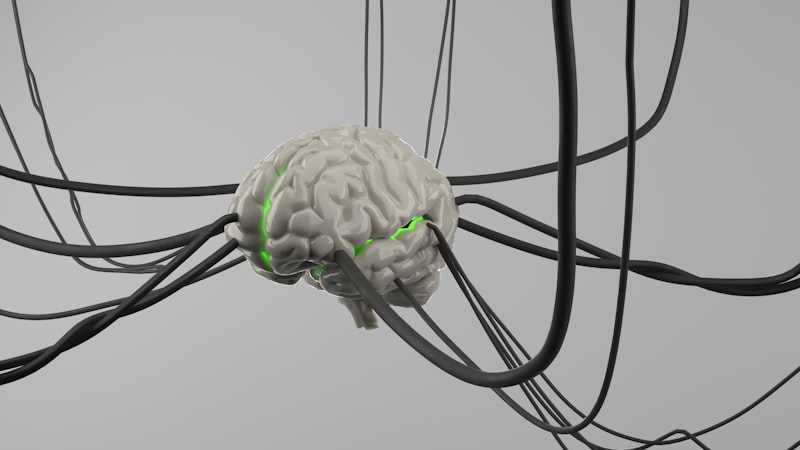
Neural Control and Motor Unit Activation
The initiation of flexion is a sophisticated neurophysiological event. Every voluntary flexor movement begins as an electrochemical signal originating in the motor cortex of the brain. This signal travels down the corticospinal tracts to the spinal cord, where it synapses with alpha motor neurons. These motor neurons exit the spinal cord and travel directly to the flexor muscle fibers, forming the neuromuscular junction.
A single alpha motor neuron and all the muscle fibers it innervates constitute a motor unit. The size of the motor unit is inversely proportional to the precision required of the flexor muscle. For example, flexors involved in fine motor control, such as those in the fingers (e.g., flexor pollicis longus), have very small motor units, where one neuron may control only a handful of fibers, allowing for highly nuanced force gradation. Conversely, powerful flexors like the iliopsoas, responsible for gross movement, possess large motor units that activate hundreds or even thousands of fibers simultaneously to generate maximum force for flexion.
The force generated by a flexor muscle is graded through two primary mechanisms: recruitment and rate coding. Recruitment involves activating an increasing number of motor units as more force is required. Smaller, more fatigue-resistant motor units (Type I fibers) are typically recruited first, followed by larger, more powerful, but faster-fatiguing units (Type II fibers). Rate coding refers to increasing the frequency of firing of the action potentials within the already recruited motor neurons. A higher firing frequency leads to temporal summation of muscle fiber tension, resulting in stronger, sustained contraction and, consequently, more powerful flexion.
Beyond voluntary control, flexor muscles are centrally involved in protective reflexes. The flexor withdrawal reflex is a rapid, involuntary response triggered by painful or noxious stimuli. If a person steps on a sharp object, sensory information rapidly travels to the spinal cord, bypassing the brain. Interneurons immediately excite the motor neurons innervating the flexor muscles of the injured limb, causing rapid and forceful flexion (withdrawal) of the limb from the source of pain, often before the sensation of pain registers consciously. This demonstrates the critical, primal role of flexor muscles in survival and injury avoidance.
Flexor Muscle Injuries, Conditioning, and Rehabilitation
Flexor muscles are susceptible to a range of injuries, particularly strains and tears, often due to sudden, explosive movements or inadequate warm-up. Hamstring strains, for example, are among the most common athletic injuries, frequently occurring during the transition phase of running when the muscle is simultaneously contracting eccentrically (controlling the forward swing of the leg) and preparing for concentric contraction. A strain, or “pulled muscle,” involves the tearing of muscle fibers, ranging from minor damage (Grade I) to complete rupture (Grade III). Immediate treatment typically involves RICE (Rest, Ice, Compression, Elevation) followed by controlled rehabilitation.
Another common clinical issue involving flexor muscles is contracture. A flexor contracture is a fixed tightening of the muscle and tendon, often resulting from prolonged immobility, neurological damage (such as stroke or cerebral palsy), or chronic inflammatory conditions. This shortening makes it extremely difficult or impossible to fully extend the joint, severely limiting mobility and function. Treatment for contractures often involves extensive physical therapy, passive stretching, splinting, and, in severe cases, surgical lengthening of the tendon.
Effective conditioning of flexor muscles requires a balanced training regimen focusing on both concentric (shortening) and eccentric (lengthening under load) strength. While concentric training builds the power necessary for initiation of movement, eccentric training is crucial for enhancing the muscle’s ability to resist external forces and control deceleration, which significantly reduces the risk of strains. For instance, exercises that specifically load the hamstrings during the lowering phase of a lift improve their eccentric capacity, making them more resilient to the stresses encountered during high-speed running.
Rehabilitation protocols following flexor injury emphasize progressive overload and restoring full range of motion. Initial phases focus on pain reduction and gentle, non-loading movements. This progresses to isometric contractions, followed by light resistance training, and finally, the incorporation of sport-specific or functional movements. The goal is not merely to restore strength, but to re-establish the precise neural coordination between the injured flexor and its antagonistic extensor, ensuring that reciprocal inhibition and synergistic stability are fully functional before the individual returns to high-intensity activity.
Evolutionary Perspective and Developmental Role
The organization and complexity of flexor muscles reflect deep evolutionary pressures, particularly those related to locomotion and dexterity. In early vertebrates, the basic segmentation of flexor and extensor groups facilitated rudimentary swimming movements. With the transition to terrestrial life, the flexor groups became increasingly specialized, moving from simple bending mechanisms to complex structures capable of supporting weight and navigating varied terrain.
The evolution of the primate hand provides a profound example of flexor specialization. The highly developed flexor muscles of the forearm and hand, coupled with fine neural control, were essential for the development of the powerful grip and precise manipulation required for arboreal life and tool use. Muscles like the flexor pollicis longus, which controls the thumb, developed exceptional independent control, a key anatomical feature distinguishing primates, and especially humans, allowing for complex fine motor tasks that would be impossible with less differentiated flexor musculature.
Developmentally, flexor muscles exhibit dominance in the fetal stage. The typical posture of a newborn, characterized by limbs drawn in and joints flexed, is often referred to as physiologic flexion. This developmental dominance reflects the necessary organization of the nervous system and musculature for survival and the transition from the constrained uterine environment. As the infant matures, the extensor muscles gain strength, gradually balancing the flexor dominance, which allows the child to overcome gravity, lift the head, sit upright, and eventually stand and walk. The entire progression of motor skill acquisition is fundamentally dependent on the increasing strength, coordination, and balanced control between the flexor and extensor muscle groups throughout the body.