Kainic Acid: Unlocking the Secrets of Neural Excitation
- Introduction to Kainic Acid
- Chemical Structure and Natural Origin
- Mechanism of Action: Receptor Binding
- Neurotoxicity and Excitotoxicity
- Applications in Research: Creating Lesion Models
- Modeling Neurological Diseases
- Pharmacological Profile and Administration Considerations
- Limitations and Ethical Considerations
Introduction to Kainic Acid
Kainic Acid (KA) is a potent, naturally occurring neuroexcitatory compound that holds immense significance in the fields of neuroscience and psychopharmacology. Chemically classified as an analogue of glutamate, the primary excitatory neurotransmitter in the mammalian central nervous system (CNS), KA is derived originally from the red marine algae, Digenea simplex. While its natural source suggests a potentially benign origin, the compound exhibits powerful and selective neurotoxic properties when administered experimentally. It is primarily recognized for its application as an experimental tool used to induce targeted brain lesions in animal models, allowing researchers to study the functional consequences of damage to specific neural circuits. This targeted destruction is mediated by its strong affinity for a specific class of glutamate receptors, leading directly to a phenomenon known as excitotoxicity.
The initial discovery and subsequent investigation into Kainic Acid revolutionized the study of neurodegenerative diseases and epilepsy. Because KA mimics the effects of excessive glutamate stimulation, it provides a reliable pharmacological method for creating acute and chronic models of neurological dysfunction that closely resemble human pathological conditions. Unlike physical lesioning techniques, chemical lesioning using KA allows for highly precise destruction of neuronal cell bodies while relatively sparing fibers of passage, making it an invaluable asset for mapping brain function. The compound’s ability to selectively target certain neuronal populations, particularly pyramidal neurons in the hippocampus and cortex, underscores its utility but also necessitates cautious handling and precise methodological control in experimental settings.
Kainic Acid is thus fundamentally defined by its dual role: it is a naturally derived substance with a distinct chemical structure and a powerful pharmacological agent capable of inducing rapid and irreversible neuronal damage through the overwhelming activation of its target receptors. Understanding the molecular mechanism of this compound is crucial for interpreting the vast body of research that relies upon KA models to dissect complex brain circuitry and pathology.
Chemical Structure and Natural Origin
Kainic Acid, chemically known as 2-carboxy-4-(1-methylethenyl)-3-pyrrolidineacetic acid, possesses a distinctive structure that allows it to interact powerfully with glutamate receptors. Its molecular configuration closely mirrors that of L-glutamate, which explains its ability to bind to and activate specific receptor subtypes. The molecule is bicyclic, incorporating a pyrrolidine ring structure that is crucial for its high binding affinity and increased metabolic stability compared to endogenous glutamate. This structural similarity is key to understanding its biological activity; it effectively hijacks the neural communication system, leading to sustained and uncontrolled excitation far exceeding physiological limits.
The natural source of Kainic Acid is primarily the red seaweed Digenea simplex, commonly found in East Asian coastal waters. Historically, extracts from this seaweed have been used in traditional medicine, particularly as an anthelminthic agent (a treatment against parasitic worms). The potent biological activity observed in these medicinal uses eventually led researchers to isolate and identify the active compound responsible for its effects in the 1950s. The isolation of KA marked a significant milestone, providing neuroscientists with a highly specific pharmacological probe for exploring excitatory neurotransmission pathways. Furthermore, other related compounds, known as kainoids, have also been isolated, but Kainic Acid remains the most widely utilized and studied derivative due to its exceptional potency and receptor selectivity.
The high potency of KA stems from its unique interaction profile with the receptor complex. Unlike the transient effects of the endogenous ligand, Kainic Acid resists normal mechanisms of enzymatic breakdown and reuptake, allowing it to remain bound to the receptor for extended periods. This persistence ensures a prolonged excitatory signal, which is the direct cause of the ensuing neurotoxicity. This stability makes it an ideal pharmacological tool for creating enduring experimental conditions that mimic chronic pathological states.
Mechanism of Action: Receptor Binding
Kainic Acid exerts its powerful neuroexcitatory effects primarily by acting as a highly specific agonist for a subset of ionotropic glutamate receptors known as kainate receptors. These receptors are ligand-gated ion channels that, upon activation, mediate rapid excitatory synaptic transmission. While glutamate itself activates all ionotropic receptor classes (AMPA, NMDA, and Kainate), Kainic Acid shows a particularly strong and persistent affinity for the kainate receptor subtype, which includes subunits such as GluK1-5. This specific binding leads to the opening of the associated ion channels, allowing a massive influx of positively charged ions, primarily sodium (Na+) and calcium (Ca2+), into the postsynaptic neuron.
The sustained activation by Kainic Acid is far more prolonged than the transient activation caused by endogenous glutamate release. Unlike glutamate, which is quickly cleared from the synaptic cleft by efficient uptake mechanisms (primarily by glial cells), KA persists, leading to prolonged depolarization that the neuron cannot resolve. This sustained ionic flux overwhelms the cell’s homeostatic mechanisms, driving the membrane potential far from its resting state. The critical feature of KA action is its ability to induce a non-desensitizing current, meaning the receptor remains open or rapidly reopens despite continuous presence of the agonist, ensuring a continuous excitatory drive.
The excessive influx of calcium ions, in particular, is the critical trigger for the subsequent cascade of intracellular events that culminate in neuronal death. The distribution of kainate receptors across different brain regions dictates the vulnerability of those regions to KA toxicity; areas such as the hippocampus, amygdala, and certain layers of the cortex are particularly rich in these receptors and are thus the primary targets of KA-induced damage. The unique kinetic properties of KA binding, combined with the anatomical distribution of its target receptors, explain the distinct pattern of cell loss observed in KA lesion models.
Neurotoxicity and Excitotoxicity
The core mechanism underlying the destructive power of Kainic Acid is excitotoxicity, a pathological process where neurons are damaged and killed by the excessive and prolonged stimulation of excitatory neurotransmitter receptors. When administered, KA causes neurons to fire rapidly and uncontrollably, leading to a state of profound hyperexcitation. This continuous firing demands enormous metabolic energy, rapidly depleting cellular adenosine triphosphate (ATP) reserves and leading to severe mitochondrial dysfunction. The resultant energy crisis compromises essential cellular processes, including the maintenance of ionic gradients by the Na+/K+-ATPase pump, further exacerbating the depolarization and ionic imbalance.
The most destructive phase is initiated by the massive intracellular calcium overload mediated by the sustained receptor activation. High levels of intracellular calcium act as a potent second messenger, inappropriately activating a host of destructive enzymes, including calcium-dependent proteases (calpains), lipases, and endonucleases. These enzymes systematically break down essential cellular components, damaging the cell membrane, the cytoskeleton, and ultimately fragmenting the nuclear DNA. This pathway typically leads to neuronal cell death via both necrosis (rapid, uncontrolled swelling and lysis due to osmotic imbalance) and apoptosis (programmed cell death, often triggered by mitochondrial release of pro-apoptotic factors).
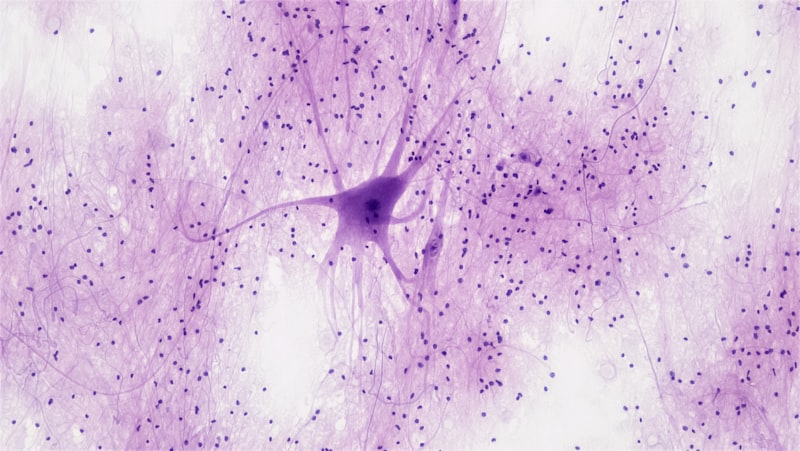
Due to the specific mechanism of action, KA selectively destroys neuronal cell bodies and dendritic fields, which are rich in glutamate receptors. Crucially, it generally spares glial cells (astrocytes and microglia) and white matter tracts (axons passing through the area) because these structures often lack the necessary density of sensitive ionotropic receptors. This selectivity makes the resulting lesion remarkably clean and functionally informative for research purposes, allowing neuroscientists to attribute behavioral deficits directly to the loss of specific neuronal processing centers rather than the disruption of connecting pathways.
Applications in Research: Creating Lesion Models
The primary utility of Kainic Acid in experimental neuroscience is the creation of highly localized, reproducible chemical lesions, offering a powerful alternative to traditional physical ablation techniques. Researchers utilize stereotaxic surgery to inject minute quantities of KA directly into a target structure, such as the dorsal hippocampus, the striatum, or the locus coeruleus. This targeted injection ensures that the excitotoxic effects are confined primarily to the intended area, allowing for precise functional mapping and the investigation of necessary versus sufficient roles of specific brain nuclei in behavior.
The resulting experimental model is crucial for understanding the behavioral and cognitive roles of the damaged structure. For instance, lesioning the CA3 region of the hippocampus allows researchers to study the neural substrates of pattern separation and memory recall, while lesions in the nucleus accumbens shed light on motivation and reward processing. The precise nature of the lesion—destroying cell bodies while sparing fibers—is a significant advantage over physical aspiration or electrolytic lesions, which are often less selective and disrupt surrounding white matter, complicating the interpretation of behavioral outcomes.
Research applications involving KA fall into distinct categories based on the route of administration:
- Systemic Administration: Used primarily to induce widespread seizure activity, mimicking generalized or temporal lobe epilepsy.
- Intracerebral Microinjection: Used to create discrete, localized lesions for functional ablation studies, allowing for high anatomical specificity.
- Intraventricular Injection: Less common, resulting in diffuse damage to structures lining the ventricles, often used for studying broader excitotoxic responses.
The consistency and reliability of KA-induced lesions have made it a standard protocol for studying circuit function and dysfunction across various mammalian species, primarily rodents.
Modeling Neurological Diseases
Kainic Acid is an indispensable tool for developing animal models of complex human neurological disorders, offering predictive validity for testing potential therapeutic agents. Its ability to induce sustained hyperexcitation makes it particularly relevant for modeling conditions characterized by neuronal overactivity and subsequent cell death, providing insights into both acute and chronic phases of disease progression.
One of the most significant applications is modeling temporal lobe epilepsy (TLE). Systemic administration of high doses of KA induces status epilepticus (prolonged, continuous seizures) in rodents. Following the initial acute phase, these animals often enter a latent period before developing chronic, spontaneous recurrent seizures, mirroring the progression of TLE in humans. This model allows detailed study of epileptogenesis—the process by which a normal brain becomes epileptic—and is the standard platform for the evaluation of novel anticonvulsant and neuroprotective drugs. Furthermore, the KA model is instrumental in investigating the associated pathology, such as mossy fiber sprouting and selective neuronal loss in the CA1 and CA3 regions of the hippocampus, which are characteristic neuropathological hallmarks of human TLE.
Beyond epilepsy, KA is used to model aspects of Huntington’s disease (HD). Intrastriatal injection of KA produces lesions in the striatum that selectively mimic the loss of medium spiny neurons, the primary cell population affected in early-stage HD. This model, along with the use of related toxins like quinolinic acid, helps researchers understand the role of excitotoxicity in neurodegenerative processes and provides a platform for testing neuroprotective strategies aimed at mitigating neuronal death in the basal ganglia. The versatility of KA in creating disease-relevant pathology underscores the widespread agreement that excitotoxicity is a common final pathway for neuronal death in many debilitating neurological conditions, including stroke and traumatic brain injury.
Pharmacological Profile and Administration Considerations
The pharmacological profile of Kainic Acid requires extremely careful consideration during experimental design due to its high potency and capacity to induce systemic toxicity. KA is typically administered via several routes, depending on the research objective. For generalized seizure induction, intraperitoneal (IP) or subcutaneous (SC) injections are common, leading to systemic distribution and widespread, but heterogeneous, excitation. For targeted lesioning, the intracerebral or intrahippocampal route, guided by high-precision stereotaxic coordinates, is mandatory to ensure precise localization of the damage and prevent systemic effects.
Key factors influencing the outcome of KA administration include the dose, the concentration, the injection rate, and the vehicle solution used. High doses or rapid injection rates significantly increase the likelihood of widespread damage and secondary systemic effects, often leading to severe systemic toxicity, hyperthermia, and profound weight loss. Precise control over these variables is essential for generating reliable and interpretable data.
- Dose Precision: Microgram quantities or even nanograms are often sufficient for creating defined, localized lesions; doses must be titrated carefully to achieve the desired level of selectivity.
- Injection Rate: A slow, controlled injection (often delivered over five to ten minutes) is necessary to minimize backflow along the needle track and better confine the drug to the intended target area.
- Age and Strain Variability: The vulnerability of experimental animals to KA toxicity varies significantly based on age, genetic strain, and species, requiring extensive pilot studies to optimize protocols and ensure consistent lesion boundaries.
The resulting neuroanatomical changes are typically monitored using standard histological techniques, such as Nissl staining (to visualize cell loss) or immunohistochemistry (to identify specific cellular markers), usually several days to weeks after injection to allow the full extent of the excitotoxic cascade to complete.
Limitations and Ethical Considerations
While Kainic Acid is an invaluable research tool, its use is associated with several methodological limitations and significant ethical considerations that must be rigorously addressed. Methodologically, the precise extent of the lesion can sometimes be difficult to control perfectly, often resulting in damage that extends slightly beyond the intended target boundary, particularly if high concentrations are used or if the injection is too rapid. Furthermore, systemic administration intended to induce epilepsy can result in highly variable seizure severity among animals, introducing unwanted variability into the data and complicating analysis.
Ethically, the induction of severe, prolonged seizures (status epilepticus) and subsequent permanent brain damage requires robust justification under international animal welfare regulations. Researchers must demonstrate that the scientific gains derived from the study significantly outweigh the suffering inflicted upon the animals. Mitigating strategies are mandatory, including the use of anesthetic agents or benzodiazepines (e.g., diazepam) to control the most severe acute seizure activity without interfering with the long-term neuroplastic changes being studied, thus minimizing unnecessary distress.
The principle of reduction, refinement, and replacement (the 3Rs) is critical in all studies involving Kainic Acid. This necessitates careful titration of the minimum effective dose required to produce the desired pathology, rigorous refinement of surgical procedures to minimize invasiveness, and continuous monitoring and supportive care (e.g., hydration and temperature control) to alleviate suffering. The long-term physiological and behavioral changes induced by KA, while useful for modeling disease, represent a serious permanent deficit for the experimental subject, emphasizing the moral imperative for meticulous adherence to ethical guidelines.