Neuroimaging: How Brain Scans Reveal Our Hidden Thoughts
- The Core Definition of Magnetic Resonance Imaging (MRI)
- The Scientific Principles Behind MRI
- Historical Development and Key Pioneers
- A Detailed Look at the MRI Procedure
- Clinical Applications and Diagnostic Utility
- Significance, Impact, and Advantages in Modern Medicine
- Limitations, Challenges, and Contraindications of MRI
- Connections to Broader Scientific and Medical Fields
The Core Definition of Magnetic Resonance Imaging (MRI)
Magnetic Resonance Imaging (MRI) is a sophisticated medical imaging technique that provides exceptionally detailed views of the body’s internal structures, including organs, soft tissues, bone, and virtually all other internal body structures. Unlike X-rays or computed tomography (CT) scans, MRI is a non-invasive and generally safe procedure that does not utilize ionizing radiation, making it a preferred choice for repeated examinations and for sensitive populations. This advanced diagnostic tool has fundamentally transformed modern medicine, offering physicians an unparalleled ability to visualize anatomical details and detect a wide array of pathological conditions without the need for surgical exploration.
At its essence, MRI functions by harnessing a powerful magnetic field and radio waves to generate intricate cross-sectional images. The human body is predominantly composed of water molecules, which contain hydrogen nuclei, or protons. These protons act like tiny magnets. When a patient is placed inside the MRI scanner, the strong magnetic field causes these protons to align in a specific direction. Subsequently, brief pulses of radio waves are emitted, temporarily knocking the protons out of alignment. When the radio waves are turned off, the protons relax back into alignment with the main magnetic field, releasing energy in the process. This emitted energy, or signal, is detected by the MRI scanner’s coils and then processed by a powerful computer to construct highly detailed, three-dimensional images of the tissues under examination.
The ability of MRI to differentiate between various types of soft tissue, such as brain matter, muscles, tendons, and ligaments, is what sets it apart from other imaging modalities. This exceptional contrast resolution allows for the detection of subtle abnormalities that might be missed by other techniques. It plays a critical role in the diagnosis of numerous conditions, ranging from neurological disorders like stroke, multiple sclerosis, and brain tumors, to musculoskeletal injuries, cardiovascular diseases, and various forms of cancer. The information gleaned from an MRI scan is invaluable for precise medical diagnostics, guiding treatment planning, and monitoring disease progression or response to therapy.
The Scientific Principles Behind MRI
The fundamental principle underpinning MRI is Nuclear Magnetic Resonance (NMR), a phenomenon discovered in the 1940s that describes how atomic nuclei absorb and re-emit radiofrequency energy when placed in a magnetic field. In the context of medical imaging, the hydrogen protons within the water molecules prevalent in the human body serve as the primary source of the NMR signal. When a patient enters the bore of the MRI scanner, these protons, which normally spin randomly, align themselves with the direction of the strong external magnetic field. This alignment creates a net magnetization vector within the patient’s body, analogous to a compass needle aligning with the Earth’s magnetic field.
Following this alignment, a precisely timed radiofrequency pulse, tuned to the specific resonant frequency of the hydrogen protons (known as the Larmor frequency), is briefly applied. This pulse temporarily perturbs the aligned protons, causing them to absorb energy and flip their alignment away from the main magnetic field. When the radiofrequency pulse is switched off, the excited protons begin to “relax” back to their original alignment, releasing the absorbed energy in the form of a radiofrequency signal. The rate at which these protons relax and the characteristics of the emitted signal vary significantly depending on the type of tissue they are within, as different tissues have different compositions of water and macromolecules. This differential relaxation time is what allows MRI to distinguish between healthy and diseased tissues, providing exceptional contrast.
To create a spatially resolved image, the MRI system employs additional, weaker gradient fields alongside the main static magnetic field. These gradient fields cause the magnetic field strength to vary linearly across different parts of the body. This variation means that protons at different locations within the body will resonate at slightly different frequencies. By carefully manipulating these gradient fields, the scanner can encode the spatial location of the emitted signals. Sophisticated computer algorithms then process these frequency-encoded signals, combining them to reconstruct highly detailed two-dimensional slices or three-dimensional volumes of the scanned area. The ability to acquire images in any plane (axial, sagittal, coronal, or oblique) without moving the patient further enhances MRI’s versatility and diagnostic power.
Historical Development and Key Pioneers
The journey to modern MRI began with the discovery of Nuclear Magnetic Resonance (NMR) in the 1940s by independent research groups led by Felix Bloch and Edward Purcell, who were jointly awarded the Nobel Prize in Physics in 1952 for their foundational work. Initially, NMR was primarily used by chemists and physicists for studying the molecular structure of materials. However, the application of NMR principles to biological systems and, eventually, to medical imaging, required significant conceptual leaps and technological advancements. The challenge lay in transforming a technique that produced a single signal from a bulk sample into one that could generate spatially resolved images of complex biological tissues.
The crucial breakthrough that bridged NMR spectroscopy to diagnostic imaging came in the early 1970s. In 1971, Raymond Damadian demonstrated that different types of cancerous tissues had distinct NMR relaxation times compared to healthy tissues, suggesting the potential for disease detection. Building upon this, Paul Lauterbur published a seminal paper in 1973 demonstrating the use of gradient fields to create the first 2D NMR images, which he termed “zeugmatography.” This innovation was pivotal, as it showed how to spatially encode the NMR signal, effectively creating a “picture.” Around the same time, Peter Mansfield further refined the mathematical techniques for rapid image acquisition and reconstruction, developing the concept of echo-planar imaging (EPI), which significantly reduced scan times and laid the groundwork for modern fast MRI sequences.
For their groundbreaking contributions that made MRI a practical medical imaging method, Paul Lauterbur and Peter Mansfield were jointly awarded the Nobel Prize in Physiology or Medicine in 2003. Their work transformed NMR from a laboratory curiosity into an indispensable clinical tool. The subsequent decades saw rapid advancements in scanner technology, computing power, and pulse sequence development, leading to the sophisticated, high-resolution MRI systems we use today. This journey from fundamental physics to clinical application underscores the interdisciplinary nature of scientific progress, combining insights from physics, chemistry, engineering, and medicine to revolutionize medical diagnostics.
A Detailed Look at the MRI Procedure
Undergoing an MRI scan typically begins with thorough preparation to ensure patient safety and optimize image quality. Patients are usually asked to remove all metallic objects, including jewelry, watches, hearing aids, and clothing with metal fasteners, as the powerful magnetic field can interact with these items, potentially causing injury or degrading image quality. A comprehensive screening for metallic implants or foreign bodies, such as pacemakers, certain types of aneurysm clips, cochlear implants, or shrapnel, is critical, as these can be absolute contraindications for an MRI due to the risk of movement or malfunction within the strong magnetic environment. Patients with claustrophobia may be offered mild sedation, and contrast agents containing gadolinium may be administered intravenously to enhance the visibility of specific tissues or pathologies, particularly in cases of inflammation, tumors, or vascular abnormalities.
Once prepared, the patient lies on a movable examination table that slides into the bore of the cylindrical MRI scanner. During the scan, it is paramount for the patient to remain absolutely still to prevent motion artifacts that can blur the images and necessitate repeat sequences. The technologist communicates with the patient from an adjacent control room, providing instructions and reassurance through an intercom system. The scanning process itself is characterized by loud banging and knocking noises, which are generated by the rapid switching of the gradient fields. Patients are provided with earplugs or headphones to mitigate this noise. A typical MRI exam can last anywhere from 30 minutes to over an hour, depending on the number of areas being scanned and the complexity of the required sequences.
For instance, consider a patient experiencing persistent knee pain, suspected of having a ligament tear. An MRI of the knee would be ordered. The patient would lie supine, with their knee positioned within the scanner. The MRI would then acquire multiple sequences, each designed to highlight different tissue characteristics. These images would reveal the intricate details of the knee joint, including the menisci, anterior cruciate ligament (ACL), posterior cruciate ligament (PCL), collateral ligaments, cartilage, and surrounding musculature. A torn ACL, for example, would appear as a discontinuity or abnormal signal intensity within the ligament fibers. The detailed images would allow the radiologist to precisely locate the injury, assess its severity, and identify any associated damage to other structures, providing the orthopedic surgeon with crucial information for treatment planning, whether it involves conservative management or surgical repair. This precise visualization is a cornerstone of modern musculoskeletal care, significantly improving diagnostic accuracy and patient outcomes.
Clinical Applications and Diagnostic Utility
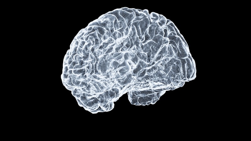
The diagnostic utility of MRI spans nearly every medical specialty, making it an indispensable tool for clinicians. In neurology, MRI is the gold standard for visualizing the brain and spinal cord, detecting conditions such as strokes, brain tumors, multiple sclerosis plaques, epilepsy, and spinal cord injuries with exceptional clarity. Its ability to depict subtle changes in brain tissue, often before symptoms become severe, allows for early intervention and improved patient prognoses. For example, in the case of a suspected stroke, an MRI can differentiate between ischemic and hemorrhagic strokes and determine the age of the stroke, guiding urgent treatment decisions.
In musculoskeletal medicine, MRI offers unparalleled insights into joints, muscles, tendons, ligaments, and bone marrow. It is routinely used to diagnose sports injuries, degenerative joint diseases, spinal disc herniations, bone infections, and soft tissue tumors. The high contrast resolution between different types of soft tissue allows for precise identification of tears, inflammation, and edema, which are often invisible on X-rays or CT scans. This detailed anatomical information is critical for orthopedists and rheumatologists in developing tailored treatment plans, from physical therapy regimens to surgical interventions.
Furthermore, MRI has significant applications in oncology for the detection, staging, and monitoring of various cancers. It is particularly valuable for imaging soft tissue tumors in the brain, breast, liver, prostate, and female pelvic organs, where its superior contrast and multi-planar imaging capabilities outperform other modalities. By providing detailed information about tumor size, location, and spread to adjacent tissues or lymph nodes, MRI assists oncologists in determining the most effective course of treatment, such as surgery, radiation therapy, or chemotherapy. Beyond these areas, MRI is also used in cardiac imaging to assess heart function and detect cardiac abnormalities, in abdominal imaging for organ evaluation, and in vascular imaging (MRA) to visualize blood vessels without invasive angiography, highlighting its vast and continually expanding diagnostic utility.
Significance, Impact, and Advantages in Modern Medicine
The advent of Magnetic Resonance Imaging has irrevocably revolutionized the landscape of modern medicine, fundamentally transforming medical diagnostics and research. Its significance lies in its ability to provide exquisitely detailed anatomical and physiological information that was previously unattainable without invasive procedures. Before MRI, diagnosing many conditions involving soft tissue pathologies, such as brain tumors or ligament tears, often required exploratory surgery or less precise imaging methods like X-rays, which offered limited contrast for these structures. MRI changed this paradigm by offering a non-invasive window into the body’s most intricate structures, empowering clinicians to make highly accurate diagnoses with unprecedented confidence.
One of the foremost advantages of MRI is its complete absence of ionizing radiation, a stark contrast to X-rays and CT scans. This makes it an exceptionally safe option, particularly for patients requiring multiple follow-up scans, children, and pregnant women (after careful consideration). The ability to repeatedly image a patient without cumulative radiation exposure is invaluable for monitoring the progression of chronic diseases, assessing the effectiveness of ongoing treatments, or tracking the growth of tumors over time. This safety profile, combined with its superior soft tissue contrast, positions MRI as a cornerstone technology for longitudinal studies and personalized medicine, where continuous, detailed assessment is critical to patient care and research endeavors.
Beyond its diagnostic prowess, MRI has profoundly impacted various clinical applications. In surgical planning, high-resolution MRI images guide neurosurgeons in precisely locating tumors or lesions before intervention, minimizing collateral damage to healthy tissue. In radiation therapy, MRI provides detailed anatomical maps for highly targeted radiation delivery, improving efficacy while reducing side effects. Furthermore, the development of functional MRI (fMRI) has opened new avenues in neuroscience, allowing researchers to observe brain activity in real-time by detecting changes in blood flow. This has advanced our understanding of cognitive processes, neurological disorders, and the effects of psychological interventions, demonstrating MRI’s far-reaching influence beyond purely anatomical imaging into the realm of functional assessment and research.
Limitations, Challenges, and Contraindications of MRI
Despite its numerous advantages and profound impact, Magnetic Resonance Imaging is not without its limitations and challenges. One of the most significant drawbacks is the substantial cost associated with MRI technology. The acquisition of an MRI scanner, its installation, ongoing maintenance, and the specialized personnel required to operate and interpret the scans represent a considerable financial investment. This high cost can limit accessibility, particularly in developing regions, and contributes to the higher expense of an MRI examination compared to other imaging modalities, potentially creating barriers to patient care.
Another major challenge relates to patient comfort and contraindications. Many patients experience claustrophobia when placed inside the narrow, enclosed bore of a traditional MRI scanner, leading to anxiety or the inability to complete the scan. The loud banging noises produced during image acquisition can also be distressing. More critically, the extremely powerful magnetic field of an MRI scanner poses significant risks for individuals with certain metallic implants or foreign bodies. Absolute contraindications include pacemakers, implantable cardioverter-defibrillators (ICDs), specific types of cerebral aneurysm clips, cochlear implants, and certain neurostimulators, as the magnetic field can cause these devices to malfunction, heat up, or move, potentially leading to severe injury or death. Even non-ferromagnetic metallic implants can sometimes cause significant image artifacts, obscuring the area of interest.
Furthermore, while MRI excels in soft tissue contrast, it has certain inherent limitations in imaging specific areas or conditions. For instance, imaging of structures containing air, such as the lungs, or bone, can be challenging due to the scarcity of hydrogen protons or rapid signal decay, respectively. The long scan times required for high-resolution images can also be problematic for patients who are unable to remain still for extended periods, such as young children, uncooperative patients, or those experiencing severe pain. Motion artifacts remain a persistent issue, degrading image quality and sometimes necessitating repeat scans, which prolongs the procedure and adds to patient discomfort. While contrast agents like gadolinium improve diagnostic yield, their use is contraindicated in patients with severe kidney disease due to the risk of nephrogenic systemic fibrosis, necessitating careful patient screening.
Connections to Broader Scientific and Medical Fields
Magnetic Resonance Imaging is deeply integrated into a multitude of scientific and medical disciplines, serving as a critical bridge between fundamental physics and clinical practice. Its roots lie firmly in physics, specifically in the principles of Nuclear Magnetic Resonance (NMR), quantum mechanics, and electromagnetism. The continuous development of MRI technology relies heavily on advancements in engineering, particularly in superconducting magnet technology, radiofrequency coil design, and high-performance computing for image reconstruction and processing. Thus, MRI stands as a testament to the power of interdisciplinary collaboration, drawing upon expertise from across the physical sciences to create a powerful biomedical tool.
Within the medical domain, MRI is a cornerstone of Radiology, the medical specialty dedicated to medical imaging. Radiologists are highly trained physicians responsible for interpreting MRI scans and other diagnostic images to diagnose diseases and injuries. Beyond general radiology, MRI has forged strong connections with various clinical subspecialties. In Neuroscience, it is indispensable for studying brain structure, function, and connectivity, with techniques like diffusion tensor imaging (DTI) mapping neural pathways and functional MRI (fMRI) investigating brain activity during cognitive tasks. This has profoundly advanced our understanding of neurological and psychiatric disorders, from Alzheimer’s disease to depression.
Moreover, MRI forms crucial links with biomedical engineering, which focuses on the design and improvement of medical devices and diagnostic tools. Engineers continually work to enhance MRI scanner capabilities, improve image resolution, reduce scan times, and develop new pulse sequences for specific clinical applications. Its utility also extends to pharmacology, where MRI can be used in preclinical research to monitor drug efficacy and tissue response. In a broader sense, MRI is a vital component of the overarching field of Medical Diagnostics, working in concert with other imaging modalities, laboratory tests, and clinical assessments to provide a comprehensive picture of a patient’s health. Its continuous evolution and integration across these diverse fields underscore its pivotal role in advancing both scientific knowledge and patient care in the 21st century.