The Medulla: Decoding the Brain’s Vital Command Center
- Introduction to the Concept of the Medulla
- The Medulla Oblongata: Anatomical Localization and Structure
- Primary Functions: Autonomic and Reflexive Control
- Sensory and Motor Pathway Decussation
- Clinical Significance and Associated Disorders
- The Medulla in Other Organ Systems: Renal and Adrenal Examples
- Comparative Anatomy and Evolutionary Development
- Summary of the Medulla’s Central Importance
Introduction to the Concept of the Medulla
The term medulla derives from the Latin word meaning “marrow” or “pith,” and in biological contexts, it consistently refers to the central, innermost region of an organ, often distinct in structure and function from the outer layer, known as the cortex. This fundamental anatomical distinction is pervasive throughout the body, defining the core operational area of numerous vital structures. While the general definition applies broadly—as seen in the medulla of the kidney or the medulla of a hair shaft—the term holds particular significance in neuroscience, where the Medulla Oblongata constitutes one of the most ancient and essential parts of the central nervous system. Understanding the medulla requires appreciating this duality: its role as a generalized anatomical descriptor for an internal core and its specific, paramount importance as the caudal-most segment of the brainstem, controlling life-sustaining involuntary functions.
Historically, early anatomists recognized the necessary organizational principle of having a protective outer shell (cortex) surrounding a highly functional, centralized core (medulla). This structure often facilitates complex physiological processes, such as filtration and fluid regulation in the renal system, or the rapid, involuntary coordination of autonomic responses in the nervous system. The medulla is typically characterized by a concentration of specialized cells, dense vascular networks, or crucial neural tracts that must be centrally located for optimal efficiency and protection. It is the anatomical nexus where foundational physiological activity occurs, insulated by the surrounding cortical layers. Therefore, any detailed physiological analysis must differentiate the functions originating in this central zone versus those handled by the periphery.
In the context of human physiology and psychology, the Medulla Oblongata receives the most substantial attention due to its indispensable role as the primary relay station between the spinal cord and the upper brain centers, including the pons and the midbrain. This segment manages reflex activities critical for immediate survival, operating entirely below the level of conscious awareness. Its integrity is so paramount that damage to the Medulla Oblongata often results in immediate death or severe, irreversible neurological impairment. Thus, while the term medulla is anatomically versatile, its association with the brainstem highlights its status as a core regulator of life itself, embodying the ultimate control center for basal functions.
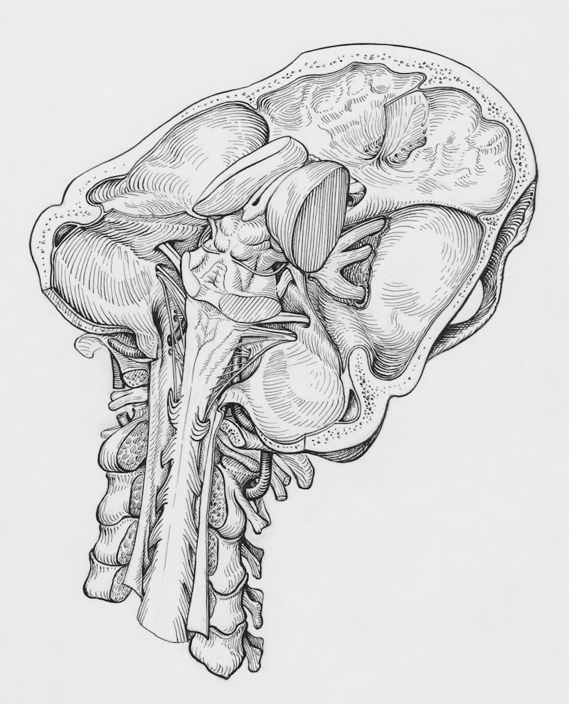
The Medulla Oblongata: Anatomical Localization and Structure
The Medulla Oblongata is the lower half of the brainstem, situated immediately inferior to the pons and superior to the spinal cord, serving as the crucial transition point between these major segments of the central nervous system. Approximately three centimeters in length, this structure resides within the posterior cranial fossa, nestled securely within the bony confines of the skull base, a location necessitated by its vital role. Its external surface is marked by distinct topographical features, most notably the anteriorly located pyramids, which are two large bulges containing the descending corticospinal tracts responsible for voluntary motor control. The crucial landmark known as the decussation of the pyramids occurs at the junction of the medulla and the spinal cord, where approximately 80 to 90 percent of the motor fibers cross over to the contralateral side, explaining why movement control for one side of the body originates in the opposite cerebral hemisphere.
Lateral to the pyramids lie the olives, or olivary bodies, prominent oval structures containing the inferior olivary nuclei, which play a significant role in motor learning and coordination, particularly in relaying information to the cerebellum. The dorsal surface of the open medulla forms the floor of the fourth ventricle, connecting cerebrospinal fluid circulation routes. Internally, the medulla is a complex tapestry of white matter tracts and gray matter nuclei. The white matter consists of ascending sensory pathways (such as the medial lemniscus, carrying touch and proprioception information) and the descending motor pathways previously mentioned. The gray matter is organized into discrete nuclei that serve as the origins or terminations for several key cranial nerves, specifically the glossopharyngeal (IX), vagus (X), accessory (XI), and hypoglossal (XII) nerves, underscoring its role in controlling functions related to the head, neck, and vital thoracic and abdominal organs.
Furthermore, the internal architecture includes a crucial network known as the reticular formation, which extends throughout the core of the brainstem and is particularly dense within the medulla. This diffuse collection of neurons is indispensable for integrating sensory, motor, and visceral information, forming the foundation of the vital centers. The integrity of the medulla’s organization—with its strategically placed tracts and nuclei—allows for the instantaneous, reflexive processing necessary for survival. Any structural compromise, whether due to trauma, ischemia, or tumor growth, disrupts this highly integrated machinery, leading to profound and often catastrophic physiological instability.
Primary Functions: Autonomic and Reflexive Control
The Medulla Oblongata is often described as the most critical region of the brain for immediate survival because it houses the primary vital centers that regulate fundamental autonomic functions necessary for maintaining homeostasis. These centers operate entirely without conscious input, managing the body’s most essential internal processes. The three most important vital centers are the cardiac center, the respiratory center, and the vasomotor center. The cardiac center regulates heart rate and force of contraction, receiving input from baroreceptors and chemoreceptors located in the major arteries, allowing it to rapidly adjust cardiac output in response to changes in blood pressure or oxygen levels. For instance, if blood pressure drops too low, the cardiac center stimulates sympathetic outflow, increasing heart rate and contractility to stabilize circulation.
The respiratory center is perhaps the most well-known functional cluster within the medulla, controlling the rate, rhythm, and depth of breathing. This center is highly sensitive to carbon dioxide and pH levels in the cerebrospinal fluid and blood. Specific groups of neurons, including the Ventral Respiratory Group (VRG) and the Dorsal Respiratory Group (DRG), generate the basic rhythm of respiration. The DRG primarily controls inspiration, while the VRG is involved in both inspiration and forced expiration. The medulla’s constant monitoring and adjustment of ventilation ensure adequate gas exchange, preventing hypoxia and regulating the body’s acid-base balance—a process so fundamental that its failure leads rapidly to death.
Finally, the vasomotor center is responsible for controlling the diameter of blood vessels, thereby regulating systemic blood pressure and the distribution of blood flow to various organs. By adjusting the degree of vasoconstriction (narrowing) or vasodilation (widening) of arterioles, the medulla ensures that blood pressure remains within a narrow, healthy range. For example, during strenuous activity, the vasomotor center redirects blood flow away from non-essential organs (like the digestive tract) and toward working muscles. Beyond these vital centers, the medulla also mediates several essential non-vital reflexes, including coughing, sneezing, vomiting, and swallowing. These protective reflexes are integrated through distinct medullary nuclei, underscoring the structure’s comprehensive role in maintaining physiological integrity against environmental challenges.
Sensory and Motor Pathway Decussation
A defining characteristic of the Medulla Oblongata is its role as the major site of pathway crossing, or decussation, for both crucial sensory and motor tracts traveling between the brain and the spinal cord. This anatomical organization dictates the contralateral control of the body. On the motor side, the aforementioned decussation of the pyramids is paramount. These pyramids contain the upper motor neuron axons of the corticospinal tracts, which originate in the cerebral cortex and are responsible for initiating voluntary movement. As these tracts descend through the medulla, the majority of fibers cross the midline in the caudal medulla, allowing the right hemisphere to control the voluntary muscles on the left side of the body, and vice versa. This cross-over ensures that motor commands are correctly routed to the descending spinal tracts.
The sensory system also utilizes the medulla as a primary relay point and decussation site. The ascending sensory information related to fine touch, conscious proprioception (awareness of body position), and vibration is carried via the dorsal column-medial lemniscus pathway. Axons carrying this highly detailed sensory input travel up the spinal cord to the lower medulla, where they synapse in the gracile nucleus (for lower body information) and the cuneate nucleus (for upper body information). After synapsing, the second-order neurons emerge from these nuclei and immediately cross the midline, forming the medial lemniscus. This crossed pathway then ascends through the pons, midbrain, and eventually terminates in the thalamus. This sensory decussation ensures that the somatosensory cortex in each hemisphere receives sensory data originating from the opposite side of the body.
The strategic positioning of these crossing points within the medulla means that damage localized to this structure can result in specific and often devastating neurological deficits that affect both motor and sensory functions contralaterally to the injury, or ipsilaterally depending on whether the nuclei (pre-decussation) or the tract (post-decussation) is affected. For instance, lesions affecting the medial medulla can produce alternating hemiplegia, where motor paralysis occurs on the opposite side of the body (due to damage to the crossed pyramidal tracts) while cranial nerve deficits occur on the same side as the lesion (due to damage to the cranial nerve nuclei before their exit). This intricate organization highlights the medulla’s function not merely as a conduit, but as a sophisticated central processing and routing hub essential for coordinated neural communication.
Clinical Significance and Associated Disorders
Given the concentration of vital centers and essential pathways within the Medulla Oblongata, any pathology affecting this region carries an extremely high risk of mortality or profound disability. Strokes, tumors, or traumatic injuries that compromise the medullary blood supply—primarily derived from the vertebral and posterior inferior cerebellar arteries (PICA)—can induce acute neurological crises. A classic presentation of medullary damage is Wallenberg syndrome, or lateral medullary syndrome, typically caused by occlusion of the PICA. This syndrome presents with a complex array of symptoms due to the involvement of various nuclei, including ipsilateral ataxia, vertigo, nystagmus, difficulty swallowing (dysphagia), and loss of pain and temperature sensation on the contralateral side of the body, illustrating the mixture of crossed and uncrossed deficits.
Furthermore, because the medulla controls the basic rhythm of breathing, certain pathological conditions directly impact respiratory drive. Central Hypoventilation Syndrome (CHS), often referred to as Ondine’s Curse, is a rare and severe disorder characterized by the failure of the autonomic respiratory control centers in the medulla to adequately respond to rising carbon dioxide levels, especially during sleep. Patients with CHS must rely on mechanical ventilation to sustain life when unconscious, starkly demonstrating the inability of higher brain centers to override the necessity of the medullary respiratory center. Damage to the reticular formation within the medulla can also severely impair arousal and consciousness, contributing to brain death definitions, where the permanent cessation of all medullary functions is a primary diagnostic criterion.
The location of the medulla at the base of the skull also makes it vulnerable to structural problems, such as Chiari malformations, where the cerebellar tonsils herniate downward into the foramen magnum, compressing the medulla and upper cervical spinal cord. This compression can lead to hydrocephalus, chronic headaches, and progressive loss of medullary functions, including balance, coordination, and sometimes respiratory stability. The profound clinical consequences of medullary lesions reinforce its status not only as an anatomical landmark but as the functional cornerstone of the autonomic nervous system. Treatment for medullary disorders often requires rapid, highly specialized intervention to preserve the remaining viable tissue and stabilize the core vital functions that define life.
The Medulla in Other Organ Systems: Renal and Adrenal Examples
Beyond the neurological context, the term medulla retains its original anatomical definition as the central core in several other crucial organ systems, notably the kidney and the adrenal gland. The Renal Medulla forms the inner region of the kidney, situated deep to the renal cortex. Its structure is highly specialized, consisting of renal pyramids—cone-shaped tissue masses that contain essential components of the nephron, the functional unit of the kidney. The primary physiological role of the renal medulla is the concentration of urine through the maintenance of an osmotic gradient. This gradient is established by the highly permeable loops of Henle and the collecting ducts, which run vertically through the medullary pyramids. The unique hypertonic environment created within the renal medulla allows for the reabsorption of water, enabling the body to conserve fluids effectively and produce concentrated urine, a necessity for maintaining fluid balance.
Similarly, the Adrenal Medulla constitutes the innermost layer of the adrenal gland, completely surrounded by the adrenal cortex. Unlike the cortex, which produces steroid hormones, the adrenal medulla is essentially a modified sympathetic ganglion, derived from neural crest cells. Its function is neuroendocrine; it serves as the primary site for the synthesis and release of catecholamines, specifically epinephrine (adrenaline) and norepinephrine (noradrenaline). These hormones are released directly into the bloodstream in response to signals from the sympathetic nervous system (preganglionic neurons). The rapid secretion of catecholamines is the body’s instantaneous response to acute stress, initiating the “fight or flight” response by increasing heart rate, blood pressure, blood glucose levels, and diverting blood flow to skeletal muscles. This functional specialization highlights how the central, medullary structure in this gland is dedicated to immediate, systemic regulation.
The structural and functional independence of the medulla in these organs—the renal medulla focused on osmotic regulation and the adrenal medulla focused on acute stress response—reinforces the principle that the core of an organ often houses the most specialized or rapid-response machinery. In both cases, the cortex provides support, protection, and complementary functions (e.g., filtration in the renal cortex, steroid synthesis in the adrenal cortex), while the medulla executes the high-stakes, core physiological actions that maintain dynamic equilibrium within the body.
Comparative Anatomy and Evolutionary Development
The Medulla Oblongata is recognized across the vertebrate lineage as an evolutionarily ancient structure, reflecting its crucial role in maintaining basic life functions that predate complex cognitive abilities. In comparative anatomy, the brainstem, and specifically the medulla, demonstrates a high degree of structural and functional conservation from fish and amphibians to reptiles, birds, and mammals. This conservation suggests that the centers governing respiration, circulation, and primitive motor reflexes were among the earliest neural structures to evolve, providing the necessary autonomic stability for complex life to emerge.
In simpler vertebrates, the medulla often forms the largest and most prominent part of the brain relative to the cerebrum, which is less developed. For example, in many fish, the medulla contains centers that integrate sensory information from the lateral line system and control intricate swimming movements, in addition to basic respiratory and cardiac regulation. While the overall size and complexity of the tracts passing through the medulla increase significantly in mammals, particularly primates, due to the expansion of the cerebral cortex and the corresponding corticospinal tracts, the fundamental organization of the vital nuclei remains remarkably consistent across species. The presence of the same cranial nerve nuclei (IX, X, XII) controlling equivalent functions (e.g., gill or lung ventilation, heart rate) illustrates this deep evolutionary stability.
The conservation of medullary function underscores its foundational importance. It provides a robust, fail-safe system for managing immediate physiological needs, allowing higher brain centers to evolve specialized cognitive and executive functions without the burden of micromanaging breathing or heart rate. Therefore, studying the medulla across species provides invaluable insights into the evolutionary pressures that prioritized autonomic stability and the hierarchical organization of the nervous system, confirming the medulla as the indispensable biological interface between the brain and the body’s operational mechanisms.
Summary of the Medulla’s Central Importance
In summary, the term medulla serves as a critical descriptor for the central, functional core of various organs throughout human physiology, ranging from the renal medulla’s role in fluid conservation to the adrenal medulla’s management of acute stress. However, its most profound significance lies in the Medulla Oblongata, the caudal segment of the brainstem. This small structure is disproportionately vital, acting as the non-negotiable headquarters for involuntary life functions. It houses the cardiac, respiratory, and vasomotor centers that ensure continuous, stable operation of the cardiovascular and pulmonary systems—processes that cannot be consciously suspended.
Furthermore, the medulla functions as the essential crossroads for major neural traffic. It is the mandatory site where the vast majority of motor control pathways (the pyramids) and crucial sensory pathways (the medial lemniscus) cross the midline, establishing the contralateral control of the body by the cerebral hemispheres. This intricate anatomical routing ensures efficient communication between the brain and the periphery. The integrity of these tracts and nuclei is paramount; damage to the medulla results in severe, often fatal, disruptions to homeostasis and coordinated movement, as exemplified by complex syndromes like Wallenberg syndrome.
Ultimately, whether referring to the core of a gland or the base of the brain, the medulla represents the innermost stratum where highly specialized, fundamental operations are concentrated and protected. The Medulla Oblongata, in particular, stands as a testament to the evolutionary necessity of a centralized, autonomous regulator, underpinning consciousness, movement, and the very continuation of life itself. Its constant, silent control allows for the higher cognitive functions that define human experience, making the medulla an essential subject of study in neuroscience and physiology.