The Midbrain: Your Hidden Sensory Gateway
Scientifically Reviewed · Dr. Marwa Abd-Alazim · August 21, 2026
Review Criteria & Clinical Standards
This content undergoes rigorous scientific peer-review and medical editorial standards at Arab Psychology Network to ensure clinical accuracy, validity, and compliance with evidence-based guidelines from leading psychological and healthcare authorities (APA / WHO).
The Core Definition and Anatomical Location
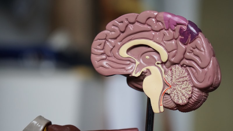
The Mesencephalon, commonly referred to as the midbrain, is a fundamental structure within the central nervous system, representing the most superior segment of the brainstem. Its primary role is to act as a vital relay station for sensory and motor information traveling between the forebrain (cerebrum and diencephalon) and the hindbrain (pons and cerebellum). Structurally, it is a relatively small but highly dense region, measuring only about two centimeters in length in humans, yet housing crucial nuclei and fiber tracts responsible for essential homeostatic and behavioral functions, including basic visual and auditory reflexes, as well as the regulation of motor movement.
Anatomically, the mesencephalon is situated immediately below the thalamus and pineal gland, and directly above the pons. This central position ensures that virtually all ascending sensory pathways and descending motor commands must pass through or synapse within its boundaries. The fundamental mechanism underlying the midbrain’s function is the rapid processing of non-conscious stimuli, allowing the body to respond reflexively to sudden environmental changes, such as unexpected sounds or lights, before the cerebral cortex has time to fully analyze the threat. This rapid action is facilitated by specialized structures within the midbrain roof, known as the tectum.
The mesencephalon is fundamentally divided into three major sections: the tectum (or roof), the tegmentum (the floor), and the paired cerebral peduncles, which serve as the massive tracts of white matter descending from the cerebrum. The overall architecture is built around the cerebral aqueduct (of Sylvius), a narrow channel connecting the third and fourth ventricles, which allows for the circulation of cerebrospinal fluid. This strategic anatomical arrangement underscores the midbrain’s importance not only in neural conduction but also in the maintenance of intracranial pressure and fluid dynamics within the brain.
Functional Role in Neural Processing
The functions of the mesencephalon are diverse, encompassing critical aspects of sensory integration, motor coordination, and consciousness. One of its most famous roles relates to immediate, reflexive responses to external stimuli. For instance, the superior portion of the tectum, the Superior Colliculi, processes visual information, specifically detecting the location of objects in the visual field and initiating rapid orienting movements of the head and eyes towards those objects, a process known as the visual grasp reflex. This ensures immediate attention to potentially significant visual events, regardless of cortical interpretation.
Complementing the visual role, the inferior portion of the tectum, the Inferior Colliculi, serves as the primary auditory relay center for the brainstem. It receives input from multiple lower auditory nuclei and processes the spatial localization of sound, initiating auditory startle reflexes. Both sets of colliculi work in concert to ensure the organism can quickly locate and react to sudden changes in the sensory environment, forming the foundation of non-conscious defensive behavior. This intricate processing highlights the midbrain’s role as a sophisticated sensory integrator that operates outside the realm of voluntary thought.
Furthermore, the tegmentum of the mesencephalon is indispensable for voluntary movement and posture. It houses the Substantia Nigra (black substance), which is rich in dopaminergic neurons that project to the basal ganglia, playing a critical role in initiating and smoothing out motor commands. Adjacent to this is the Red Nucleus, a large, vascular structure involved in coordinating movements, particularly in the upper limbs, by sending fibers down the rubrospinal tract. Damage to either of these motor centers results in profound deficits, demonstrating the midbrain’s central position in the motor hierarchy of the nervous system.
Key Substructures of the Mesencephalon
A detailed understanding of the mesencephalon requires recognizing its specific nuclear groups and fiber pathways, which define its functional subdivisions. The Tectum, situated dorsally, contains the two pairs of colliculi—the corpora quadrigemina—which are specialized for sensory reflexes. Ventral to the tectum lies the Tegmentum, a complex region hosting several critical nuclei. These include the previously mentioned Substantia Nigra and Red Nucleus, essential for motor control, as well as the nuclei for the third (Oculomotor) and fourth (Trochlear) cranial nerves, which control eye movements.
The Periaqueductal Gray (PAG) is another crucial component of the tegmentum, surrounding the cerebral aqueduct. The PAG is an area of gray matter heavily involved in descending pain modulation and defensive behaviors. It receives input from higher brain centers and projects to lower brainstem nuclei to suppress pain signals through the release of endogenous opioids, representing a powerful, built-in analgesic system. The integrity of the PAG is essential for regulating emotional responses, particularly those related to fear and anxiety, and governing the fight-or-flight response.
The most ventral portion of the mesencephalon consists of the two massive bundles of axons known as the Cerebral Peduncles (or crus cerebri). These peduncles house the corticospinal and corticobulbar tracts, which carry the motor commands originating in the primary motor cortex down to the spinal cord and lower brainstem nuclei, respectively. The sheer volume of these descending tracts emphasizes the mesencephalon’s role as the primary conduit for voluntary motor control, connecting the cognitive planning centers of the forebrain with the executive motor structures of the lower nervous system.
Historical Discovery and Early Neuroanatomy
The historical identification and understanding of the mesencephalon evolved slowly, paralleling advancements in overall neuroanatomy. Early anatomists, dating back to figures like Galen in the second century, recognized the brainstem as a functional bridge, but detailed localization was impossible without sophisticated dissection techniques. The term “midbrain” or mesencephalon came into clearer focus during the Renaissance, particularly with the meticulous work of Andreas Vesalius in the 16th century, who provided accurate drawings of the brain’s internal architecture, though the functional significance of the internal nuclei remained largely unknown.
The 17th and 18th centuries saw incremental progress in identifying specific landmarks, such as the corpora quadrigemina (colliculi). However, it was not until the late 19th century, with the refinement of histological staining techniques by researchers like Camillo Golgi and Santiago Ramón y Cajal, that the cellular architecture of the midbrain was truly appreciated. These techniques allowed neuroscientists to map the dense network of nuclei and fiber tracts, leading to the functional localization of structures like the Substantia Nigra and Red Nucleus, linking them empirically to movement disorders.
The early 20th century cemented the midbrain’s role in reflex arcs. Experiments involving lesioning and electrical stimulation in animal models confirmed the functions of the Superior and Inferior Colliculi in orienting behaviors, establishing the mesencephalon as the primary center for non-cortical sensory reflexes. This historical progression illustrates a shift from simply mapping the structure to understanding the dynamic, integrative functions of this crucial brain region.
A Practical Example: The Startle Reflex
To illustrate the mesencephalon’s function in everyday life, the auditory startle reflex provides a clear, practical example. Imagine a person walking down a quiet street when suddenly a car horn blares loudly right next to them. The immediate, involuntary reaction is a full-body flinch, rapid tensing of the neck muscles, and a quick turn of the head toward the source of the sound, all occurring within milliseconds before the person consciously recognizes the sound as a car horn.
The “how-to” of this reflex demonstrates the efficiency of the midbrain. The sound waves travel through the auditory pathway, reaching the cochlear nuclei, which then relay the signal directly to the Inferior Colliculus. This nucleus, acting as a rapid acoustic processing center, immediately sends signals via the tectospinal tract down to the motor neurons in the spinal cord, initiating the defensive muscle contraction (the flinch). Simultaneously, the Inferior Colliculus relays information to the Superior Colliculi, which coordinates the involuntary eye and head movements that orient the person toward the sound source.
Crucially, this entire sequence is executed without the involvement of the cerebral cortex, meaning the reaction is faster than conscious thought. The mesencephalon handles the immediate survival response, ensuring rapid detection and orientation, while the forebrain subsequently processes the context (“It was just a horn”) and modulates the emotional reaction (e.g., stopping the heightened heart rate). This division of labor exemplifies the midbrain’s role as the body’s rapid-response safety system.
Significance and Impact
The mesencephalon’s importance to the field of psychology and neurology cannot be overstated, primarily because of its intimate involvement in fundamental motor control and the pathology of movement disorders. The discovery of the critical role of the Substantia Nigra in producing dopamine has had a revolutionary impact on understanding and treating Parkinson’s Disease. This neurodegenerative condition is characterized by the progressive death of dopaminergic neurons in the Substantia Nigra pars compacta, leading to the debilitating symptoms of tremor, rigidity, and bradykinesia. The treatment protocols, largely based on L-DOPA replacement therapy, are a direct result of understanding this midbrain mechanism.
Beyond motor pathology, the midbrain is critical in clinical psychology and psychiatry due to its role in the reticular formation and the modulation of arousal, attention, and sleep-wake cycles. Damage or dysfunction in the midbrain can lead to severe disturbances in consciousness, ranging from hypersomnia to coma, underscoring its role in regulating the brain’s overall state of alertness. Furthermore, the Periaqueductal Gray’s involvement in pain processing and defensive behaviors makes it a target for pain management research and an important area of study for understanding panic and anxiety disorders.
In the broader context of neurobiology, the mesencephalon serves as a model for understanding the evolutionary history of the vertebrate brain. Since it houses ancient, highly conserved nuclei responsible for survival reflexes, studying its structure provides insight into the basic neural circuits that preceded the development of the complex mammalian cortex. Its continued research drives pharmacological development, surgical interventions for movement disorders, and therapeutic approaches for managing chronic pain.
Connections and Relations within the Central Nervous System
The mesencephalon does not operate in isolation; rather, it functions as a highly integrated node connecting various segments of the nervous system. As the superior segment of the brainstem, it establishes crucial relationships with both the diencephalon above and the pons and medulla oblongata below. It is the major thoroughfare for the vast descending motor tracts originating in the cortex, ensuring that cognitive intentions are translated into physical movement via the cerebral peduncles.
Its closest functional relationships are with the basal ganglia and the cerebellum. The basal ganglia rely heavily on the dopaminergic projections from the Substantia Nigra for movement initiation and habit formation. Similarly, the Red Nucleus receives extensive input from the cerebellum, contributing to the fine-tuning and correction of ongoing movements, particularly locomotion and arm swing. Therefore, the midbrain serves as a feedback loop mechanism, integrating sensory data from the periphery with motor plans from the forebrain and coordinating them with cerebellar corrections.
The mesencephalon also belongs to the broader subfield of **Biological Psychology** or **Physiological Psychology**, as it fundamentally deals with the anatomical structures underlying behavior. Its components are key parts of the Reticular Activating System (RAS), which is housed partly in the midbrain tegmentum and is responsible for regulating wakefulness and sleep-wake transitions. Understanding the midbrain’s role in these fundamental physiological processes is essential for comprehending states of consciousness, arousal, and the basic neural architecture that supports all psychological phenomena.