Polymicrogyria: Understanding Complex Cortical Development
The Core Definition of Micropolygyria
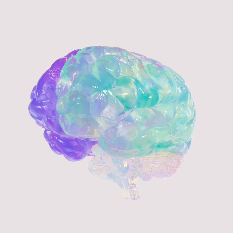
Micropolygyria, often referred to more commonly as Polymicrogyria, is a complex neurological disorder characterized by an abnormal development of the brain’s cerebral cortex. This condition manifests as an excessive number of unusually small and convoluted folds, known as gyri, on the surface of the brain. Instead of the typical broad, smooth gyri separated by sulci (grooves), the brain affected by micropolygyria presents a disorganized appearance with numerous tiny, irregular wrinkles, often described as a “cobblestone” or “bumpy” surface. This structural malformation is a critical indicator of disrupted neuronal development and migration during the early stages of brain formation. The term itself, derived from Greek roots, literally means “many small gyri,” precisely describing the macroscopic anatomical anomaly.
The fundamental mechanism underlying micropolygyria involves a severe disruption in the normal process of neuronal migration and cortical organization, which typically occurs during the second and third trimesters of pregnancy. During this crucial developmental window, newly formed neurons migrate from their birthplace deep within the brain to their final destinations in the cerebral cortex, where they organize into distinct layers and form the characteristic folds and grooves. In individuals with micropolygyria, this intricate process goes awry, leading to neurons stopping prematurely or migrating incorrectly. This misplacement and disorganization result in a thinned, often four-layered cortex (instead of the normal six layers) and the formation of numerous small, shallow gyri that lack the functional complexity of a healthy cortical architecture. The degree and pattern of these abnormalities can vary significantly, influencing the range and severity of clinical symptoms experienced by affected individuals.
This neurological condition is typically congenital, meaning it is present at birth, although its clinical manifestations may not become apparent until later in infancy or childhood. The exact cause remains largely unknown in many cases, making it a challenging disorder to predict or prevent. It is not a progressive degenerative disease; rather, it represents a static encephalopathy, meaning the structural abnormalities are fixed. However, the neurological consequences and the impact on a child’s development can be profound and evolve over time as developmental milestones are missed or seizures emerge. Understanding the core definition of micropolygyria is the first step toward appreciating the widespread impact this structural anomaly has on neurological function and, consequently, on an individual’s life.
Historical Context and Discovery
The recognition and detailed understanding of cortical malformations, including micropolygyria, have evolved significantly over centuries, paralleling advancements in neuropathology and, more recently, neuroimaging techniques. Early observations of brain anomalies date back to ancient times, but precise anatomical descriptions were limited by the tools available. The concept of disordered cortical folding began to take shape in the late 19th and early 20th centuries as neurologists and pathologists meticulously examined post-mortem brain specimens. Scientists like Paul Flechsig and Santiago Ramón y Cajal, through their pioneering work on brain architecture and neuronal organization, laid the groundwork for understanding normal cortical development, implicitly highlighting deviations from this norm. However, specific identification and categorization of conditions like micropolygyria as distinct entities were still nascent.
The true characterization of micropolygyria as a specific entity gained momentum in the mid-20th century, particularly with improved histological staining techniques and a growing focus on developmental neuropathology. Researchers began to consistently identify the distinctive microscopic features of the condition: an abnormally thick cortex with disorganized layers, often fused pial surfaces, and the characteristic small, numerous gyri. These early neuropathological studies were critical in establishing micropolygyria as a distinct form of cortical dysgenesis, differentiating it from other malformations such as lissencephaly (smooth brain) or pachygyria (thickened gyri). However, diagnosing these conditions during life remained a significant challenge, often only confirmed through autopsy.
The advent of advanced neuroimaging technologies, especially Magnetic Resonance Imaging (MRI) in the late 20th century, revolutionized the diagnosis and study of micropolygyria. MRI allowed clinicians to visualize the intricate details of brain structure in living individuals with unprecedented clarity, making it possible to identify the characteristic small, irregular gyri and abnormal cortical thickness non-invasively. This technological leap enabled earlier diagnosis, correlation of imaging findings with clinical symptoms, and the identification of different patterns and distributions of micropolygyria (e.g., bilateral, unilateral, focal, diffuse). This period marked a pivotal shift from post-mortem pathological identification to ante-mortem clinical diagnosis, significantly advancing research into its etiology, clinical spectrum, and potential management strategies. The understanding continues to evolve with genetic discoveries and refined imaging protocols.
Etiology and Pathogenesis
The precise cause of micropolygyria remains largely unknown in a significant number of cases, presenting a considerable challenge for diagnosis, counseling, and prevention. However, current research points towards a multifactorial etiology, implicating a complex interplay of genetic mutations, various prenatal insults, and occasionally, perinatal events. The common thread among these diverse causative factors is their capacity to disrupt the highly orchestrated processes of neuronal proliferation, migration, and cortical organization during critical stages of fetal brain development. It is this delicate window of neurodevelopment, particularly during the second and third trimesters, that is most vulnerable to disturbances leading to the formation of abnormal cortical folds.
Genetic factors represent a significant and increasingly recognized category of causes. A growing number of specific gene mutations have been identified and linked to different forms of micropolygyria. These genes are typically involved in crucial pathways related to brain development, including those governing neuronal proliferation, cell adhesion, migration, and differentiation. For instance, mutations in genes such as SRPX2, GPR56, WDR62, and others, have been associated with various syndromic and non-syndromic forms of polymicrogyria. Some of these genetic conditions follow autosomal recessive or dominant inheritance patterns, while others may be sporadic. The identification of these genetic underpinnings is crucial for understanding the molecular mechanisms of the disorder and offers potential avenues for genetic counseling and future targeted therapies, although the exact mechanism by which these mutations lead to the characteristic cortical malformation is still an active area of research.
Beyond genetic predisposition, a range of prenatal insults can also contribute to the development of micropolygyria. These environmental or acquired factors occur during fetal life and can severely impact the developing brain. Examples include intrauterine infections, such as those caused by cytomegalovirus (CMV) or Zika virus, which can directly damage brain cells or interfere with their migratory pathways. Ischemic events, such as strokes or reduced blood flow to the fetal brain, particularly during the critical period of cortical development, can also lead to focal or diffuse areas of micropolygyria. Exposure to certain toxins or drugs during pregnancy, though less commonly confirmed as a direct cause, is also hypothesized to play a role in some instances. While perinatal events, such as complications during birth leading to hypoxia-ischemia, are more often associated with white matter injuries, severe prolonged oxygen deprivation around the time of birth can potentially exacerbate or contribute to already vulnerable developmental processes in rare circumstances, though direct causation of classic micropolygyria by isolated perinatal events is less common than prenatal factors.
Clinical Manifestations and Diagnostic Approaches
The clinical features of micropolygyria are highly variable, depending significantly on the extent, location, and severity of the abnormal cortical folding. Because the cerebral cortex is responsible for a vast array of higher-order functions, disruptions to its structure can lead to a wide spectrum of neurological symptoms. One of the most common and often devastating manifestations is epilepsy, with seizures varying from focal to generalized, sometimes becoming refractory to medication. The abnormal neuronal architecture in micropolygyric regions creates an environment of hyperexcitability, making individuals prone to recurrent seizures. The age of seizure onset can vary, from early infancy to later childhood, and their specific characteristics often correlate with the particular brain regions affected by the malformation.
Beyond seizures, developmental delays are almost universally observed in individuals with significant micropolygyria, impacting various domains of functioning. These delays can include significant impairments in cognitive development, leading to intellectual disabilities ranging from mild to severe. Motor impairments are also prevalent, manifesting as difficulties with gross motor skills (e.g., walking, balance) and fine motor skills (e.g., grasping, manipulation). This can range from mild clumsiness and poor coordination to severe spasticity and cerebral palsy-like symptoms, especially if the motor cortex is involved. Speech and language delays are common, with some individuals experiencing severe communication deficits, while others may only have mild articulation issues. The overall developmental trajectory is often slowed, requiring intensive therapeutic interventions to optimize functional outcomes.
Furthermore, micropolygyria can impact a range of other sensory and higher cortical functions. Depending on the specific brain regions affected, individuals may experience visual impairments, including cortical blindness or problems with visual processing, if the occipital lobes are involved. Hearing deficits, though less common, can occur if auditory processing areas are compromised. Learning disabilities are frequent, even in those with average intelligence, due to difficulties with attention, memory, executive functions, and specific academic skills. Behavioral and emotional challenges, such as anxiety, attention-deficit/hyperactivity disorder (ADHD) symptoms, or autism spectrum disorder features, are also more commonly observed in children with micropolygyria, likely due to the widespread cortical disorganization affecting neural networks involved in social cognition and emotional regulation. The diagnostic process primarily relies on detailed clinical evaluation, electroencephalography (EEG) to assess brain electrical activity, and crucially, high-resolution Magnetic Resonance Imaging (MRI) of the brain, which can definitively visualize the characteristic abnormal cortical folding patterns.
Management and Therapeutic Strategies
Currently, there is no cure for the underlying structural brain malformation of micropolygyria. Therefore, treatment is primarily symptomatic and supportive, aimed at managing the diverse array of neurological symptoms and maximizing the individual’s quality of life and developmental potential. A multidisciplinary team approach is essential, involving neurologists, developmental pediatricians, therapists (physical, occupational, speech), educators, and social workers. The cornerstone of medical management often revolves around controlling seizures, which are a prominent and debilitating feature for many affected individuals.
Anticonvulsant medications are the primary pharmacological intervention for managing seizures associated with micropolygyria. The choice of medication depends on the seizure type, frequency, severity, and individual patient factors. It often involves a trial-and-error process to find the most effective regimen with the fewest side effects. In cases of severe, refractory epilepsy that does not respond to multiple medications, other treatment options may be considered, such as the ketogenic diet, vagus nerve stimulation (VNS), or, in highly selected cases with focal and resectable lesions, epilepsy surgery. Additionally, other medications such as sedatives might be used to manage associated symptoms like severe spasticity or sleep disturbances, which can significantly impact daily functioning and care.
Beyond pharmacological interventions, comprehensive rehabilitative therapies play a crucial role in addressing the developmental delays and motor impairments often seen in micropolygyria. Physical therapy is vital for improving gross motor skills, muscle tone, balance, and mobility, helping individuals achieve developmental milestones like sitting, crawling, and walking, or maintaining existing functional abilities. Occupational therapy focuses on enhancing fine motor skills, activities of daily living (ADLs), sensory processing, and adaptive strategies to promote independence in tasks like feeding, dressing, and writing. Speech and language therapy is indispensable for addressing communication deficits, ranging from improving articulation and vocabulary to developing alternative and augmentative communication (AAC) strategies for those with severe speech impairments. Educational support and special education programs are also critical to address learning disabilities and ensure access to appropriate educational opportunities tailored to the individual’s cognitive profile and learning style. The goal of these therapies is not to “fix” the brain malformation but to facilitate optimal neurodevelopmental outcomes and improve functional independence through targeted interventions and adaptive strategies.
A Practical Example of Living with Micropolygyria
Consider the case of a child named Alex, who was diagnosed with bilateral perisylvian micropolygyria at 18 months of age after presenting with frequent focal seizures and significant delays in speech and motor development. His parents initially noticed that Alex wasn’t babbling like other children his age, and he struggled with sitting independently and reaching for toys. The seizures, which manifested as brief staring spells and subtle twitching on one side of his face, prompted a neurological evaluation, including an MRI scan that revealed the characteristic small, irregular gyri predominantly in the regions surrounding the Sylvian fissure on both sides of his brain. This specific pattern of involvement often affects areas critical for speech and motor control.
The “how-to” of applying psychological and medical principles in Alex’s life involved a coordinated, ongoing, multidisciplinary effort. First, a pediatric neurologist prescribed anticonvulsant medication to control his seizures. This required careful monitoring and dose adjustments to find the optimal balance between seizure control and minimizing side effects like drowsiness. Simultaneously, Alex began an intensive regimen of early intervention therapies. His physical therapist worked with him on core strength, balance, and coordination, using playful exercises to encourage sitting, crawling, and eventually walking. The occupational therapist focused on fine motor skills, helping him grasp objects, manipulate toys, and developing early self-feeding skills, also addressing sensory sensitivities that made certain textures or sounds overwhelming for him.
Crucially, a speech and language pathologist became an integral part of his team. Recognizing his severe expressive language delay due to the perisylvian involvement, they started with basic vocalization exercises, followed by teaching him to use a few simple signs. As he grew, the focus shifted to picture-exchange communication systems (PECS) and eventually an augmentative and alternative communication (AAC) device, which allowed him to communicate his needs and wants through a tablet. Psychologically, his parents received counseling to cope with the diagnosis and learned strategies for creating a stimulating yet supportive home environment. This comprehensive approach, continuously adapting to Alex’s evolving needs, allowed him to make gradual but significant progress, enabling him to attend a specialized preschool and develop foundational skills despite his ongoing challenges. This example highlights the practical, intensive, and individualized application of therapeutic principles in managing micropolygyria.
Significance and Impact
The study and understanding of micropolygyria hold profound significance for the broader field of psychology and neuroscience, extending far beyond the immediate clinical management of affected individuals. This condition serves as a critical model for investigating fundamental processes of human brain development, particularly the intricate mechanisms of neuronal migration and cortical folding. By studying the genetic mutations and environmental insults that lead to micropolygyria, researchers gain invaluable insights into the genes and pathways essential for normal brain formation. This knowledge not only illuminates the etiology of this specific disorder but also contributes to a deeper understanding of the genetic and developmental underpinnings of other neurodevelopmental disorders, potentially leading to novel therapeutic targets or preventive strategies for a wider range of conditions affecting brain architecture and function.
In clinical psychology and neuropsychology, the impact of micropolygyria is substantial. It underscores the critical link between brain structure and function, vividly demonstrating how subtle yet widespread cortical disorganization can lead to a spectrum of cognitive, motor, and behavioral impairments. Neuropsychologists play a vital role in assessing the specific cognitive profiles of individuals with micropolygyria, identifying areas of strength and weakness, and guiding educational and therapeutic interventions. This understanding informs the development of specialized educational programs and rehabilitation strategies tailored to the unique learning styles and challenges presented by individuals with such structural brain anomalies. Furthermore, the psychosocial impact on families, including coping with chronic illness, navigating complex medical systems, and advocating for their child’s needs, highlights the importance of psychological support and counseling for caregivers, emphasizing the holistic nature of care required.
The application of knowledge gleaned from micropolygyria extends into various practical domains. In developmental psychology, it informs our understanding of atypical development trajectories and the factors that can derail normal cognitive and motor skill acquisition. In rehabilitation medicine, insights into the functional consequences of specific malformation patterns help optimize physical, occupational, and speech therapy protocols. The ongoing research into the genetics of micropolygyria is also crucial for genetic counseling, allowing families to understand recurrence risks and make informed reproductive decisions. Ultimately, the study of micropolygyria contributes significantly to our collective effort to unravel the mysteries of the developing brain, improve diagnostic accuracy, and enhance the quality of life for individuals living with complex neurological conditions.
Connections and Relations
Micropolygyria, as a disorder of cortical development, is intimately connected to a broader array of neurological and psychological concepts. It falls under the overarching category of cortical malformations, a group of conditions characterized by structural abnormalities of the cerebral cortex resulting from disturbances during embryonic and fetal brain development. Other prominent members of this category include lissencephaly (smooth brain, characterized by a lack of gyri and sulci), pachygyria (thickened gyri, where gyri are fewer and broader than normal), and heterotopia (clusters of neurons in abnormal locations, typically in the white matter). While each has distinct features, they share common pathogenic mechanisms involving abnormal neuronal proliferation, migration, or post-migratory organization. Micropolygyria is often considered a milder form of cortical disorganization compared to severe lissencephaly but can still lead to significant neurological deficits.
The concept of micropolygyria is also closely linked to the field of developmental neuroscience, which explores the processes by which the nervous system forms and develops. Understanding micropolygyria requires knowledge of neurogenesis (the birth of neurons), neuronal migration (neurons moving to their correct locations), and synaptogenesis (formation of synaptic connections). Disruptions in any of these fundamental processes can lead to structural anomalies like micropolygyria. Furthermore, it relates to clinical neurology, particularly in the subspecialties of pediatric neurology and epilepsy. The clinical presentation of seizures, developmental delays, and motor impairments necessitates a comprehensive neurological evaluation and ongoing management, placing it firmly within the purview of clinical neurological practice. Its genetic underpinnings also connect it to medical genetics and genetic counseling, as an increasing number of cases are attributed to specific gene mutations.
Within the broader subfield of neurodevelopmental disorders, micropolygyria stands alongside conditions such as autism spectrum disorder, intellectual disability, and cerebral palsy, often sharing overlapping symptoms and requiring similar multidisciplinary therapeutic approaches. While micropolygyria is a structural brain malformation, its functional consequences often manifest as cognitive and behavioral challenges that are central to the study of developmental psychology and neuropsychology. The impact on learning, communication, and social interaction necessitates an understanding of psychological principles for effective intervention. Therefore, micropolygyria serves as a powerful example of how a specific anatomical anomaly can have widespread functional implications, forging connections across neuroanatomy, neurophysiology, developmental biology, genetics, and various branches of clinical and cognitive psychology.