Microsplanchnic Type: Anatomy and Psychological Impact
- Introduction to Microsplanchinic Type
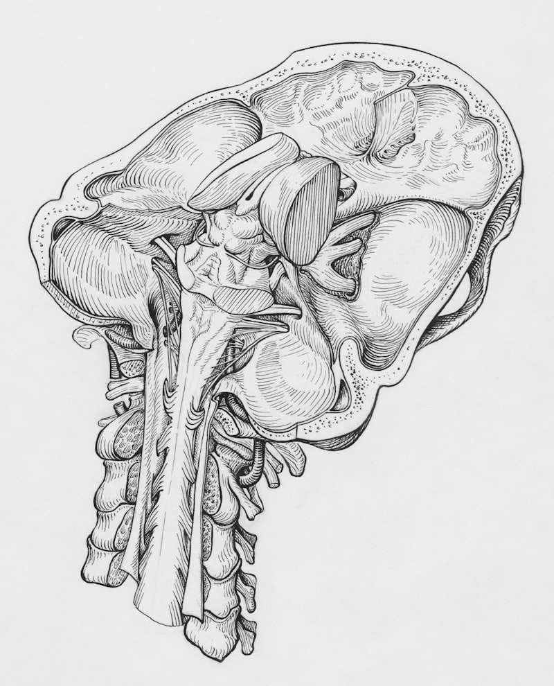
- Detailed Anatomical Characteristics
- Historical Recognition and Early Observations
- Clinical Implications and Associated Risks
- Comorbidities and Related Medical Conditions
- Psychological Impact and Patient Quality of Life
- Diagnosis and Management Strategies
- Connections to Broader Medical and Psychological Fields
- Future Research and Understanding
Introduction to Microsplanchinic Type
The term Microsplanchinic type refers to a rare and distinct anatomical variation characterized by a notably smaller abdominal cavity, accompanied by a narrow mesentery, a shortened mesenteric artery, and a proportionally short mesenteric vein. This specific constellation of features deviates significantly from typical human anatomy, presenting unique challenges and considerations within the medical field. It is not a disease in itself but rather an inherent structural predisposition that can influence physiological processes and clinical outcomes, impacting an individual’s physical health and subsequently, their psychological well-being.
This anatomical variant is considered exceptionally uncommon, with current estimates suggesting its prevalence in less than 1% of the general population. Its rarity often means that medical professionals may encounter it infrequently, making its recognition and understanding particularly important for accurate diagnosis and effective management of related conditions. The fundamental principle behind understanding Microsplanchinic type lies in recognizing how structural deviations, even subtle ones, can dramatically alter the functional dynamics of the visceral organs and the surrounding supportive tissues within the abdominal region, necessitating a holistic approach to patient care.
The unique configuration of a constricted abdominal space, combined with an attenuated mesentery and shortened vascular supply, has profound implications for the organs housed within, particularly the intestines. This structural peculiarity can lead to mechanical stresses, altered blood flow dynamics, and increased susceptibility to certain medical complications. Understanding this type transcends mere descriptive anatomy, extending into the realm of clinical physiology, surgical planning, and the psychological support required for individuals living with such a rare and impactful condition.
Detailed Anatomical Characteristics
At the core of Microsplanchinic type lies the distinctive characteristic of a significantly smaller abdominal cavity than typically observed. This reduction in volume means that the visceral organs, including the stomach, intestines, liver, and spleen, are housed within a more confined space. Such spatial constraints can affect organ positioning, mobility, and the overall physiological efficiency of the digestive and associated systems. The restricted environment inherently limits the natural movement and expansion of these organs, which can become particularly problematic during processes like digestion, pregnancy, or in conditions involving organ enlargement or inflammation, leading to a higher propensity for discomfort and dysfunction.
Complementing the small abdominal cavity is a remarkably narrow mesentery. The mesentery is a crucial double layer of peritoneum that anchors the small intestine and parts of the large intestine to the posterior abdominal wall, providing a conduit for blood vessels, nerves, and lymphatic vessels. In individuals with Microsplanchinic type, this supporting structure is not only narrower but also often shorter, leading to a more compact arrangement of the intestines. This lack of expansive support can predispose the bowel loops to crowding and potentially compromise their natural peristaltic movements, affecting nutrient absorption and waste elimination, thereby impacting overall gastrointestinal health.
Further contributing to this unique anatomical profile are a shortened mesenteric artery and a correspondingly short mesenteric vein. These vessels are vital for supplying oxygenated blood to the intestines and draining deoxygenated blood and nutrients away. The reduced length of these major vessels necessitates a more compact arrangement of the intestinal loops to maintain vascular continuity. This shortened vascular tree can impact the overall blood supply dynamics to the extensive length of the intestines, potentially making them more vulnerable to ischemic events or compromised perfusion under certain physiological stresses or surgical interventions, which can have cascading effects on organ viability and function.
The cumulative effect of these anatomical deviations—a constrained abdominal space, a compact mesentery, and shortened primary mesenteric vessels—presents substantial challenges for medical imaging and surgical procedures. Visualizing individual abdominal organs can be difficult due to their close proximity and altered positioning, making diagnostic interpretations more complex and requiring specialized radiological expertise. Moreover, performing abdominal surgery in such a confined and anatomically varied environment demands exceptional skill and precision, as the usual landmarks and expected spatial relationships are often distorted, increasing the technical difficulty and potential risks associated with interventions.
Historical Recognition and Early Observations
While the specific term “Microsplanchinic type” may be a more recent designation in medical literature, the broader concept of anatomical variations has been a subject of medical inquiry for centuries. Early anatomy, dating back to ancient Egyptian, Greek, and Roman periods, focused on descriptive human structure, often noting deviations from the norm during dissections. However, it was during the Renaissance, with figures like Leonardo da Vinci and Andreas Vesalius, that systematic and detailed anatomical studies truly flourished, laying the groundwork for understanding both typical and atypical human forms. The meticulous dissections of this era began to catalog a wider range of individual differences, though rare variants like this specific type would have been challenging to categorize without modern imaging and surgical techniques.
The systematic documentation of rare anatomical variations, particularly those impacting visceral organs and their vascular supply, gained significant momentum in the 19th and 20th centuries. Advances in surgical techniques and post-mortem examinations allowed for more detailed observations of internal structures. As medical understanding progressed, the importance of these variations, not merely as curiosities but as factors influencing pathology and surgical outcomes, became increasingly recognized. The development of specialized medical fields, such as surgical anatomy and clinical pathology, provided frameworks for identifying and classifying such rare types, moving beyond anecdotal observations to more structured medical reporting and scientific inquiry.
The recognition of Microsplanchinic type as a distinct clinical entity likely emerged from accumulated observations in surgical practice and diagnostic imaging, particularly as techniques like angiography and later CT and MRI scans became prevalent. These technologies allowed clinicians to visualize the internal architecture of the abdominal cavity and its vascular network with unprecedented detail, revealing consistent patterns of this specific rare variation. The detailed characterization of the small abdominal cavity, narrow mesentery, and shortened mesenteric vessels would have been crucial for its definition, distinguishing it from other more common anatomical anomalies or acquired conditions that affect abdominal structures, eventually leading to its formal description in the medical literature.
Clinical Implications and Associated Risks
The unique anatomical configuration of Microsplanchinic type carries several significant clinical significance, primarily stemming from the constrained space within the abdominal cavity and the altered mechanics of the mesenteric structures. One major consequence is an increased pressure within the abdomen, known as intra-abdominal pressure. In a typical abdomen, there is sufficient space for organs to shift and expand naturally. However, in Microsplanchinic type, the reduced volume means that even slight increases in organ size, gas accumulation, or fluid retention can rapidly elevate pressure, leading to a cascade of potential complications that impact organ function and patient well-being, necessitating careful monitoring and intervention.
This elevated intra-abdominal pressure significantly contributes to an increased risk of abdominal hernias. A hernia occurs when an organ, or fatty tissue, protrudes through a weak spot or opening in the muscle or tissue wall that normally contains it. In Microsplanchinic type, the persistent high pressure on the abdominal wall, combined with potential congenital weaknesses, can force intestinal loops or other abdominal contents through fascial defects, leading to various types of hernias, such as inguinal, umbilical, or incisional hernias, especially after surgery. The structural integrity of the abdominal wall is constantly challenged by the internal forces exerted by the confined viscera, making surgical repair often complex and prone to recurrence.
Furthermore, individuals with Microsplanchinic type often present considerable challenges during abdominal surgeries. The small abdominal cavity and the narrow, often short, mesentery make surgical access difficult and restrict the surgeon’s working space. Manipulating organs, identifying structures, and performing anastomoses (surgical connections) become more complex and time-consuming. This increased technical difficulty can prolong operative times, increase blood loss, and elevate the risk of intraoperative complications, necessitating highly experienced surgical teams and meticulous preoperative planning to navigate these anatomical intricacies safely and effectively, often requiring specialized techniques.
Another critical clinical concern is the observed increased risk of intra-abdominal infections in patients diagnosed with Microsplanchinic type. While the exact mechanisms are still under investigation, it is hypothesized that the reduced mobility of the intestines in a confined space, coupled with potentially compromised vascular supply due to shortened mesenteric vessels, could contribute to areas of stagnation or ischemia. These factors can create an environment conducive to bacterial overgrowth, translocation, or impaired immune response, making individuals more susceptible to infections such as peritonitis or abscess formation, particularly following surgical interventions or in the presence of inflammatory bowel conditions like Crohn’s disease.
Comorbidities and Related Medical Conditions
The presence of Microsplanchinic type has been noted in association with several distinct medical conditions, suggesting a potential underlying genetic or developmental link, or at least a propensity for these conditions to manifest more severely in the context of this specific anatomical variation. Two prominent examples include Crohn’s disease and Ehlers-Danlos syndrome. The interplay between the anatomical predisposition and the pathology of these conditions often exacerbates clinical challenges and complicates management strategies, requiring a nuanced understanding from healthcare providers.
Crohn’s disease is a chronic inflammatory bowel disease (IBD) that can affect any part of the digestive tract from mouth to anus, though it most commonly affects the small intestine and the beginning of the large intestine. In patients with Microsplanchinic type, the constricted mesentery and crowded intestinal loops might influence the pattern and severity of Crohn’s inflammation. The reduced mesenteric fat, which typically provides cushioning and support, could alter the inflammatory response or predispose segments of the bowel to increased mechanical stress, potentially leading to more severe strictures, fistulas, or abscesses in an already compromised anatomical setting, thereby intensifying disease burden and complicating treatment.
Another significant association is with Ehlers-Danlos syndrome (EDS), a group of inherited connective tissue disorders primarily affecting collagen. EDS is characterized by joint hypermobility, skin hyperextensibility, and tissue fragility. While the vascular type of EDS (vEDS) is particularly known for vascular and organ fragility, other types can also impact the gastrointestinal tract, leading to issues like chronic abdominal pain, digestive motility problems, and increased risk of hernias and organ rupture. The fragile connective tissues in EDS patients, when combined with the structural peculiarities of Microsplanchinic type, such as the narrow mesentery and increased intra-abdominal pressure, could synergistically heighten the risk of severe complications, including spontaneous bowel perforations or challenging hernia repairs.
Psychological Impact and Patient Quality of Life
Beyond the direct physiological and surgical challenges, living with a rare anatomical variation like Microsplanchinic type, especially when coupled with chronic conditions such as Crohn’s disease or Ehlers-Danlos syndrome, can have profound psychological impact on an individual’s quality of life. The journey often begins with a lengthy diagnostic process, during which patients may experience frustration, anxiety, and a sense of being misunderstood due to the rarity of their condition. This diagnostic odyssey itself can be a significant source of psychological distress, as individuals seek answers for their symptoms without immediate resolution, sometimes enduring years of uncertainty and misdiagnosis.
The chronic nature of associated conditions and the potential for recurrent hospitalizations, surgeries, and ongoing medical management can lead to significant emotional burdens. Patients may grapple with chronic pain, dietary restrictions, and limitations on daily activities, all of which can contribute to feelings of isolation, depression, and generalized anxiety disorder. The uncertainty surrounding their health, the fear of future complications, and the challenges of managing a complex medical regimen can further exacerbate these psychological stressors, requiring comprehensive care that extends beyond purely physical treatment, encompassing mental health support and psychosocial interventions.
Furthermore, the visible and invisible aspects of their condition can affect body image and self-esteem. Surgical scars, changes in body function (e.g., ostomies), or the need for assistive devices can alter an individual’s perception of their body, leading to self-consciousness, shame, or a diminished sense of well-being. The rarity of Microsplanchinic type also means that specific peer support networks might be less available or harder to find, potentially isolating patients further from those who truly understand their unique challenges. Therefore, a holistic approach that integrates psychological counseling, support groups, and patient education is crucial to help individuals cope with the multifaceted challenges imposed by this complex anatomical variant and its related health issues, enhancing their overall quality of life and fostering resilience.
Diagnosis and Management Strategies
The diagnosis of Microsplanchinic type primarily relies on advanced diagnostic imaging techniques, as its rarity and internal nature preclude simple physical examination for definitive identification. Cross-sectional imaging modalities such as Computed Tomography (CT) scans and Magnetic Resonance Imaging (MRI) are instrumental in visualizing the internal architecture of the abdominal cavity, assessing its dimensions, and detailing the length and configuration of the mesentery and its associated vascular structures. Angiography, either CT-angiography or MR-angiography, can provide crucial information regarding the length and branching patterns of the mesenteric arteries and veins, confirming the shortened nature characteristic of this type and guiding surgical planning.
Management strategies are predominantly focused on addressing the clinical implications and associated conditions rather than “curing” the anatomical variation itself. For instance, the increased risk of abdominal hernias necessitates vigilant monitoring and, if symptomatic or complicated, surgical repair. However, such repairs must consider the underlying anatomical constraints and potential tissue fragility, requiring specialized surgical expertise. Similarly, the management of Crohn’s disease or Ehlers-Danlos syndrome in these patients follows standard protocols for those conditions but with heightened awareness of how Microsplanchinic type might influence disease progression, treatment efficacy, or surgical outcomes, often requiring personalized treatment plans.
Surgical planning for individuals with Microsplanchinic type requires meticulous attention to detail and a thorough understanding of the patient’s specific anatomy. Surgeons must anticipate challenges related to limited operative space, altered organ positioning, and potentially fragile tissues or vessels. Minimally invasive techniques might be more difficult or contraindicated in some cases, necessitating open surgery to ensure adequate visualization and manipulation. Furthermore, given the significant psychological impact, a multidisciplinary team approach is often beneficial, including gastroenterologists, surgeons, geneticists, and importantly, mental health professionals to provide psychological support, coping strategies, and to address concerns related to quality of life, thereby offering comprehensive, patient-centered care.
Connections to Broader Medical and Psychological Fields
The study of Microsplanchinic type naturally connects to several broader medical disciplines, highlighting the interconnectedness of human anatomy, physiology, and pathology. It falls under the umbrella of developmental anomalies and congenital variations, a field explored within medical genetics and embryology. Understanding the genetic basis or developmental missteps that lead to such a rare configuration is a critical area of ongoing research, especially in light of its associations with other genetic conditions. Furthermore, its clinical implications place it firmly within general surgery, gastroenterology, and vascular surgery, as these specialists are most likely to diagnose and manage its associated complications, demanding cross-disciplinary collaboration.
From a psychological perspective, Microsplanchinic type, like other chronic and rare medical conditions, is highly relevant to the subfield of health psychology. Health psychology investigates how biological, psychological, and social factors influence health and illness. Patients with this condition face unique stressors, including the rarity of their diagnosis, the complexity of their medical management, and the potential for chronic pain or disability. These factors contribute significantly to their mental well-being, coping mechanisms, and overall quality of life, making psychological assessment and intervention integral to their care.
Moreover, the concept touches upon biopsychology, which explores the biological bases of behavior and mental processes. While Microsplanchinic type is an anatomical rather than a purely neurological condition, the way the body’s physical structure influences physiological function, and subsequently, an individual’s psychological state and coping abilities, is a direct application of biopsychological principles. It underscores the intricate mind-body connection, where a physical variation can lead to a cascade of medical issues that then profoundly affect mental health, underscoring the necessity of integrated care models that acknowledge both the anatomical reality and its extensive psychological ramifications for truly comprehensive patient support.
Future Research and Understanding
Despite the existing understanding of Microsplanchinic type, there remains a substantial need for further research to fully elucidate its etiology, long-term prognosis, and optimal management strategies. One primary area of future inquiry involves genetics. Identifying specific genetic markers or mutations associated with this anatomical variant could provide insights into its developmental origins and potentially lead to early diagnostic tools or even preventive strategies. Understanding the precise genetic underpinnings could also help to explain its association with conditions like Crohn’s disease and Ehlers-Danlos syndrome, clarifying whether these are coincidental associations or indicative of shared developmental pathways and predispositions.
Another critical area for future investigation is the development of standardized diagnostic imaging criteria and advanced imaging techniques that are specifically tailored to detect and characterize Microsplanchinic type more effectively. Given its rarity, enhanced diagnostic protocols could improve early identification, allowing for proactive management and counseling for affected individuals. Longitudinal studies are also essential to track the natural history of individuals with this anatomical variant, to better understand the progression of associated risks such as abdominal hernias and intra-abdominal infections, and to evaluate the long-term efficacy of various medical and surgical interventions.
Finally, from a biopsychosocial perspective, there is a clear need for research into the specific psychological impact of living with Microsplanchinic type. Studies focusing on quality of life, mental health outcomes, coping strategies, and the effectiveness of psychological interventions for these patients would be invaluable. Developing specialized patient education materials and support networks could also significantly improve patient well-being and empower individuals to navigate their complex health journeys. Ultimately, a collaborative, multidisciplinary research effort across anatomical, genetic, surgical, and psychological fields will be key to advancing our comprehensive understanding and improving the lives of individuals affected by this rare anatomical variation.