Motor Neurons: The Architecture of Human Movement
- Definition and Fundamental Function
- Classification and Types of Motor Neurons
- Anatomical Location and Motor Unit Organization
- The Neuromuscular Junction: Mechanism of Action
- The Final Common Path: Integration and Control
- Motor Neuron Activation and Force Generation
- Clinical Significance and Related Disorders
Definition and Fundamental Function
A motor neuron is an essential component of the human neuromuscular system, functioning as the primary efferent pathway that links the central nervous system (CNS) directly to the effector organs, specifically the muscle fibers. These highly specialized nerve cells are indispensable for virtually all movement, encompassing everything from subtle shifts in posture and automatic reflexes to complex, voluntary motor actions like walking or writing. Without the effective transmission of signals carried by motor neurons, the intricate musculoskeletal structure would be rendered inert, highlighting their critical necessity in maintaining life functions and interaction with the environment. They act as the final executors of commands generated by billions of processing neurons located in the brain and spinal cord, translating electrical signals into mechanical force.
The core function of the motor neuron is to propagate an action potential from its cell body, typically residing within the spinal cord or brainstem, along its often lengthy axon to the target muscle. Upon reaching the muscle fiber, this signal triggers a cascade of chemical events that culminate in muscle contraction. This singular responsibility for direct muscle activation has earned the motor neuron the crucial designation: the final common path (FCP). This term underscores the fact that regardless of where a motor command originates—be it the cerebral cortex, the cerebellum, the basal ganglia, or local reflex circuits—all such information must ultimately converge upon and be integrated by the motor neuron before any movement can occur. The motor neuron thus serves as the ultimate gatekeeper for physical action.
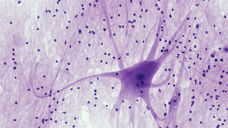
Anatomically, motor neurons exhibit a typical multipolar structure, featuring a large cell body (soma), numerous dendrites which receive thousands of synaptic inputs, and a single, myelinated axon. The length of this axon can be remarkably extensive, particularly in the case of motor neurons innervating distal muscles, such as those in the feet, where the axon may stretch a meter or more. This substantial physical connection ensures that the signal transmission is rapid, efficient, and highly reliable. The integrity of the axon, the myelin sheath surrounding it, and the terminal branches that synapse onto the muscle are all prerequisites for robust and coordinated movement, placing the motor neuron at the intersection of neurological command and physiological execution.
Classification and Types of Motor Neurons
Motor neurons are broadly categorized based on their anatomical location and the type of muscle fiber they innervate, leading to important functional distinctions. The most fundamental classification separates them into Upper Motor Neurons (UMNs) and Lower Motor Neurons (LMNs). UMNs originate in the primary motor cortex or brainstem and carry signals down to the spinal cord, where they synapse upon the LMNs. Conversely, LMNs are the true efferent neurons that constitute the final common path; their axons exit the CNS entirely to directly innervate skeletal muscle. Damage to UMNs results in different clinical signs (spasticity, exaggerated reflexes) than damage to LMNs (flaccidity, muscle atrophy), illustrating their distinct functional roles within the motor hierarchy.
Within the group of LMNs, several specialized subtypes exist, each tailored to specific roles in muscle control and sensation. The most numerous and functionally significant are the Alpha motor neurons. These large, fast-conducting neurons innervate the extrafusal muscle fibers—the main fibers responsible for generating the force of muscle contraction. A single Alpha motor neuron, along with all the extrafusal fibers it controls, constitutes a motor unit. The precise control of movement and the grading of muscle force are directly dependent on the activity and recruitment patterns of these Alpha motor units, which vary greatly in size depending on whether the muscle is designed for fine control (e.g., eye muscles) or gross power (e.g., quadriceps).
A second critical subtype includes the Gamma motor neurons. Unlike Alpha motor neurons, Gamma motor neurons do not directly contribute to the force generating contraction of the muscle. Instead, they innervate the intrafusal muscle fibers located within the muscle spindle, which is a specialized sensory receptor embedded parallel to the extrafusal fibers. The primary role of the Gamma motor system is to adjust the sensitivity of the muscle spindle. By causing the intrafusal fibers to contract slightly, the Gamma motor neurons ensure that the spindle remains taut and sensitive to changes in muscle length, especially during active muscle shortening. This mechanism is crucial for maintaining muscle tone and providing vital sensory feedback to the CNS, which is necessary for refined motor coordination.
Anatomical Location and Motor Unit Organization
The cell bodies of lower motor neurons are precisely organized within the CNS, specifically residing in the ventral (anterior) horn of the spinal cord gray matter or in the motor nuclei of the brainstem for cranial nerves. This arrangement is highly topographical, meaning that neurons controlling specific muscle groups are clustered together in distinct motor pools. For instance, motor neurons controlling axial and proximal limb muscles are typically located medially in the ventral horn, while those controlling the more distal muscles of the limbs are located laterally. This systematic organization ensures that motor commands traveling down the descending tracts can efficiently activate the correct combination of motor neurons required for a coordinated movement pattern.
The concept of the motor unit is central to understanding the functional output of the motor system. A motor unit consists of one single motor neuron and all the muscle fibers it innervates. The size of the motor unit—the number of muscle fibers per neuron—is inversely related to the degree of precision required of that muscle. Muscles designed for highly delicate movements, such as the intrinsic muscles of the hand or the extraocular muscles, possess small motor units (e.g., 10-20 fibers per neuron). In contrast, large, powerful muscles like the gastrocnemius possess motor units containing thousands of fibers per neuron. This structural variability allows the nervous system to select the appropriate level of motor granularity needed for any given task.
The transition from CNS processing to peripheral action necessitates the precise integration of information through the spinal cord. The descending pathways, such as the massive corticospinal tract, synapse directly or indirectly onto the LMNs. These LMNs then consolidate the final command and project their axons out of the ventral root, forming the peripheral nerves. This structural continuity ensures that conscious intent, generated in the cortex, is channeled efficiently through the spinal cord circuitry and delivered reliably to the muscles, confirming the LMN’s undisputed role as the final common path for musculoskeletal activation.
The Neuromuscular Junction: Mechanism of Action
The operational point where the electrical signal of the motor neuron is transduced into a chemical signal and then back into an electrical signal in the muscle fiber is the highly specialized synapse known as the Neuromuscular Junction (NMJ). This junction is structurally distinct from typical neuronal synapses due to its large size, high density of receptors, and unparalleled reliability; almost every action potential that reaches the presynaptic terminal successfully triggers a muscle action potential. The NMJ ensures that the command issued by the CNS via the motor neuron is executed faithfully and immediately by the muscle, making it a pivotal point in motor control.
The transmission process begins when the action potential arrives at the axon terminal of the motor neuron. This depolarization causes the opening of voltage-gated calcium channels, leading to an influx of calcium ions. The subsequent rise in intracellular calcium concentration triggers the rapid fusion of synaptic vesicles with the presynaptic membrane, releasing the neurotransmitter acetylcholine (ACh) into the synaptic cleft. ACh is the exclusive neurotransmitter utilized by Alpha motor neurons at the NMJ, making this specific chemical signal central to all voluntary muscular contraction. The swift and substantial release of ACh is paramount for initiating effective movement.
Once released, ACh rapidly diffuses across the narrow cleft and binds to specific receptors located on the muscle fiber membrane, which are known as nicotinic acetylcholine receptors. The binding of two ACh molecules opens these ligand-gated ion channels, allowing a massive influx of sodium ions into the muscle cell. This influx causes a significant local depolarization of the muscle membrane, termed the End Plate Potential (EPP). The EPP is typically large enough to reliably reach threshold, triggering a true muscle action potential which then propagates across the entire muscle fiber, initiating the release of intracellular calcium and leading directly to muscle contraction. Because the integrity of the motor neuron determines the health of the NMJ, failure at this interface, often due to autoimmune disease or toxins, results in severe weakness or paralysis.
The Final Common Path: Integration and Control
The designation of the motor neuron as the final common path is functional rather than purely anatomical, reflecting its critical role as the integrator of all descending, ascending, and local spinal cord inputs. Prior to firing, the motor neuron’s dendrites and cell body must process an enormous barrage of signals originating simultaneously from various sources, including excitatory commands from the primary motor cortex (via the UMNs), modulatory signals from the basal ganglia and cerebellum, and crucial inhibitory and excitatory feedback from sensory afferents and interneurons within the spinal cord. It is the algebraic summation of all these postsynaptic potentials—both spatial and temporal—that ultimately determines whether the motor neuron reaches its firing threshold.
This complex integration is vital for ensuring smooth, coordinated, and purposeful movement. For instance, during a simple movement like extending the arm, the motor neurons controlling the triceps (extensor) receive excitatory input from the UMNs, while the motor neurons controlling the antagonistic biceps (flexor) simultaneously receive inhibitory signals via spinal interneurons. This phenomenon, known as reciprocal innervation, prevents simultaneous contraction of opposing muscle groups, ensuring the efficiency and precision of the movement. The motor neuron, therefore, is not merely a passive conduit, but an active computational center that coordinates these opposing forces before issuing the final, unified command to the muscle.
Furthermore, the firing rate of the motor neuron is meticulously controlled to grade the force produced by the muscle. A low firing frequency results in muscle twitches, while increasing the frequency leads to a summation of contractions, culminating in a smooth, sustained contraction known as tetanus. The ability of the motor neuron to finely tune its firing rate based on the integrated input it receives allows for the incredible range and subtlety of human movement, from maintaining static balance against gravity to executing rapid, ballistic movements. This ability to synthesize disparate signals into a single, modulated efferent signal solidifies its standing as the indispensable final common path.
Motor Neuron Activation and Force Generation
The nervous system utilizes two primary mechanisms, both dependent on the motor neuron, to grade the force exerted by a muscle: motor unit recruitment and rate coding. Recruitment involves increasing the number of active motor units within the muscle. This process follows the influential Size Principle (Henneman’s Principle), which states that motor units are activated in an orderly sequence based on the size of the motor neuron cell body and axon. Smaller motor neurons, which innervate fewer muscle fibers and have lower thresholds for excitation, are recruited first during minimal effort. As the demand for force increases, larger motor neurons with higher thresholds are progressively recruited, bringing more powerful motor units into play.
The systematic recruitment ensures that force increases smoothly and incrementally, minimizing sudden, jerky movements. This mechanism is physiologically advantageous because the smaller, easily fatigable motor units handle low-level, sustained tasks (like posture maintenance), while the large, powerful, but quickly fatiguing units are reserved for movements requiring maximum force, offering an efficient energy conservation strategy. The ability of the motor neuron to adhere to this size principle, governed by the intrinsic properties of its membrane and the summation of incoming signals, is critical for achieving refined motor control.
Rate coding refers to the second mechanism, which involves increasing the frequency of action potentials fired by an already recruited motor neuron. At low frequencies, each action potential produces a brief twitch. As the firing frequency increases, the subsequent twitches occur before the muscle has fully relaxed, leading to temporal summation and increased force output. Eventually, at very high frequencies, the individual contractions fuse completely, resulting in a smooth, sustained, maximal contraction (fused tetanus). The continuous interplay between recruiting more motor units and increasing the firing rate of the active motor neurons allows the central nervous system to precisely adjust muscle tension to meet the specific requirements of any task.
Clinical Significance and Related Disorders
Given their role as the final common path, the health and function of motor neurons are vital; their degeneration or damage leads to severe, often fatal, neurological disorders. Motor neuron diseases (MNDs) specifically target these cells, resulting in progressive muscle weakness, atrophy, and eventual paralysis, without typically affecting sensory function or cognitive abilities. The most widely recognized and devastating condition is Amyotrophic Lateral Sclerosis (ALS), also known as Lou Gehrig’s disease, which involves the progressive death of both upper motor neurons in the cortex and lower motor neurons in the brainstem and spinal cord.
The clinical presentation differs based on the location of the lesion. Lower motor neuron lesions, where the LMN cell body or axon is damaged, result in muscle flaccidity, hypotonia (reduced tone), hyporeflexia (reduced reflexes), and visible muscle wasting (atrophy) and involuntary twitching (fasciculations), because the muscle is detached from the nervous system’s input. Conversely, Upper motor neuron lesions, where the descending pathways are damaged, typically result in spasticity, hypertonia (increased tone), and hyperreflexia, as the inhibitory control exerted by the brain over the LMNs is lost. Understanding which type of motor neuron is primarily affected is crucial for diagnosis and prognosis.
Other significant pathologies include Spinal Muscular Atrophy (SMA), a genetic disorder primarily affecting LMNs in the spinal cord, particularly in infants and children, leading to severe generalized muscle weakness. Additionally, infectious diseases such as Poliomyelitis specifically target and destroy the cell bodies of Alpha motor neurons, resulting in permanent flaccid paralysis in affected muscle groups. The vulnerability of the motor neuron, particularly its long axon and high metabolic demand, makes it a frequent target for both neurodegenerative processes and environmental insults, underscoring the delicate nature of the musculoskeletal structure’s dependence on this single line of command.