MS Lesion Imaging: Mapping the Mind’s Invisible Scars
- The Core Definition: Understanding Multiple Sclerosis Lesion Imaging
- Historical Context and Evolution of MSLI
- Technical Principles and Advanced Techniques
- Applications and Clinical Uses of MSLI
- A Practical Example: Navigating an MSLI Scan
- Significance and Impact on Psychology and Neurology
- Advantages and Disadvantages of MSLI
- Connections to Related Concepts and Broader Categories
The Core Definition: Understanding Multiple Sclerosis Lesion Imaging
Multiple Sclerosis Lesion Imaging (MSLI) refers to the specialized application of Magnetic Resonance Imaging (MRI) techniques specifically designed to detect, characterize, and monitor lesions associated with Multiple Sclerosis (MS) within the central nervous system. This advanced diagnostic tool is indispensable in the diagnosis, prognosis, and management of MS, a chronic autoimmune disease affecting the brain and spinal cord. MSLI provides highly detailed anatomical and pathological information, revealing the hallmark inflammatory and demyelinating lesions that define the disease.
The fundamental mechanism behind MSLI relies on the principles of MRI, which uses strong magnetic fields and radio waves to generate detailed images of organs and soft tissues within the body. In the context of MS, this technology is particularly adept at distinguishing between healthy brain tissue and areas affected by demyelination, inflammation, and axonal loss—the pathological processes characteristic of MS lesions. By employing various MRI sequences, such as T1-weighted, T2-weighted, and Fluid-Attenuated Inversion Recovery (FLAIR), neuroradiologists can visualize these lesions, assess their burden, and track their evolution over time.
A critical component of MSLI often involves the intravenous administration of a gadolinium-based contrast agent. This paramagnetic substance enhances the visibility of active inflammatory lesions by accumulating in areas where the blood-brain barrier, normally a protective interface, has been disrupted due to acute inflammation. The presence of gadolinium enhancement is a strong indicator of current disease activity, providing crucial insights into the dynamic nature of MS and guiding treatment decisions. Without MSLI, the precise localization and quantification of these lesions would be significantly more challenging, impeding both clinical care and research efforts into this complex neurological disorder.
Historical Context and Evolution of MSLI
The journey of MSLI is intrinsically linked to the broader development of Magnetic Resonance Imaging technology itself, which saw its conceptual foundations laid in the mid-20th century and its practical application in medicine beginning in the 1970s and 1980s. Before the advent of MRI, diagnosing Multiple Sclerosis was a protracted and often uncertain process, relying heavily on clinical symptoms, neurological examinations, and invasive procedures such as lumbar punctures to analyze cerebrospinal fluid. The ability to directly visualize brain and spinal cord pathology was a revolutionary leap forward.
Early MRI systems, while crude by today’s standards, quickly demonstrated their superior capability over computed tomography (CT) in detecting white matter lesions, which are characteristic of MS. Researchers and clinicians began to appreciate the sensitivity of MRI in identifying these discrete areas of demyelination and inflammation. Key figures in neuroimaging and neurology, across various academic institutions, spearheaded the application of this emerging technology to understand the pathophysiology of MS. Their pioneering work established the initial protocols for imaging the brain and spinal cord in MS patients, setting the stage for what would become MSLI.
The formal integration of MRI into diagnostic criteria for Multiple Sclerosis, most notably through the evolving McDonald Criteria, marked a pivotal moment. These criteria, first published in 2001 and subsequently revised, incorporated specific MRI findings—such as dissemination in space (lesions in multiple brain regions) and dissemination in time (new lesions over time or simultaneously enhancing and non-enhancing lesions)—as essential elements for a definitive diagnosis. This historical progression underscores how advancements in imaging technology directly influenced and refined the clinical understanding and diagnostic pathways for MS, transforming it from a largely clinical diagnosis to one heavily reliant on precise radiological evidence.
Technical Principles and Advanced Techniques
The effectiveness of Multiple Sclerosis Lesion Imaging stems from its utilization of various specialized Magnetic Resonance Imaging sequences, each designed to highlight different aspects of MS pathology. T2-weighted and FLAIR (Fluid-Attenuated Inversion Recovery) sequences are particularly sensitive to areas of increased water content, which are characteristic of edema and demyelination within MS lesions. FLAIR sequences are often preferred because they suppress the signal from cerebrospinal fluid, making periventricular lesions—a common location for MS lesions—much more conspicuous and easier to detect than on standard T2-weighted images.
In contrast, T1-weighted sequences are crucial for identifying “black holes,” which are hypointense (dark) areas that can represent severe tissue damage, such as axonal loss and atrophy, particularly when they persist after the acute inflammatory phase. The administration of a gadolinium-based contrast agent, which appears bright on T1-weighted images, is essential for detecting active lesions where the blood-brain barrier is compromised. These enhancing lesions signify acute inflammation and are a key indicator of current disease activity, providing valuable information for assessing treatment response and identifying relapses.
Beyond these conventional sequences, advanced MSLI techniques are increasingly employed to gain deeper insights into MS pathology. These include Diffusion Tensor Imaging (DTI), which measures the diffusion of water molecules to infer white matter tract integrity and detect subtle axonal damage; Magnetization Transfer Ratio (MTR), which quantifies demyelination; and Magnetic Resonance Spectroscopy (MRS), which can measure metabolite concentrations, providing biochemical information about tissue health and inflammation. Functional MRI (fMRI) is also being explored to understand brain reorganization and plasticity in response to MS. These advanced methods aim to provide a more comprehensive picture of both focal lesions and diffuse tissue damage, contributing to a more nuanced understanding of disease progression and cognitive impairment in MS patients.
Applications and Clinical Uses of MSLI
Multiple Sclerosis Lesion Imaging is a cornerstone in the comprehensive management of MS, impacting every stage from initial diagnosis to long-term monitoring. Its primary application lies in the diagnosis of MS, where it provides objective evidence of central nervous system involvement. According to the McDonald Criteria, MRI findings demonstrating dissemination in space (lesions in at least two characteristic MS locations) and dissemination in time (evidence of lesions occurring at different points in time, either new lesions or simultaneously enhancing and non-enhancing lesions) are critical for confirming an MS diagnosis, often allowing for earlier and more definitive identification of the disease.
Beyond diagnosis, MSLI is invaluable for monitoring disease activity and progression. Regular MRI scans help clinicians track the development of new lesions, the enlargement of existing ones, and the presence of new enhancing lesions, all of which indicate ongoing inflammation and demyelination. This longitudinal tracking is essential for assessing the effectiveness of disease-modifying therapies (DMTs). A reduction in new or enhancing lesions typically signifies a positive response to treatment, while continued lesion activity may prompt a change in therapeutic strategy. The ability to visualize these changes allows for personalized and adaptive management plans, optimizing patient outcomes.
Furthermore, MSLI plays a crucial role in differentiating MS from other neurological conditions that may present with similar symptoms, such as stroke, migraine, or other autoimmune diseases. The characteristic distribution, morphology, and evolution of MS lesions on MRI are often distinct, aiding in accurate differential diagnosis. In research settings, MSLI is fundamental for understanding the natural history of the disease, identifying prognostic markers, and evaluating novel therapeutic agents in clinical trials. By providing a quantifiable measure of disease burden and activity, MSLI continues to drive advancements in both clinical practice and scientific discovery related to Multiple Sclerosis.
A Practical Example: Navigating an MSLI Scan
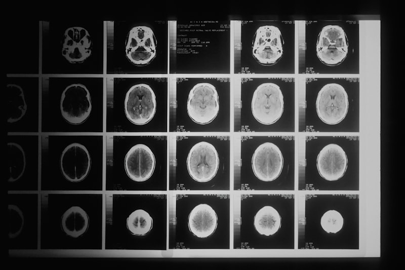
Consider Sarah, a 35-year-old woman who has recently experienced episodes of blurred vision, numbness in her left arm, and unexplained fatigue. Her neurologist suspects Multiple Sclerosis and refers her for a comprehensive Magnetic Resonance Imaging (MRI) scan of her brain and spinal cord, which constitutes the core of Multiple Sclerosis Lesion Imaging (MSLI). This practical scenario illustrates how MSLI is applied in a real-world clinical setting, from the patient’s perspective to the diagnostic interpretation.
Upon arrival at the imaging center, Sarah is prepped for her MRI. She lies on a narrow table that slides into the MRI scanner, a large, cylindrical machine. She is given earplugs to mitigate the loud knocking noises produced during the scan. The MRI technologist explains that the scan will involve several different sequences, some lasting a few minutes each, totaling approximately 45-60 minutes. Midway through the scan, an intravenous line is used to administer a gadolinium-based contrast agent. This agent is crucial because it highlights active inflammatory lesions by accumulating where the blood-brain barrier has been compromised, indicating acute disease activity.
Following the scan, a neuroradiologist reviews the vast number of images generated. In Sarah’s case, the MSLI reveals several distinct lesions in her brain, appearing bright on T2-weighted and FLAIR images, consistent with demyelination. Crucially, one of these lesions also shows enhancement after gadolinium administration on the T1-weighted images, indicating an active inflammatory process. Furthermore, the scan identifies a separate, older lesion in her cervical spinal cord that does not enhance. These findings—multiple lesions in different locations (dissemination in space) and evidence of both active and older lesions (dissemination in time)—meet the radiological criteria for a diagnosis of Multiple Sclerosis. The detailed information from the MSLI allows Sarah’s neurologist to confirm her diagnosis and immediately initiate a personalized treatment plan aimed at reducing inflammation and preventing further lesion development, significantly impacting her prognosis and future quality of life.
Significance and Impact on Psychology and Neurology
The advent and continuous refinement of Multiple Sclerosis Lesion Imaging have profoundly impacted both the fields of neurology and, indirectly, clinical psychology. For neurology, MSLI has transformed MS from a disease primarily diagnosed through clinical symptoms and spinal taps into one where precise anatomical and pathological visualization is paramount. It allows for earlier and more accurate diagnosis, which is critical because early intervention with disease-modifying therapies can significantly slow disease progression and minimize long-term disability. The ability to monitor lesion activity and brain atrophy over time provides objective measures of disease burden and treatment efficacy, guiding clinical decisions and improving patient care.
The impact extends to psychology by providing a biological basis for understanding the cognitive and emotional symptoms often experienced by individuals with MS. While MS is traditionally viewed as a motor and sensory disorder, a significant proportion of patients experience cognitive impairment (e.g., memory, attention, processing speed deficits) and psychological distress (e.g., depression, anxiety). MSLI, especially with advanced techniques like fMRI and DTI, helps researchers correlate specific lesion locations and patterns of brain damage with particular cognitive deficits or mood disturbances. This connection allows neuropsychologists to better understand the underlying neural correlates of these symptoms, leading to more targeted cognitive rehabilitation strategies and psychological interventions.
Furthermore, MSLI has been instrumental in advancing our understanding of the pathophysiology of Multiple Sclerosis, driving research into new diagnostic biomarkers and therapeutic targets. It has facilitated large-scale clinical trials for new disease-modifying drugs, providing quantifiable endpoints for treatment success. By offering a window into the living brain and spinal cord, MSLI has not only revolutionized patient care but also fostered a deeper scientific understanding of central nervous system diseases, bridging the gap between macroscopic pathology and the lived experiences of individuals affected by complex neurological conditions.
Advantages and Disadvantages of MSLI
Multiple Sclerosis Lesion Imaging, primarily using Magnetic Resonance Imaging, offers numerous compelling advantages that have cemented its role as the gold standard for diagnosing and monitoring MS. Foremost among these is its non-invasive nature, meaning it does not require surgical procedures, ionizing radiation, or the collection of tissue samples, making it safe for repeated use over a patient’s lifetime. MSLI provides exceptionally high spatial resolution, allowing for the detection of even small lesions (a few millimeters in size) in complex anatomical regions of the brain and spinal cord. Its superior soft-tissue contrast enables excellent differentiation between white matter lesions, gray matter lesions, and healthy neural tissue, which is critical for accurate diagnosis and assessment of disease burden.
Moreover, MSLI is uniquely capable of providing dynamic information about disease activity through the use of gadolinium contrast, which highlights acutely inflamed lesions. This feature is invaluable for identifying relapses, monitoring treatment efficacy, and differentiating new lesions from old ones. The ability to perform longitudinal studies with MSLI allows clinicians to track disease progression over years, adjust treatment strategies as needed, and gain insights into the natural history of MS. Furthermore, advancements in MRI sequences have made it possible to characterize different types of lesions, such as those indicating active inflammation versus chronic demyelination or axonal loss, contributing to a more nuanced understanding of the disease state.
Despite its significant advantages, MSLI also presents certain disadvantages and limitations. One major challenge is patient movement during the scan, which can lead to motion artifacts and compromise image quality, potentially resulting in inaccurate lesion detection or characterization. The MRI environment itself can be a source of discomfort for some patients due to the enclosed space (claustrophobia) and loud noises, sometimes requiring sedation. There are also contraindications for MRI, such as the presence of certain metallic implants (e.g., pacemakers, some surgical clips) that can interfere with the magnetic field or pose safety risks. The use of gadolinium contrast, while generally safe, carries a rare risk of adverse reactions, including nephrogenic systemic fibrosis in individuals with severe kidney dysfunction. Finally, while MSLI is highly sensitive, its specificity can sometimes be a concern, as other conditions can mimic MS lesions, necessitating careful clinical correlation and sometimes further diagnostic tests to ensure an accurate diagnosis.
Connections to Related Concepts and Broader Categories
Multiple Sclerosis Lesion Imaging is intimately connected to a wide array of psychological and neurological concepts and belongs firmly within the broader category of Neuroimaging and Neurology. Within neuroimaging, MSLI stands alongside other techniques like Positron Emission Tomography (PET) and computed tomography (CT), though MRI is uniquely suited for MS due to its superior soft tissue contrast and ability to detect demyelinating lesions. It complements clinical assessments, such as neurological examinations, evoked potentials (which measure electrical activity in response to sensory stimuli), and cerebrospinal fluid analysis, all of which contribute to the holistic diagnosis and understanding of MS.
The insights gained from MSLI are directly relevant to the pathophysiology of MS, particularly concerning concepts like demyelination (the loss of the myelin sheath protecting nerve fibers), inflammation (the body’s immune response causing damage), and axonal degeneration (damage to the nerve fibers themselves). MSLI provides visual evidence of these processes occurring in vivo, allowing researchers and clinicians to correlate imaging findings with disease progression and disability. Furthermore, MSLI plays a critical role in validating and applying diagnostic frameworks such as the McDonald Criteria, which have standardized the diagnostic process for MS globally.
From a psychological perspective, MSLI helps connect observable brain pathology to the cognitive and emotional symptoms often experienced by MS patients. Concepts like cognitive impairment, fatigue, and mood disorders in MS can be better understood by correlating them with lesion location, volume, and atrophy patterns seen on MSLI. This connection facilitates research into brain-behavior relationships in chronic neurological disease and supports the development of targeted neurorehabilitation and psychological support strategies. Thus, MSLI serves as a vital bridge between the macroscopic pathological changes in the central nervous system and the complex clinical manifestations of Multiple Sclerosis, influencing diagnosis, treatment, and our fundamental understanding of the disease across multiple disciplines.