Cellular Growth: The Psychology of Biological Patterns
A neoplasm, derived from Greek words meaning “new formation,” is fundamentally defined in pathology and medicine as an abnormal mass of tissue resulting from the uncontrolled, excessive proliferation of cells. This proliferation persists even after the cessation of the stimuli that initially triggered the growth, distinguishing it from normal tissue hyperplasia or repair processes. The key characteristic of a neoplasm, as initially described, is its novelty and deviation from standard biological structure and function. While the term is often used synonymously with a tumor or mass, it specifically emphasizes the underlying pathology of abnormal cellular growth rather than merely the physical presence of a lump. The original definition highlights that these growths typically lack the coordinated cellular structure found in healthy tissue, instead presenting as an ill-regulated bundle of cells that often exhibit aggressive behaviors within the host organism. Understanding the distinction between neoplastic growth and other forms of tissue enlargement is crucial for accurate diagnosis and prognostic determination in fields ranging from oncology to developmental biology.
The significance of a neoplasm lies in its potential to disrupt the normal physiological environment. As the original definition suggests, a neoplasm is typically used to describe a new, abnormal, and often malignant tumor, especially in the context of cancer. This implies a critical level of biological aggression and a profound threat to adjacent tissues and overall systemic health. The abnormal cellular mass exhibits properties of invasiveness, meaning it does not respect the boundaries of the tissue of origin. This invasion leads to the destruction or significant damage of the cells and surrounding matrix adjacent to the tumor mass, compromising organ function and initiating the cascade of events associated with advanced disease. The study of neoplasms, known as oncology, focuses on elucidating the molecular mechanisms that drive this uncontrolled growth and subsequent tissue destruction.
In essence, neoplastic development represents a critical failure in the body’s intrinsic regulatory systems governing cell division, differentiation, and apoptosis (programmed cell death). Normal cells adhere to strict growth limits dictated by genetic programming and environmental signals, maintaining tissue homeostasis. Neoplastic cells, however, acquire mutations that grant them growth autonomy, resistance to inhibitory signals, and limitless replicative potential, often through the reactivation of telomerase. This disregulation results in a population of cells that are not only proliferating rapidly but are also functionally and morphologically aberrant. This profound biological shift necessitates a detailed classification system to predict the clinical course and guide intervention strategies for patients affected by these abnormal growths.
- Definition and Fundamental Characteristics
- Classification: Benign versus Malignant Neoplasms
- Etiology and Mechanisms of Carcinogenesis
- Pathological Features and Histological Structure
- Invasiveness, Metastasis, and Angiogenesis
- Clinical Presentation and Diagnostic Procedures
- Risk Factors and Prevention
- Therapeutic Approaches to Neoplastic Disease
Definition and Fundamental Characteristics
The term neoplasm encompasses a broad spectrum of cellular abnormalities, but its defining feature remains the dysregulated and autonomous proliferation of tissue. Unlike inflammatory responses or regenerative processes, neoplastic growth is irreversible and non-physiological in its persistence. Pathologically, a neoplasm arises from a single cell (monoclonal origin) that has undergone genetic alteration, allowing it to escape the normal checks and balances of the cell cycle. This founding cell subsequently gives rise to a population of daughter cells that share these acquired genetic defects, leading to the formation of a macroscopic mass. The fundamental characteristics include excessive proliferation, reduced differentiation, and, critically, a degree of autonomy from host regulatory mechanisms.
The structure of a typical neoplasm is inherently disorganized, supporting the observation that there is often a distinct lack of cohesive cellular structure when compared to the highly organized architecture of the tissue of origin. This disorganization is referred to as dysplasia or anaplasia, depending on the severity of the cellular deviation. Anaplasia represents the most severe loss of differentiation, where neoplastic cells bear little resemblance to their mature counterparts, often displaying variations in nuclear size and shape (pleomorphism), increased mitotic activity, and abnormal mitotic figures. This structural breakdown is directly linked to the invasiveness often associated with malignant neoplasms, as the lack of normal cellular adhesion facilitates movement into the surrounding extracellular matrix and vasculature. The rate of growth is highly variable, ranging from slow, decades-long expansion in some benign tumors to exponential, rapid growth characteristic of highly aggressive malignancies.
Furthermore, the growth of a neoplasm is resource-intensive, often diverting necessary nutrients and blood supply from healthy tissues. A critical step in the progression of many neoplasms is angiogenesis, the formation of new blood vessels, which the tumor induces to supply its rapidly growing mass with oxygen and nutrients, and to remove metabolic waste. This newly formed vasculature is often structurally abnormal, leaky, and disorganized, but it is essential for the tumor’s sustained viability and subsequent potential for metastatic spread. The understanding that the neoplasm is an active entity, capable of manipulating its microenvironment, underscores the complexity of cancer biology and the challenge of therapeutic intervention.
Classification: Benign versus Malignant Neoplasms
Neoplasms are traditionally classified based on their potential for destruction and spread, categorized broadly into two primary groups: benign and malignant. While both types represent abnormal growths, their clinical behaviors and prognoses are vastly different. A benign neoplasm remains localized to its site of origin, typically grows slowly, is often encapsulated by a fibrous rim, and does not invade surrounding tissue. While benign tumors can cause significant morbidity through mass effect (compression of vital organs) or hormonal secretion (e.g., pituitary adenomas), they are generally not life-threatening unless located in critical, confined spaces like the brain or spinal cord. Their cells closely resemble the normal tissue from which they originated, indicating a higher degree of differentiation.
In stark contrast, a malignant neoplasm, commonly referred to as cancer, is characterized by its capacity for aggressive local invasion and the potential for metastasis—the spread to distant sites in the body. The original text correctly emphasizes that the term neoplasm is frequently used to describe a new, abnormal, malignant tumor. Malignant cells display poor differentiation (anaplasia), high mitotic indices, and exhibit cellular pleomorphism. The defining biological features of malignancy include the ability to breach basement membranes, invade adjacent structures, and gain access to the circulatory or lymphatic systems. This invasive behavior is the primary reason why malignant neoplasms pose a serious threat, as they actively destroy or damage adjacent cells, leading to systemic failure and death if left untreated.
The nomenclature of neoplasms is systematic, often incorporating the tissue of origin and the suffix indicating its biological nature. For instance, a benign tumor of glandular tissue is an adenoma, while its malignant counterpart is an adenocarcinoma. Tumors arising from mesenchymal tissue (e.g., bone, muscle, fat) are termed sarcomas when malignant (e.g., osteosarcoma) and are generally termed benign with the suffix ‘-oma’ (e.g., lipoma). However, there are exceptions; for example, melanoma, lymphoma, and seminoma are malignant despite ending in ‘-oma’. The precise histological classification is paramount for staging the disease and determining the most effective course of therapy, utilizing grading systems that assess the degree of cellular differentiation and mitotic rate.
Etiology and Mechanisms of Carcinogenesis
The development of a neoplasm, or carcinogenesis, is a complex, multi-step process driven by accumulated genetic damage and epigenetic alterations. It is fundamentally a disease of the genome. The initial event typically involves a somatic mutation in critical regulatory genes, specifically proto-oncogenes, tumor suppressor genes, and genes responsible for DNA repair. Proto-oncogenes, when mutated or overexpressed, become oncogenes that promote unchecked cell growth and division. Conversely, tumor suppressor genes, such as p53 and Rb (Retinoblastoma), normally inhibit cell proliferation or induce apoptosis; their inactivation or loss removes critical brakes on the cell cycle, leading to autonomous growth.
The process often requires multiple hits or sequential mutations over time, a concept known as clonal evolution. As the initial mutated cell divides, its descendants accumulate further genetic defects, leading to increased cellular heterogeneity within the tumor mass and escalating malignant potential. External factors, known as carcinogens, play a crucial role by inducing this genetic damage. Carcinogens can be chemical (e.g., components of tobacco smoke, asbestos), physical (e.g., ultraviolet radiation, ionizing radiation), or biological (e.g., certain viruses like Human Papillomavirus or Hepatitis B and C viruses). These agents directly or indirectly damage DNA, overwhelming the cell’s repair mechanisms and leading to permanent mutations that drive the neoplastic transformation.
Beyond simple genetic mutations, epigenetic modifications—heritable changes in gene expression that do not involve alterations to the underlying DNA sequence—are also critically involved in carcinogenesis. These include DNA methylation, histone modification, and microRNA dysregulation, which can silence tumor suppressor genes or activate oncogenes without structural genomic change. The interplay between genetic susceptibility, environmental exposures, and these epigenetic shifts determines the likelihood and trajectory of neoplastic development. Comprehensive genomic sequencing is now a standard tool used to map the specific molecular signatures driving an individual’s neoplasm, allowing for increasingly personalized therapeutic interventions.
Pathological Features and Histological Structure
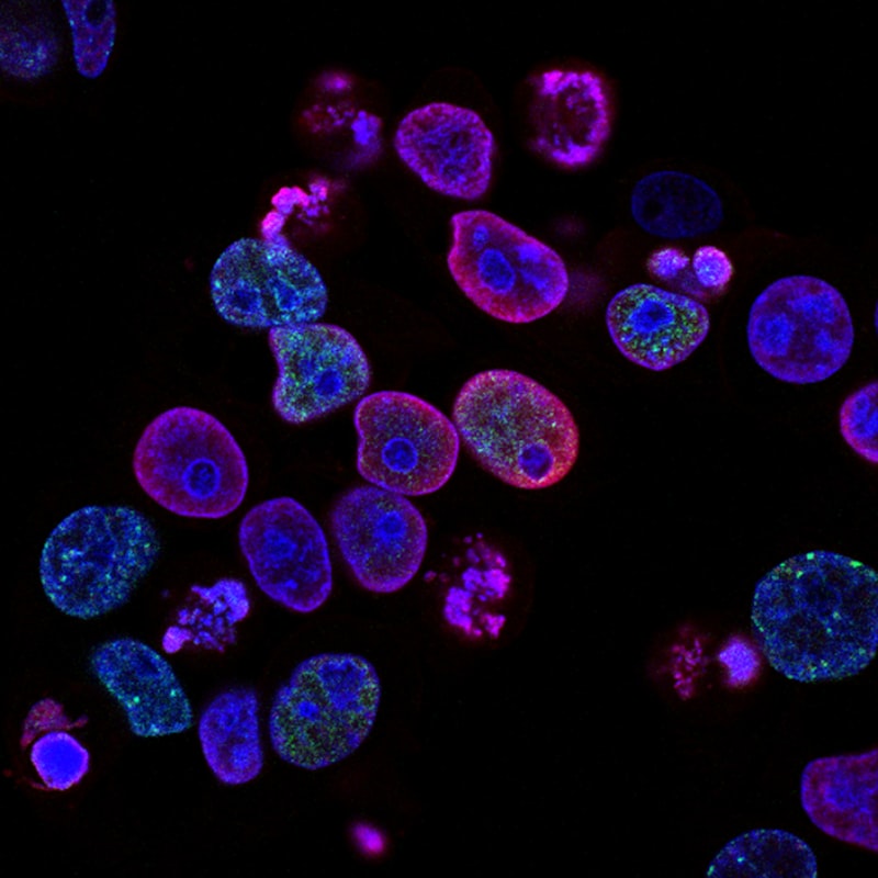
Pathological analysis is essential for confirming the presence of a neoplasm, determining its type, and assessing its grade. When examining neoplastic tissue under a microscope (histology), pathologists look for several key features that indicate abnormal proliferation and differentiation. These include alterations in cell morphology, such as an increased nuclear-to-cytoplasmic ratio, where the nucleus is disproportionately large and often hyperchromatic (darkly stained due to excess DNA). Furthermore, the organization of cells into tissues is profoundly disturbed; the normal polarity and architectural pattern are lost, replaced by disorganized sheets, clusters, or cords of cells that lack functional integration.
A critical pathological feature distinguishing malignancy is the breach of the basement membrane. In normal epithelial tissues, the basement membrane acts as a physical barrier preventing cells from invading deeper layers. Malignant cells acquire the ability to secrete lytic enzymes (e.g., matrix metalloproteinases) that degrade this barrier, facilitating local invasion into the stroma. This invasive growth pattern is the hallmark that confirms a diagnosis of carcinoma (malignant epithelial neoplasm). The presence of abnormal and frequent mitotic figures—cells actively dividing—is also a strong indicator of high proliferative activity and high-grade malignancy, confirming the aggressive nature of the abnormal growth.
The surrounding microenvironment, often called the tumor stroma, is also profoundly affected by the neoplasm. The stroma consists of extracellular matrix, connective tissue, inflammatory cells, and blood vessels. Neoplastic cells actively communicate with the stroma, inducing a desmoplastic reaction—the proliferation of dense fibrous connective tissue—that can sometimes be felt clinically as a hard, fixed mass. This interaction is reciprocal; the stroma provides structural support, growth factors, and immune evasion mechanisms that enhance tumor growth and survival. Pathological grading systems (e.g., Gleason score for prostate cancer, Nottingham grade for breast cancer) quantify these histological features to predict the biological aggression and likely clinical outcome of the specific neoplasm.
Invasiveness, Metastasis, and Angiogenesis
The most dangerous characteristic of a malignant neoplasm is its capacity for invasiveness and subsequent metastasis. Invasiveness refers to the local spread into adjacent tissues, a process where the bundle of cells actively penetrates and destroys or damages surrounding normal cells, as noted in the initial description. This local destructive behavior is mediated by altered cellular adhesion molecules and the secretion of enzymes that break down the extracellular matrix. Once local invasion is established, the tumor cells gain access to the circulatory (hematogenous spread) or lymphatic (lymphatic spread) systems, which serve as pathways to distant sites.
Metastasis is the process by which malignant cells travel through the vasculature or lymphatics, arrest in capillary beds of distant organs (e.g., lung, liver, bone, brain), and successfully establish secondary tumor colonies. This is an inefficient process, but the few cells that survive the journey and successfully colonize a secondary site determine the stage and prognosis of the cancer. The ability to metastasize requires that the neoplastic cells acquire several specialized biological capabilities, collectively known as the metastatic cascade, which include epithelial-to-mesenchymal transition (EMT), survival in the circulation, and successful colonization of a foreign microenvironment. The failure to treat or control metastasis is the primary cause of cancer-related mortality.
Crucially intertwined with invasiveness and metastasis is the process of tumor angiogenesis. A tumor larger than 1-2 millimeters requires its own blood supply to sustain growth and prevent central necrosis (cell death due to lack of oxygen and nutrients). Neoplastic cells secrete pro-angiogenic factors, notably Vascular Endothelial Growth Factor (VEGF), which stimulates the growth of new capillaries from existing host vessels toward the tumor mass. This neo-vasculature not only ensures sustained growth but also provides the necessary conduit for tumor cells to enter the bloodstream, thereby enabling hematogenous metastasis. Targeting tumor angiogenesis through therapies designed to block VEGF signaling is a major strategy in modern oncological treatment.
Clinical Presentation and Diagnostic Procedures
The clinical presentation of a neoplasm is highly varied, depending on its location, size, biological type (benign or malignant), and stage of progression. Symptoms can range from vague systemic complaints (e.g., unexplained weight loss, fatigue, fever) to specific local signs (e.g., palpable lump, abnormal bleeding, pain, or compression symptoms such as neurological deficits or intestinal obstruction). The development of a neoplasm in children or adults can become serious because they are essentially cancerous tumours, however they tend to be malignant, requiring immediate and often aggressive intervention. Early detection is paramount, as the prognosis is strongly correlated with the stage at diagnosis, highlighting the importance of screening programs.
Diagnosis typically involves a multi-modal approach combining imaging studies, laboratory tests, and pathological confirmation. Imaging techniques such as X-rays, Computed Tomography (CT) scans, Magnetic Resonance Imaging (MRI), and Positron Emission Tomography (PET) scans are used to localize the mass, determine its size, assess local invasion, and detect potential metastatic spread. Laboratory tests may reveal tumor markers (substances produced by the tumor or by the body in response to the tumor) or evidence of organ dysfunction related to the neoplastic process. However, imaging and markers provide circumstantial evidence; definitive diagnosis relies solely on obtaining tissue for microscopic examination.
The gold standard for diagnosing a neoplasm is biopsy. This procedure involves surgically removing a small sample of the abnormal tissue (or the entire mass) for histological analysis. Pathologists examine the biopsy specimen to confirm if the tissue is neoplastic, determine if it is benign or malignant, identify the tissue of origin, and assign a grade (degree of differentiation). Furthermore, modern pathology utilizes immunohistochemistry and molecular diagnostics to identify specific protein expressions and genetic mutations, which are critical for characterizing the specific subtype of the neoplasm and predicting its response to targeted therapies. This detailed pathological assessment is the foundation upon which all subsequent clinical management decisions are built.
Risk Factors and Prevention
Identifying and mitigating risk factors is central to the prevention of neoplastic disease. Risk factors are broadly categorized into hereditary predispositions and acquired environmental or lifestyle factors. While inherited germline mutations (e.g., BRCA1/2, Lynch syndrome genes) confer a high lifetime risk for specific cancers, the vast majority of neoplasms are sporadic, resulting from accumulated somatic mutations influenced by external exposures. Lifestyle factors such as chronic tobacco use, excessive alcohol consumption, obesity, poor diet, and lack of physical activity are major contributors to global cancer incidence, demonstrating the significant modifiability of overall neoplastic risk.
Environmental and infectious agents represent another major category of risk. Prolonged exposure to known chemical carcinogens (e.g., benzene, arsenic), occupational hazards, and chronic infections are highly correlated with increased risk. For instance, chronic infection with Helicobacter pylori increases the risk of gastric carcinoma, while chronic inflammation from inflammatory bowel disease increases the risk of colorectal cancer. Preventative strategies therefore emphasize reducing exposure to established carcinogens, promoting healthy lifestyle choices, and utilizing vaccines against oncogenic viruses, such as the HPV vaccine for preventing cervical and head and neck cancers.
Preventative measures also include proactive surveillance and screening for high-risk populations. Early detection through procedures like mammography (breast cancer), colonoscopy (colorectal cancer), and Pap smears (cervical cancer) allows for the identification and removal of precancerous lesions (dysplasia) or early-stage neoplasms before they develop the capacity for invasive growth and metastasis. Public health initiatives focused on education regarding sun protection, smoking cessation, and maintaining a healthy body weight are crucial components of primary prevention, aiming to reduce the incidence of the underlying genetic damage that initiates the process of neoplastic transformation.
Therapeutic Approaches to Neoplastic Disease
The management of a neoplastic disease is highly individualized, depending on the type, stage, grade, molecular profile, and overall health of the patient. Treatment goals range from curative intent (complete eradication of the disease) to palliative care (control of symptoms and maintenance of quality of life). The three classical pillars of cancer therapy are surgery, radiation therapy, and chemotherapy, which are often used in combination or sequence to achieve optimal outcomes.
Surgery remains the cornerstone for treating many solid tumors, aiming for the complete physical removal of the primary neoplasm and, often, regional lymph nodes. For malignant tumors, achieving clear surgical margins (removing all cancer cells visible microscopically) is critical for minimizing local recurrence. Radiation therapy utilizes high-energy rays to damage the DNA of neoplastic cells, leading to cell death. It can be used pre-operatively (neoadjuvant) to shrink the tumor, post-operatively (adjuvant) to eliminate residual microscopic disease, or as a primary treatment for localized tumors that are surgically inaccessible.
Systemic therapies, including chemotherapy, hormonal therapy, and targeted therapy, are necessary for treating metastatic or widespread disease. Chemotherapy employs cytotoxic drugs that target rapidly dividing cells, although this often results in side effects due to collateral damage to healthy proliferating cells (e.g., bone marrow, hair follicles). In the last two decades, advances in molecular biology have introduced targeted therapies (drugs that inhibit specific signaling pathways critical for tumor growth, like tyrosine kinase inhibitors) and, most recently, immunotherapy. Immunotherapies, such as checkpoint inhibitors, harness the patient’s own immune system to recognize and attack neoplastic cells, marking a paradigm shift in the successful long-term management of many aggressive malignancies. The integration of these diverse therapeutic modalities requires multidisciplinary teams working collaboratively to tailor the treatment plan to the specific characteristics of the neoplasm.