Cellular Nucleus: The Architect of Your Mental Life
- Definition and Foundational Biology
- Structure and Composition of the Nuclear Envelope
- Functions of the Nucleus: Genetic Regulation
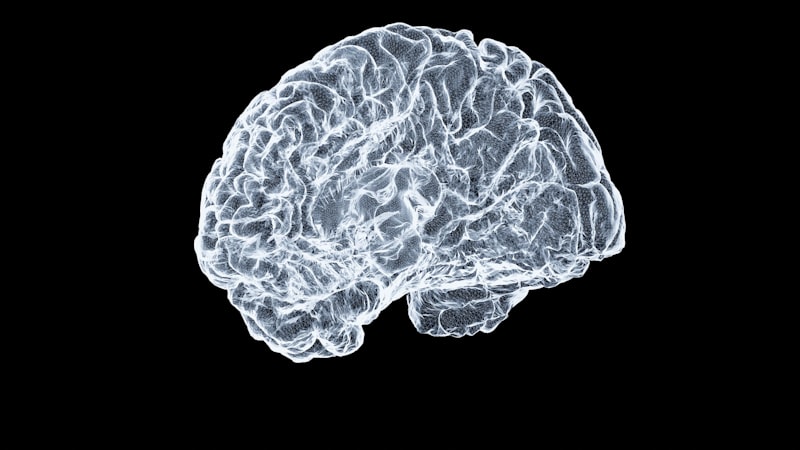
- Transition to Neuroscience: The Concept of Brain Nuclei
- Key Brain Nuclei: The Basal Ganglia System
- Brainstem and Thalamic Nuclei: Sensory and Motor Integration
- Nuclear Involvement in Disease and Pathology
Definition and Foundational Biology
The term nucleus (plural: nuclei) refers fundamentally to a prominent, membrane-bound organelle observed within the cells of all eukaryotic life forms, distinguishing them definitively from prokaryotic organisms such as bacteria and archaea, which lack this specialized compartment. Serving as the definitive control center of the cell, the nucleus houses the vast majority of the cell’s genetic material, primarily organized as long strands of deoxyribonucleic acid, or DNA, complexed with various proteins to form chromatin. This structure is not merely a storage vault; it is the site of essential processes including DNA replication, gene transcription, and the regulation of gene expression, all of which dictate the cell’s function, structure, and overall lifecycle. The presence of the nucleus allows for spatial and temporal separation of transcription (occurring within the nucleus) from translation (occurring in the cytoplasm), a critical mechanism for the complexity and adaptability inherent in multicellular organisms.
In terms of scale, the nucleus is typically the largest organelle within the eukaryotic cell, often occupying approximately ten percent of the cell’s total volume, and is generally the first component identified during initial microscopic examination, as noted in foundational biology education. Its size and central location underscore its biological importance; its structural integrity is vital for maintaining genomic stability. The nucleus essentially organizes the cell’s inherited traits and regulatory machinery, ensuring that the necessary proteins are synthesized at the appropriate times and in the required quantities. Furthermore, the nucleus plays a crucial role in initiating programmed cell death, or apoptosis, responding to cellular damage or developmental signals, thereby maintaining tissue homeostasis.
While the primary biological definition refers to this cellular organelle, the term nucleus is also employed extensively in neurobiology and psychology to describe a specialized cluster or mass of neuron cell bodies located deep within the central nervous system (CNS), such as the brainstem or basal ganglia. These neuronal nuclei perform specific, coordinated functions, acting as relay stations or processing centers for sensory, motor, or cognitive information. It is imperative to differentiate between the cellular nucleus, which is present within every nucleated cell, and the anatomical brain nucleus, which refers to a functional aggregation of neurons, often representing a critical component of a complex neural circuit responsible for behavior or cognition. Understanding the dual application of this term is essential for interdisciplinary study in biological psychology and neuroscience.
Structure and Composition of the Nuclear Envelope
The structural integrity of the cellular nucleus is maintained by the nuclear envelope, a highly specialized double-membrane system that physically separates the nucleus from the cytoplasm. This envelope is continuous with the endoplasmic reticulum (ER), which emphasizes the interconnectedness of cellular compartments. The outer nuclear membrane is often studded with ribosomes and engages in protein synthesis, while the space between the inner and outer membranes, known as the perinuclear space, is contiguous with the lumen of the ER. This dual membrane system is punctuated by numerous intricate protein channels called nuclear pore complexes (NPCs), which are indispensable for regulating the selective transport of macromolecules—including RNA molecules leaving the nucleus and regulatory proteins entering it—a process critical for gene expression control and cellular signaling.
The inner surface of the nuclear envelope is supported by the nuclear lamina, a dense meshwork composed primarily of intermediate filaments known as lamins. This lamina provides mechanical stability to the nucleus and serves as attachment points for chromatin fibers. Disruptions or mutations in the genes encoding lamins are associated with a class of rare genetic disorders termed laminopathies, which affect tissues requiring high mechanical stress, such as muscle and connective tissue, demonstrating the critical structural role of this underlying network. The dynamic association between the nuclear lamina and chromatin also suggests that the lamina plays a significant, though complex, role in organizing the three-dimensional architecture of the genome within the nuclear space, influencing which genes are accessible for transcription.
Internally, the nucleus contains the nucleoplasm, a viscous fluid analogous to the cytoplasm, which suspends the chromatin and other internal structures. The most prominent internal structure besides chromatin is the nucleolus, a non-membrane-bound dense region responsible for the synthesis of ribosomal RNA (rRNA) and the assembly of ribosomal subunits. The size and activity of the nucleolus are often direct indicators of the cell’s protein synthesis demand; highly active cells, such as those involved in rapid growth or secretion, typically possess large, highly visible nucleoli. Chromatin, the complex of DNA and histone proteins, exists in two main states: euchromatin, which is less condensed and transcriptionally active, and heterochromatin, which is highly condensed, typically silenced, and often found aggregated near the nuclear periphery or the nucleolus.
Functions of the Nucleus: Genetic Regulation
The primary and most critical function of the nucleus is the management and execution of genetic processes, including the faithful replication of the genome before cell division and the controlled expression of genetic information via transcription. Replication ensures that daughter cells receive an exact copy of the genetic blueprint, a process requiring precise coordination of numerous enzymes and checkpoint mechanisms. Transcription, the synthesis of RNA from a DNA template, is the first step in gene expression and is tightly regulated by an array of transcription factors and epigenetic modifications, which determine whether a specific gene is turned on or off in a particular cell type or at a specific stage of development. This intricate regulatory capacity allows a single genome to produce the diverse cell types necessary for a complex organism.
The nucleus effectively acts as a regulatory hub, receiving numerous signaling molecules from the cytoplasm and the extracellular environment. These signals often lead to the activation or inhibition of transcription factors that then translocate into the nucleus via the nuclear pores, binding to specific regulatory sequences on the DNA. This precise control over gene expression is fundamental to cellular differentiation, enabling a pluripotent stem cell to commit to becoming a neuron, a muscle cell, or an epithelial cell, for example. Errors in this signaling pathway or in the subsequent transcription process can lead to aberrant protein production, which is a hallmark of numerous diseases, including cancer and developmental disorders, highlighting the vulnerability inherent in such complex regulation.
Furthermore, the nucleus is the site where pre-messenger RNA (pre-mRNA) transcripts undergo extensive processing before being exported to the cytoplasm for translation. This processing includes splicing, where non-coding introns are removed and coding exons are ligated together, capping, and polyadenylation. Alternative splicing is a particularly important mechanism, allowing a single gene to encode multiple distinct protein isoforms, significantly increasing the complexity and diversity of the proteome without increasing the number of genes. The efficiency and accuracy of these post-transcriptional modifications are rigorously controlled within the nucleus, ensuring that only fully mature and functional mRNA molecules are exported, thus serving as a final quality control layer before protein synthesis commences.
Transition to Neuroscience: The Concept of Brain Nuclei
Moving beyond the cellular level, the term nucleus takes on a distinct anatomical meaning within the field of neuroscience, referring not to a subcellular compartment but to a centrally located, functionally related group of neuronal cell bodies (somas) within the central nervous system (CNS). This contrasts with the organization in the peripheral nervous system (PNS), where similar clusters of cell bodies are termed ganglia. Brain nuclei are integral components of neural circuits, serving as processing centers that integrate incoming synaptic input and generate coordinated output signals, often relaying information across major functional systems, such as the motor pathways or the limbic system. The precise organization and connectivity of these nuclei are what define specific neural functions and behaviors.
The identification and mapping of these nuclei are foundational to understanding brain organization. They are typically delineated based on cytoarchitecture (the arrangement and type of neurons), specific neurotransmitter profiles, and their distinct afferent (input) and efferent (output) connections. For instance, the collections of cell bodies that form the cranial nerve nuclei in the brainstem are responsible for controlling sensory and motor functions of the head and neck, each nucleus dedicated to a specific nerve and function, such as eye movement or facial sensation. The meticulous study of these nuclei is essential for clinical neurology, allowing physicians to localize neurological damage based on functional deficits observed in patients.
The functional complexity of the mammalian brain is largely attributable to the intricate interplay between these aggregated neuronal nuclei. While individual neurons within a nucleus process information, the nucleus as a whole performs a specific computational role, whether it is filtering sensory data, generating rhythmic motor patterns, or integrating emotional context into decision-making. The integrity of the connections between different nuclei is just as critical as the integrity of the nuclei themselves; disorders often arise not from the destruction of a single nucleus but from the failure of communication pathways connecting multiple nuclei across different brain regions, illustrating the distributed nature of complex cognitive functions.
Key Brain Nuclei: The Basal Ganglia System
Among the most functionally critical aggregations of neuronal cell bodies are those constituting the basal ganglia, a set of subcortical nuclei intimately involved in the initiation and execution of voluntary movements, procedural learning, and habit formation. The primary components of this system include the striatum (comprising the Caudate Nucleus and the Putamen), the Globus Pallidus (internal and external segments), the Subthalamic Nucleus, and the Substantia Nigra. These nuclei form a series of parallel, interlinked cortico-basal ganglia-thalamo-cortical loops that modulate cortical activity, ensuring smooth, purposeful motor output while suppressing unwanted movements. The balance of excitatory and inhibitory signaling within these loops is extraordinarily delicate.
The striatum serves as the major input structure, receiving glutamatergic excitatory signals from nearly all areas of the cerebral cortex. This input is then processed through two major pathways: the direct pathway, which facilitates movement, and the indirect pathway, which inhibits movement. The dynamic equilibrium between these two pathways, controlled largely by the dopaminergic input originating from the Substantia Nigra pars compacta (SNc), determines the motor state of the organism. Dysfunction in the basal ganglia is the underlying pathophysiology of several major neurological disorders. For example, the degeneration of dopaminergic neurons in the SNc leads to the characteristic motor symptoms of Parkinson’s Disease, including bradykinesia (slowness of movement) and resting tremor, directly demonstrating the crucial role of these nuclei in motor control.
Beyond motor function, specific loops involving the basal ganglia are dedicated to non-motor functions, including cognitive and affective processing. The associative loop, involving the prefrontal cortex, and the limbic loop, involving emotional centers, link the basal ganglia to decision-making, working memory, and motivation. For instance, the Nucleus Accumbens, often considered part of the ventral striatum, is a key nucleus within the brain’s reward circuit, playing a central role in reinforcement learning, addiction, and goal-directed behavior. Damage or dysregulation within these specific non-motor nuclei can contribute significantly to psychiatric conditions such as obsessive-compulsive disorder and major depressive disorder, underscoring the broad functional reach of the basal ganglia system.
Brainstem and Thalamic Nuclei: Sensory and Motor Integration
Another profound concentration of nuclei lies within the thalamus, often described as the brain’s primary relay station for sensory and motor information destined for the cerebral cortex. The thalamus is not a single structure but a collection of dozens of functionally distinct nuclei, each dedicated to processing specific types of information. For example, the Lateral Geniculate Nucleus (LGN) processes visual information originating from the retina before relaying it to the visual cortex, while the Medial Geniculate Nucleus (MGN) handles auditory information. Other nuclei, such as the Ventral Lateral Nucleus (VL), are critical components of the motor circuit, receiving input from the cerebellum and basal ganglia and projecting to the motor cortex, thus finalizing the motor command signal.
The brainstem—comprising the midbrain, pons, and medulla oblongata—houses numerous vital nuclei responsible for controlling basic life functions and mediating cranial nerve activity. These nuclei regulate essential autonomic processes that maintain homeostasis, such as respiration, heart rate, and blood pressure. Specific nuclei within the brainstem, such collectively known as the reticular formation, are crucial for regulating states of consciousness, arousal, and sleep-wake cycles. For example, the Nucleus Locus Coeruleus, located in the pons, is the primary source of norepinephrine projections to the rest of the forebrain, playing a critical role in attention, vigilance, and stress responses, linking brainstem function directly to psychological state.
Furthermore, the brainstem contains the cell bodies of the cranial nerves (CN III through CN XII), organized into distinct motor and sensory nuclei. The motor nuclei control muscles of the face, tongue, and pharynx, while the sensory nuclei receive input from special senses like taste and balance, and general sensation from the head. The integration of these functions is highly localized; for example, the Nucleus Solitarius receives taste information and visceral sensory input, playing a key role in the regulation of swallowing, gut motility, and the complex behavioral responses associated with feeding and satiety. Damage to even small, localized brainstem nuclei can lead to devastating, life-threatening deficits due to their irreplaceable roles in vital functioning.
Nuclear Involvement in Disease and Pathology
Pathological processes affecting the nucleus, whether at the cellular or the anatomical level, are central to a vast range of human diseases. At the cellular level, dysfunctions in the eukaryotic nucleus often involve genetic instability. Errors during DNA replication or repair, or defects in the nuclear envelope structure (as seen in laminopathies), can lead to catastrophic cellular failure or uncontrolled proliferation. The breakdown of gene regulation within the nucleus is a defining feature of cancer, where mutations lead to the inappropriate activation of oncogenes or the silencing of tumor suppressor genes, driving the cell toward malignancy. Furthermore, the accumulation of abnormal proteins within the nucleoplasm can disrupt essential functions, contributing to various neurodegenerative disorders where cellular stress is high.
In the context of anatomical brain structures, the specific vulnerability of certain neuronal nuclei to neurodegenerative diseases provides critical insights into their etiology.
- The aforementioned degeneration of the Substantia Nigra is the primary cause of Parkinson’s Disease, resulting in profound motor symptoms.
- In Huntington’s Disease, there is severe selective atrophy of the neurons primarily within the Caudate Nucleus and the Putamen (the striatum), leading to characteristic involuntary movements (chorea) and severe cognitive decline.
- In Alzheimer’s Disease, while cortical atrophy is widespread, deep nuclei involved in memory and cholinergic signaling, such as the Nucleus Basalis of Meynert, often show early and significant pathological changes (neurofibrillary tangles and amyloid plaques), contributing significantly to memory loss and cognitive impairment.
These examples underscore that the localized destruction of functionally critical nuclei can precipitate systemic behavioral and cognitive collapse.
Therapeutic interventions are often designed to target the functional output of these affected nuclei. For example, deep brain stimulation (DBS) for advanced Parkinson’s Disease often involves implanting electrodes into the Subthalamic Nucleus or the internal segment of the Globus Pallidus (GPi). By delivering high-frequency electrical pulses, DBS modulates the pathological activity patterns within these motor nuclei, thereby restoring a more balanced output and alleviating debilitating motor symptoms. This intervention is a powerful demonstration of how modulating the activity of a specific anatomical nucleus can profoundly impact complex, system-wide behavior, bridging foundational neuroanatomy with cutting-edge clinical treatment in neuropsychology.