Parallel Fiber: The Brain’s Hidden Neural Superhighway
- Introduction and Definition of the Parallel Fiber
- Anatomy of the Cerebellar Cortex
- The Granule Cell and its Axonal Bifurcation
- The Molecular Layer: Environment of the Parallel Fiber
- Synaptic Connectivity and Target Cells
- The Role in Cerebellar Circuitry and Signal Processing
- Contribution to Motor Learning and Coordination
- Pathophysiological Relevance and Clinical Implications
- Experimental Techniques and Future Research
Introduction and Definition of the Parallel Fiber
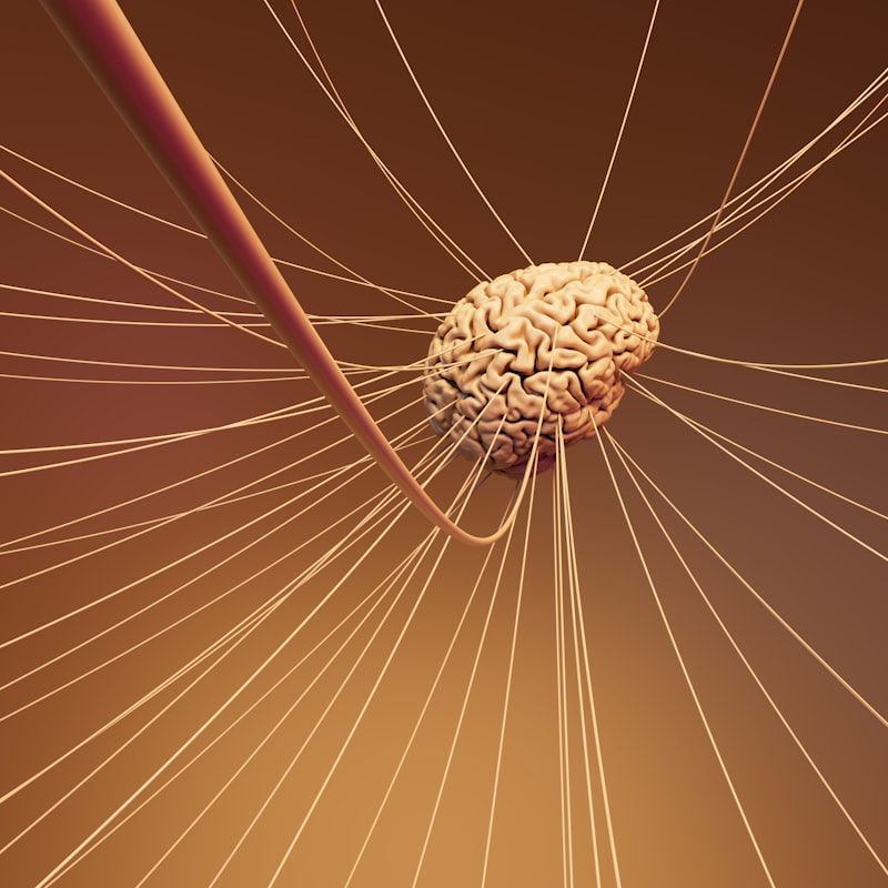
The term Parallel Fiber refers specifically to the highly distinctive axons originating from the granule cells, which constitute the most numerous neuronal population within the central nervous system. These fibers are the principal elements of the most superficial layer of the cerebellar cortex, known as the molecular layer. Characterized by their remarkably straight and parallel trajectory, these axons run longitudinally along the folia of the cerebellum, forming a vast, uniform network crucial for the temporal and spatial integration of sensory and motor information. Understanding the parallel fiber is central to comprehending the fundamental computational mechanism of the cerebellum, particularly its role in precise motor coordination, balance, and motor learning. Functionally, the parallel fibers provide the sole excitatory input to the extensive dendritic trees of the inhibitory interneurons—the stellate and basket cells—as well as the critical output neurons of the cortex, the Purkinje cells.
The naming convention, parallel fiber, is highly descriptive of its unique anatomical arrangement. After the granule cell axon ascends vertically from the granular layer, it reaches the molecular layer and bifurcates sharply into a ‘T’ shape. The two resultant branches extend horizontally, running strictly parallel to the pial surface and perpendicular to the planar orientation of the Purkinje cell dendritic arbors. This orthogonal arrangement is a defining feature of cerebellar organization, maximizing the potential synaptic contacts between a single parallel fiber and numerous Purkinje cells that lie within its path, thereby creating a highly distributed signal propagation system. This organization ensures that incoming mossy fiber input, relayed through the granule cells, is spread broadly across a narrow strip of cerebellar cortex, allowing for highly synchronized timing and coordination of complex motor outputs.
As the primary output structure of the granule cell, the parallel fiber carries excitatory signals mediated by the neurotransmitter glutamate. This excitatory input is inherently weak at the level of a single synapse; however, the massive convergence of thousands of parallel fibers onto a single Purkinje cell dendrite generates a powerful combined excitatory drive. This feature is essential for the cerebellar function because the computational power of the cerebellum relies not on the strength of isolated inputs, but on the precise timing and massive spatiotemporal summation of these numerous, weak excitatory signals. This system allows the cerebellum to perform rapid pattern recognition and prediction, enabling smooth, accurate movements by continuously adjusting and refining motor commands based on predicted sensory feedback.
Anatomy of the Cerebellar Cortex
The cerebellar cortex is a highly ordered, trilaminar structure, and the location of the parallel fiber is dictated precisely by this organization. The three distinct layers are the Granular layer (innermost), the Purkinje cell layer (middle), and the Molecular layer (outermost). The granular layer is densely packed with the small somata of the granule cells, along with Golgi cells and unipolar brush cells. The Purkinje cell layer contains the large cell bodies of the Purkinje neurons, forming a monolayer that serves as the sole output of the cortex. Finally, the molecular layer, where the parallel fibers reside, is characterized by low cellular density but high synaptic density, filled primarily with the extensive dendritic trees of the Purkinje cells and the axons of the granule cells.
The journey of the parallel fiber begins when the axon of the granule cell ascends through the Purkinje cell layer and into the molecular layer. This ascending segment, which is unmyelinated, is critical because it carries the signal vertically before the characteristic bifurcation occurs. The precise positioning and orientation of the parallel fibers within the molecular layer are critical for establishing the famous ‘cerebellar beam’ concept. Since all parallel fibers in a given area run in the same direction, activation of a group of granule cells creates a narrow, elongated zone of excitation running parallel to the fiber axis, effectively creating a functional line of excited tissue that crosses the dendritic fields of multiple Purkinje cells. This geometrical arrangement is fundamental to the ability of the cerebellum to process information in a vector-like manner.
The structural integrity and precise alignment of these layers are maintained by specialized glial cells, particularly the Bergman glia (a type of radial astrocyte). The processes of the Bergman glia envelop the parallel fibers and the Purkinje cell dendrites, providing metabolic support and regulating the extracellular environment, including the clearance of glutamate following synaptic transmission. This glial scaffolding is essential not only for the functional efficiency of the parallel fiber synapses but also for the proper migration and positioning of the neurons during development. Any disruption in the development of these glial-neuronal interactions can lead to severe disorganization of the molecular layer, impairing the parallel fiber circuitry and resulting in debilitating motor coordination disorders, known broadly as cerebellar ataxias.
The Granule Cell and its Axonal Bifurcation
The granule cell is the progenitor of the parallel fiber, and its unique morphology and immense quantity are key to understanding cerebellar computation. Granule cells are the smallest neurons in the mammalian brain, yet they outnumber all other neurons combined, providing the cerebellar cortex with an enormous capacity for pattern separation and input scaling. They receive their primary input from mossy fibers, which relay highly processed sensory and precerebellar information, such as proprioception and vestibular signals. A single granule cell typically receives inputs from only four to five mossy fibers, meaning that each granule cell acts as a sparse detector of specific combinations of inputs, enhancing the computational complexity of the cerebellar network.
Upon receiving sufficient input, the granule cell generates an action potential that travels up its single axon, termed the ascending axon. This axon traverses the depth of the molecular layer until it reaches the outer third, where the pivotal event of axonal bifurcation takes place. The ascending axon splits precisely into two equal branches, forming the “T” junction. These two branches immediately turn 90 degrees and proceed horizontally, becoming the definitive parallel fibers. The reason for this specific trajectory is structural efficiency: it allows the signal generated from a single point in the granular layer to be distributed widely and linearly across the surface of the cerebellar folium, creating the necessary line of excitation for spatial summation onto numerous output cells.
The functional significance of this bifurcation is profound. By transforming a vertical input into a linear horizontal spread, the parallel fibers effectively translate temporal coding (when the granule cell fires) into spatial coding (which Purkinje cells are affected along the line). This translation is the basis for timing movements. If a motor sequence requires activation of a specific set of Purkinje cells in a sequential manner, the parallel fiber system ensures that the firing of the initial granule cell will activate all downstream targets along the fiber’s path, but with slightly varying degrees of delay and summation, depending on the physical distance from the T-junction. This mechanism allows the cerebellum to predict the necessary timing intervals required for complex, multi-joint movements.
The Molecular Layer: Environment of the Parallel Fiber
The Molecular Layer serves as the primary processing arena for the parallel fiber system. Although sparsely populated by cell bodies, it is densely packed with synaptic connections and neuronal processes. The main components interacting with the parallel fibers are the vast dendritic trees of the Purkinje cells, which fan out perpendicular to the parallel fibers, and the inhibitory interneurons: the basket cells and the stellate cells. The environment is highly restrictive and geometric, designed to maximize the contact points between the excitatory parallel fibers and their inhibitory targets. This strict geometry is essential for maintaining the high fidelity and timing precision required for cerebellar function.
The inhibitory interneurons of the molecular layer, the stellate and basket cells, are crucial modulators of parallel fiber activity. Stellate cells, located superficially, and basket cells, located deeper, both receive strong excitatory input from the parallel fibers. In turn, they regulate the excitability of the Purkinje cells. Basket cells are particularly notable for their distinctive axons, which form a ‘basket’ around the soma and initial axon hillock of the Purkinje cells, providing powerful, localized inhibition. This arrangement ensures that the parallel fiber system does not simply excite the Purkinje cell, but also simultaneously drives the interneurons that shape and refine the Purkinje cell’s output, often gating or delaying the final firing pattern.
The environment of the molecular layer fosters significant synaptic plasticity. The parallel fiber-Purkinje cell synapse is the site of Long-Term Depression (LTD), a key cellular mechanism believed to underlie motor learning. The stability and maintenance of the parallel fibers are therefore paramount. The fibers themselves are unmyelinated and relatively thin, making them susceptible to metabolic stress. However, their physical integration with the Bergman glia ensures robust support. The overall geometry, where the excitatory input (parallel fibers) runs orthogonally to the main output (Purkinje cell dendrites), ensures that the timing of the input is critical. A slight shift in the synchronization of multiple parallel fibers can dramatically change the summation pattern on the Purkinje cell, directly influencing the accuracy of the motor command relayed to the deep cerebellar nuclei.
Synaptic Connectivity and Target Cells
The connectivity pattern established by the parallel fibers is the cornerstone of the cerebellar microcircuit. Parallel fibers form en passant synapses, meaning they form synapses along the length of the axon rather than only at the terminal end. These are generally small, asymmetric synapses, indicative of their glutamatergic, excitatory nature. A single parallel fiber may form contacts with hundreds of different Purkinje cells as it traverses the molecular layer, although the individual connection to any single Purkinje cell is typically weak and involves only one or two synaptic contacts. This diffuse, widespread connectivity pattern ensures that the motor command relayed by the parallel fibers is distributed across a large population of Purkinje cells.
The primary targets of the parallel fibers include:
- Purkinje Cell Dendritic Spines: The most critical target. Synapses occur on the spines of the immense Purkinje cell dendritic arbor. The overall firing rate and pattern of the Purkinje cell are determined by the massive convergence of potentially 150,000 to 200,000 parallel fiber inputs. This input drives the complex spike patterns essential for cerebellar timing.
- Stellate Cells: These inhibitory interneurons receive parallel fiber excitation and project their axons locally to inhibit the dendrites of nearby Purkinje cells, modulating the strength of the parallel fiber input.
- Basket Cells: Located deeper, these cells also receive parallel fiber excitation. Their powerful inhibitory output targets the Purkinje cell soma and axon hillock, controlling the Purkinje cell’s final output spike.
- Golgi Cells (indirectly): Some parallel fiber collaterals may interact with Golgi cells, which are primarily located in the granular layer but whose dendrites extend into the molecular layer. Golgi cells inhibit the granule cells themselves, creating a crucial feedback loop that controls the overall level of activity relayed by the parallel fiber system.
The efficacy of the parallel fiber synapse is highly regulated by the type of glutamate receptors present. These synapses predominantly utilize AMPA receptors for fast excitatory transmission, providing the rapid depolarization necessary for temporal precision. However, NMDA receptors are also present, and their activation is crucial for the induction of synaptic plasticity, particularly LTD. The co-activation of parallel fibers (AMPA/NMDA receptor activation) and the climbing fiber input (which causes a massive calcium influx) is the molecular signal that triggers the internalization of AMPA receptors at the parallel fiber-Purkinje cell synapse, leading to sustained depression of that synapse’s strength. This mechanism represents the cellular embodiment of learning in the cerebellar cortex.
The Role in Cerebellar Circuitry and Signal Processing
The parallel fiber system is integral to the primary feed-forward loop of the cerebellar cortex. This circuit begins with the Mossy Fibers, which convey information regarding ongoing movement and sensory state. Mossy fibers excite the granule cells, which, in turn, fire action potentials that travel via the parallel fibers across the molecular layer. The resulting excitation is then delivered to the Purkinje cells. Since Purkinje cells are GABAergic (inhibitory) and project out of the cortex to the deep cerebellar nuclei, the parallel fiber input is ultimately responsible for modulating the inhibitory output signal of the entire cortex.
The parallel fiber circuit functions as a highly sophisticated pattern separation mechanism. By spreading the input from a few mossy fibers across a vast number of granule cells and then disseminating that excitation over a wide area via the parallel fibers, the system translates a relatively coarse input into a highly refined, high-dimensional output signal. This process is essential for encoding context and fine temporal details of motor commands. For instance, the exact firing pattern of the parallel fibers across the molecular layer allows the cerebellum to distinguish between two motor actions that are otherwise very similar, ensuring that the appropriate muscle groups are activated with the correct force and timing.
Furthermore, the parallel fibers are critical for generating the complex spike patterns observed in Purkinje cells. Purkinje cells exhibit two distinct types of electrical activity: Simple Spikes, which are high-frequency, complex discharges driven primarily by the massive parallel fiber input, and Complex Spikes, which are low-frequency, high-amplitude spikes driven solely by the climbing fiber input. The parallel fiber inputs provide the raw excitatory material upon which the climbing fiber signal acts. When the climbing fiber signals an error (e.g., movement deviation), it interacts with the parallel fiber input to induce the plasticity (LTD) necessary to correct the simple spike pattern, thus refining the motor command for future iterations of the movement.
Contribution to Motor Learning and Coordination
The parallel fiber synapse is fundamentally linked to the cerebellar theory of motor learning, often encapsulated by the hypothesis proposed by Marr and Albus. This theory posits that the cerebellum acts as a learning machine, using the comparison between intended movement (signaled by mossy fibers/parallel fibers) and actual outcome (signaled by climbing fibers) to adjust the strength of the parallel fiber synapses, thereby correcting motor errors over time. The parallel fiber is thus not merely a passive conduit of excitation but a highly mutable element whose efficacy is constantly being calibrated.
The primary mechanism supporting this function is Long-Term Depression (LTD) at the parallel fiber-Purkinje cell synapse. LTD is a persistent weakening of the synaptic transmission. It is induced when the parallel fiber input (weak excitation) is precisely temporally paired with the strong, error-signaling input from the climbing fiber. This coincidence detection leads to a massive influx of calcium into the Purkinje cell spine, activating signaling cascades (including PKC) that result in the removal of AMPA receptors from the postsynaptic membrane. The weakening of the parallel fiber connection means that the same input pattern will produce a weaker response in the Purkinje cell in the future, effectively adjusting the output command to minimize the detected error.
Conversely, while less frequently studied in the context of cerebellar learning, Long-Term Potentiation (LTP)—the strengthening of the synapse—can also occur at the parallel fiber synapse under different conditions, often involving parallel fiber activity alone or specific patterns of activity lacking the climbing fiber signal. The balance between LTD and LTP in the parallel fiber system is what provides the necessary dynamic range for continuous motor adaptation. Whether learning to ride a bicycle or executing a complex musical score, the ability of the parallel fiber synapses to rapidly strengthen or weaken allows the cerebellum to store memory traces of motor skills, making the parallel fiber system the physical substrate for fine-tuning our interactions with the environment.
Pathophysiological Relevance and Clinical Implications
Disruptions to the development, function, or integrity of the parallel fiber circuitry lead directly to various forms of ataxia, the clinical hallmark of cerebellar dysfunction characterized by uncoordinated and inaccurate movements. Since the parallel fibers are the main source of cortical excitation, any damage to the granule cells or their axons severely cripples the ability of the Purkinje cells to integrate timing and spatial information, resulting in profound motor deficits, including dysmetria (inaccurate movement scaling) and intention tremor.
Specific conditions that involve parallel fiber pathology include:
- Developmental Disorders: Genetic mutations affecting granule cell proliferation or migration during embryogenesis (e.g., certain forms of medulloblastoma or granule cell layer hypoplasia) prevent the formation of properly aligned parallel fibers, leading to severe congenital ataxia.
- Toxic or Metabolic Insults: Exposure to certain toxins or chronic conditions like alcoholism can cause selective degeneration of granule cells, leading to a loss of parallel fiber input and subsequent Purkinje cell dysfunction.
- Ischemia and Trauma: The granule cell layer is metabolically highly active, and parallel fibers are vulnerable to damage from oxygen deprivation or physical trauma, leading to localized functional deficits that manifest as specific motor coordination issues.
The functional integrity of the parallel fiber is often assessed indirectly in clinical settings through measures of timing and movement accuracy. Damage to this system impairs the brain’s internal model of movement dynamics. Therefore, understanding the molecular mechanisms governing parallel fiber plasticity—LTD and LTP—is critical for developing targeted therapies for cerebellar disorders. For instance, pharmaceutical interventions aimed at modulating glutamate receptor activity or the downstream signaling cascades involved in LTD hold promise for restoring balance and coordination in patients whose parallel fiber synapses have become dysfunctional or inappropriately plastic.
Experimental Techniques and Future Research
The unique structure and accessibility of the parallel fiber system have made it a favorite target for neuroscientists studying synaptic transmission and plasticity. Classic electrophysiological techniques, particularly patch-clamp recording in cerebellar slices, have been instrumental in defining the basic properties of the parallel fiber-Purkinje cell synapse, including measuring synaptic current amplitude, kinetics, and the induction protocols for LTD. These experiments confirmed the coincidence detection mechanism involving parallel fiber and climbing fiber inputs.
More recently, advanced technologies have provided unprecedented resolution into parallel fiber function:
- Optogenetics: Using genetically encoded light-sensitive ion channels expressed specifically in granule cells, researchers can precisely control the firing of parallel fibers with millisecond accuracy, allowing for detailed investigation of how timing affects synaptic plasticity and motor output in vivo.
- Two-Photon Microscopy: This technique allows researchers to visualize calcium signaling and structural changes in individual parallel fiber synapses and Purkinje cell spines during learning tasks, offering insight into the subcellular events that drive plasticity.
- Computational Modeling: Due to the highly regular and geometric structure of the cerebellar circuitry, the parallel fiber system is ideally suited for large-scale computational modeling. These models help predict how the vast convergence of thousands of weak parallel fiber inputs translates into the specific firing patterns of the Purkinje cells and how these patterns contribute to overall motor behavior.
Future research is focused on translating the known cellular mechanisms of parallel fiber plasticity into clinical applications. Key areas of investigation include deciphering the heterogeneity among parallel fiber synapses—as not all synapses are equally plastic—and understanding how other neuromodulators, beyond glutamate, such as acetylcholine and serotonin, might fine-tune the parallel fiber transmission. A deeper comprehension of the complex interactions between the parallel fibers, inhibitory interneurons, and Bergman glia will further refine our understanding of how the cerebellum computes and learns, paving the way for novel treatments for movement disorders.