The Paraventricular Nucleus: Your Brain’s Stress Control
- Introduction and General Anatomy
- Magnocellular and Parvocellular Divisions
- Synthesis and Release of Oxytocin and Vasopressin
- Role in Stress Response (HPA Axis)
- Regulation of Fluid and Cardiovascular Homeostasis
- Afferent and Efferent Circuitry
- Regulation of Feeding and Metabolism
- Clinical Significance and Disorders
Introduction and General Anatomy
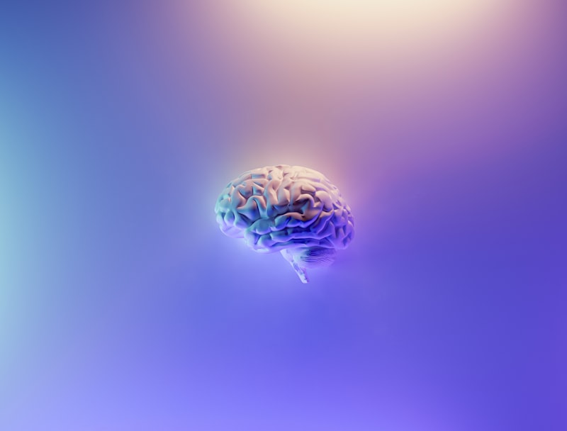
The Paraventricular Nucleus, often abbreviated as the PVN or PVH, stands as one of the most functionally critical nuclei residing within the hypothalamus. Located immediately adjacent to the third ventricle, this complex aggregation of neurons serves as a central integration hub for various physiological and behavioral processes, fundamentally bridging the nervous system and the endocrine system. Its strategic position allows it to receive extensive afferent input from numerous brain regions involved in monitoring internal states, including those related to stress, energy balance, and fluid homeostasis. The PVN is not merely a relay station; rather, it is a sophisticated modulator that processes complex sensory and hormonal signals to initiate appropriate neuroendocrine and autonomic responses, thereby maintaining the delicate internal equilibrium necessary for survival.
Anatomically, the PVN is characterized by its heterogeneous cellular composition, comprising distinct populations of neurosecretory cells, pre-autonomic neurons, and various interneurons. This structural diversity underpins its vast functional repertoire. Unlike many hypothalamic nuclei that specialize in a singular function, the PVN coordinates outputs that regulate pituitary function, activate the sympathetic and parasympathetic nervous systems, and project directly to brainstem and spinal cord centers. This intricate network ensures that when the body encounters a challenge—be it dehydration, high stress, or metabolic need—the PVN can simultaneously orchestrate multiple systemic adjustments, making it central to the concept of allostasis, or maintaining stability through change.
The definition of the PVN’s role begins with its capacity for neurosecretion. The neurons within this nucleus are renowned for their profound synthesis of two key nonapeptide hormones: vasopressin (also known as antidiuretic hormone or ADH) and oxytocin (OT). These neurohormones are packaged into vesicles and transported along axonal tracts to the posterior pituitary gland (neurohypophysis) for subsequent release into the systemic circulation. This pathway represents a classic neuroendocrine reflex, critical for regulating processes ranging from water retention and blood pressure modulation to complex social and reproductive behaviors, confirming the PVN’s indispensable role in both visceral control and higher-order functions.
Magnocellular and Parvocellular Divisions
The functional diversity of the Paraventricular Nucleus is structurally organized into two primary populations of neurosecretory cells, distinguished primarily by their size, axonal projection targets, and the types of substances they release. These are the magnocellular neurons and the parvocellular neurons. The magnocellular division, characterized by large cell bodies, primarily occupies the dorsal and medial aspects of the PVN. These large neurons possess long axons that travel down the hypothalamo-neurohypophysial tract, terminating in the posterior pituitary gland. This magnocellular projection is the classical pathway responsible for the massive, pulse-like release of neurohormones directly into the bloodstream in response to specific physiological demands.
Conversely, the parvocellular division consists of smaller neurons distributed throughout the anterior, medial, and lateral portions of the PVN. The parvocellular neurons exhibit far more diverse projection patterns than their magnocellular counterparts. While some parvocellular neurons project to the median eminence to regulate the anterior pituitary via the portal circulation, others project extensively to autonomic centers in the brainstem and spinal cord. These parvocellular projections are pivotal for controlling autonomic functions, such as heart rate, respiration, and gastrointestinal motility, providing a direct neural route for hypothalamic regulation of the peripheral nervous system.
A key distinction in function lies in their endocrine targets. The magnocellular neurons are the exclusive source for the systemic release of high concentrations of vasopressin and oxytocin from the posterior pituitary, actions which affect distant targets like the kidney and smooth muscle. The parvocellular neurons, however, are critical for the hypothalamic control of the anterior pituitary. Specifically, a large population of parvocellular neurons synthesizes and releases Corticotropin-Releasing Hormone (CRH) and Thyrotropin-Releasing Hormone (TRH) into the portal system. These releasing hormones then travel to the anterior pituitary to stimulate the release of Adrenocorticotropic Hormone (ACTH) and Thyroid-Stimulating Hormone (TSH), respectively, thus placing the PVN at the apex of the stress axis and the thyroid axis.
The organizational complexity of the PVN further includes several distinct subdivisions within both the magnocellular and parvocellular regions, each contributing to specialized functions. For instance, the medial parvocellular part is largely dedicated to the production of CRH, governing stress responses, while the lateral parvocellular part often contains pre-autonomic neurons projecting to visceral nuclei. This highly compartmentalized structure allows the PVN to integrate seemingly disparate functions—from fluid balance and endocrine regulation to autonomic outflow—within one compact anatomical area, reinforcing its role as a master regulator of neuroendocrine integration.
Synthesis and Release of Oxytocin and Vasopressin
The process of neurohormone synthesis in the PVN is a remarkable example of protein production and packaging within the central nervous system. Both vasopressin and oxytocin are synthesized as large precursor molecules within the rough endoplasmic reticulum of the magnocellular neurons. These precursors, known as propressophysin for vasopressin and prooxyphysin for oxytocin, contain the nonapeptide hormone sequence along with its specific carrier protein, neurophysin I (for oxytocin) or neurophysin II (for vasopressin). Following synthesis, these precursors are transferred to the Golgi apparatus where they are packaged into large dense-core vesicles, marking the beginning of their journey down the axon.
As the vesicles travel down the long axons of the hypothalamo-neurohypophysial tract towards the posterior pituitary, enzymatic cleavage occurs within the vesicles. This proteolytic process separates the active hormone (vasopressin or oxytocin) from its neurophysin carrier. The mature hormone and its carrier protein are then stored together in the nerve terminals located within the posterior pituitary gland, awaiting the appropriate stimulation for exocytosis. This co-storage and co-release mechanism ensures that both the active hormone and its binding protein are available upon demand, although only the peptide hormone itself exerts the physiological effects on target tissues.
The release of these hormones is triggered by afferent signals arriving at the PVN, which depolarize the magnocellular neurons and initiate action potentials that propagate all the way to the nerve terminals in the posterior pituitary. For vasopressin, the primary stimuli are increased plasma osmolality, detected by osmoreceptors in the organum vasculosum of the lamina terminalis (OVLT), and decreased blood volume or pressure, detected by baroreceptors. For oxytocin, the classic stimuli involve suckling (leading to milk ejection) and cervical distention during labor (promoting uterine contractions). These stimuli induce rapid, massive bursts of electrical activity, resulting in the synchronized release of thousands of hormone-filled vesicles into the systemic circulation within seconds.
The physiological actions resulting from this release are vital for survival. Vasopressin acts primarily on the kidney collecting ducts to increase water reabsorption, thus concentrating the urine and conserving body fluid volume. This action is paramount in maintaining fluid homeostasis. Oxytocin, while famous for its reproductive roles, is increasingly recognized for its central actions in modulating social bonding, trust, and anxiety within the brain. The dual nature of the PVN—acting as both a major endocrine gland and a critical hub for complex behaviors—underscores its significance in integrated physiology.
Role in Stress Response (HPA Axis)
The Paraventricular Nucleus is the primary control center for the initiation of the body’s neuroendocrine response to stress, serving as the capstone of the Hypothalamic-Pituitary-Adrenal (HPA) axis. When physical or psychological stressors are perceived, signals rapidly converge on the parvocellular neurons of the PVN, particularly those residing in the medial parvocellular subdivision. These neurons respond by synthesizing and releasing large quantities of Corticotropin-Releasing Hormone (CRH) into the hypothalamic-pituitary portal system. CRH is the single most important peptide initiating the cascade that mobilizes energy stores and prepares the body for fight or flight.
Upon reaching the anterior pituitary gland, CRH stimulates the corticotroph cells to synthesize and release Adrenocorticotropic Hormone (ACTH). ACTH then travels through the systemic circulation to the adrenal cortex, where it stimulates the synthesis and secretion of glucocorticoids, most notably cortisol in humans. Cortisol acts on numerous tissues to increase blood glucose levels, suppress non-essential functions like digestion and immune response, and modulate neuronal activity. The PVN’s tight control over CRH release is therefore the determinant factor in the intensity and duration of the systemic stress response, highlighting its role as the critical interface between neural stress processing and endocrine effector systems.
Furthermore, the PVN does not act in isolation; its activity is heavily regulated by both inhibitory and excitatory inputs. Inhibitory control primarily comes from the hippocampus, which contains numerous glucocorticoid receptors and provides negative feedback signals to the PVN, helping to terminate the stress response once the threat has passed. Excitatory input is received from regions like the amygdala and the nucleus of the solitary tract (NTS), which signal emotional valence and visceral feedback, respectively. The complex integration of these signals within the PVN dictates whether a stressor results in an acute, adaptive response or, if chronic, leads to maladaptive changes associated with HPA axis dysregulation and stress-related psychological disorders.
Regulation of Fluid and Cardiovascular Homeostasis
A foundational function of the Paraventricular Nucleus, mediated primarily by its magnocellular neurons, is the meticulous maintenance of fluid and cardiovascular homeostasis. The magnocellular neurons that produce vasopressin are highly sensitive to signals indicating changes in the volume and concentration of extracellular fluids. Specifically, these neurons are directly or indirectly innervated by specialized osmoreceptors located in the circumventricular organs, such as the OVLT, which monitor plasma osmolality. When the concentration of solutes in the blood increases (dehydration), the magnocellular neurons are strongly activated, leading to a massive release of vasopressin.
The released vasopressin acts on the V2 receptors in the renal collecting ducts, increasing the insertion of aquaporin channels, which dramatically enhances water reabsorption back into the circulation. This action is essential for preventing further dehydration and restoring normal plasma osmolality. Conversely, when the body is overhydrated, PVN activity is suppressed, vasopressin release decreases, and the kidney excretes dilute urine. This precise hormonal control mechanism illustrates the PVN’s direct role in regulating water balance throughout the body.
Beyond fluid concentration, the PVN also plays a central role in regulating blood pressure and volume. Low blood pressure (hypovolemia), detected by baroreceptors in the aortic arch and carotid sinus, sends signals via the NTS to the PVN. This input stimulates not only vasopressin release (which acts as a potent vasoconstrictor at high concentrations via V1 receptors) but also activates the pre-autonomic parvocellular neurons. These parvocellular neurons project to sympathetic preganglionic neurons in the spinal cord, leading to increased sympathetic outflow.
The resultant increase in sympathetic tone causes peripheral vasoconstriction, leading to an immediate increase in blood pressure. Therefore, the PVN coordinates both hormonal (vasopressin) and neural (sympathetic nervous system) strategies to rapidly stabilize blood pressure and volume during hemorrhagic shock or dehydration. This dual-system response underscores the PVN’s role as the pivotal nucleus integrating endocrine, autonomic, and behavioral components necessary for cardiovascular stability.
Afferent and Efferent Circuitry
The integrative capacity of the Paraventricular Nucleus stems from its exceptionally rich and varied neuronal connections, positioning it strategically within the neural architecture of the forebrain and brainstem. The PVN receives critical afferent input from virtually every region of the brain involved in monitoring internal states, sensory information, and emotional responses. Key inputs include:
- Circumventricular Organs (CVOs): These include the OVLT and the subfornical organ (SFO), which lack a blood-brain barrier and provide direct sensory information regarding plasma osmolality and circulating hormones, driving vasopressin release.
- Brainstem Visceral Centers: Input from the Nucleus of the Solitary Tract (NTS) relays visceral sensory information from the peripheral nervous system, including baroreceptor and chemoreceptor activity, crucial for cardiovascular regulation and sickness behaviors.
- Limbic System: Projections from the amygdala and the bed nucleus of the stria terminalis (BNST) relay emotional and anxiety-related signals, heavily influencing CRH release and the HPA axis activation during stress.
- Hypothalamic Nuclei: Connections with the arcuate nucleus (ARC) and the lateral hypothalamus (LH) relay information about energy balance and satiety signals (e.g., ghrelin, leptin), impacting feeding behavior regulation.
The efferent pathways of the PVN are equally diverse and determine its widespread physiological influence. These outputs allow the PVN to simultaneously modulate endocrine function, autonomic tone, and motivated behaviors. The major efferent targets include:
- Posterior Pituitary Gland: Magnocellular projections releasing vasopressin and oxytocin systemically.
- Median Eminence: Parvocellular projections releasing CRH and TRH into the portal system to regulate the anterior pituitary.
- Brainstem Autonomic Nuclei: Direct projections to the dorsal vagal complex (DVC) and the nucleus ambiguus, regulating parasympathetic outflow to the gut, heart, and lungs.
- Spinal Cord: Direct innervation of preganglionic sympathetic neurons in the intermediolateral cell column (IML) of the thoracic spinal cord, controlling sympathetic tone and cardiovascular function.
This complex circuitry ensures that the PVN acts as a central neuroendocrine transducer. It translates diverse neural inputs, representing physiological disturbances or psychological challenges, into synchronized hormonal and autonomic outputs designed to restore or maintain systemic stability. The integration of limbic, visceral, and humoral signals within the PVN is a prime example of how the brain governs systemic physiological responses.
Regulation of Feeding and Metabolism
In addition to its well-documented roles in stress and fluid balance, the Paraventricular Nucleus is a crucial component of the neural circuitry governing feeding behavior and energy metabolism. The PVN is densely populated with receptors for various appetite-regulating peptides and hormones, allowing it to integrate short-term satiety signals and long-term energy status indicators. Specifically, the PVN receives potent projections from the arcuate nucleus, which houses the primary sensor neurons for energy balance: the orexigenic (appetite-stimulating) NPY/AgRP neurons and the anorexigenic (appetite-suppressing) POMC/CART neurons.
Activation of the anorexigenic POMC neurons, typically in response to high levels of the satiety hormone leptin, leads to the release of alpha-melanocyte stimulating hormone (α-MSH). This peptide acts on melanocortin 4 receptors (MC4R) highly expressed within the PVN. Activation of the MC4R pathway in the PVN is one of the most powerful known signals for suppressing food intake and increasing energy expenditure. Conversely, signals from the NPY/AgRP neurons inhibit PVN activity, thereby promoting feeding behavior. The PVN thus acts as a critical bottleneck where the central nervous system determines whether to initiate or terminate a feeding bout.
The PVN’s influence on metabolism extends beyond simple caloric intake; it also regulates the body’s metabolic rate through the control of the thyroid axis. Parvocellular neurons in the PVN release Thyrotropin-Releasing Hormone (TRH), which stimulates the anterior pituitary to release TSH, leading to the production of thyroid hormones (T3 and T4). Thyroid hormones are the primary regulators of basal metabolic rate, body temperature, and overall energy expenditure. When the body requires increased metabolism, such as during cold exposure, the PVN increases TRH release, initiating the thermogenic response necessary for survival.
The interaction between stress and feeding is another domain heavily managed by the PVN. Chronic stress, mediated by prolonged CRH release from the PVN, often leads to altered feeding patterns—either hyperphagia (overeating) or hypophagia (undereating). This complex interplay means that the PVN must constantly balance competing demands: the need to conserve energy during stress (by activating the HPA axis) versus the need to maintain energy reserves (by modulating feeding signals). Malfunction in the PVN’s ability to properly integrate these signals is often implicated in the pathophysiology of obesity, anorexia, and binge eating disorders.
Clinical Significance and Disorders
Given the pivotal role of the Paraventricular Nucleus in regulating the HPA axis, fluid balance, and autonomic function, dysfunction in this region is implicated in a wide array of clinical conditions. Disturbances affecting the magnocellular neurons and their production of vasopressin lead directly to disorders of water balance. For example, damage to the PVN or the hypothalamo-neurohypophysial tract can result in Central Diabetes Insipidus, a condition characterized by deficient vasopressin secretion, leading to the inability of the kidneys to conserve water, resulting in excessive thirst and urination. Conversely, conditions leading to inappropriate high secretion of vasopressin, known as the Syndrome of Inappropriate Antidiuretic Hormone Secretion (SIADH), result in dilutional hyponatremia.
Dysregulation of the parvocellular CRH neurons is central to many psychiatric and stress-related disorders. Chronic hyperactivation of the PVN, leading to elevated CRH release and subsequent chronic hypercortisolemia, is a common finding in major depressive disorder, generalized anxiety disorder, and post-traumatic stress disorder (PTSD). In these conditions, the failure of the inhibitory feedback loops (often involving hippocampal input) allows the PVN to remain hyperactive, contributing to the persistent physiological and psychological symptoms of chronic stress. Therapeutic interventions targeting HPA axis normalization often indirectly aim to restore appropriate PVN function.
Furthermore, the PVN’s involvement in feeding circuitry and autonomic control links it to metabolic and cardiovascular diseases. Lesions or genetic mutations affecting PVN neurons or their associated receptors (such as the MC4R) are associated with severe early-onset obesity. In the cardiovascular realm, chronic over-activation of the PVN’s sympathetic outflow pathways is a significant contributor to neurogenic hypertension. The sustained increase in sympathetic tone, driven by PVN projections to the spinal cord, elevates peripheral vascular resistance and contributes to chronic high blood pressure, making the PVN a potential target for novel antihypertensive therapies focused on central sympathetic inhibition.