Parkinsonism: Beyond the Motor Symptoms
- Introduction and Definition of Parkinsonism
- Clinical Manifestations: The Cardinal Symptoms
- Pathophysiology and Neurochemical Basis
- Etiologies and Classification of Parkinsonism
- Drug-Induced Parkinsonism (DIP)
- Atypical Parkinsonism Syndromes (Parkinson-Plus Syndromes)
- Differential Diagnosis
- Management and Treatment Approaches
- Prognosis and Quality of Life Considerations
Introduction and Definition of Parkinsonism
Parkinsonism is defined broadly within the field of neurology and psychopathology as a specific clinical syndrome characterized by a constellation of motor symptoms that closely resemble those observed in idiopathic Parkinson’s Disease (PD). Crucially, Parkinsonism is not synonymous with PD; rather, it is the overarching descriptive term for any disorder whose primary indicators appear to be like those of Parkinson disease. The syndrome is fundamentally rooted in the dysfunction of the dopaminergic pathways in the brain, particularly the nigrostriatal system, leading to a marked disruption in the control of voluntary movement. The recognition of Parkinsonism is vital, as the underlying etiology dictates the appropriate therapeutic strategy and determines the long-term prognosis, which can vary significantly from the relatively slow progression typical of PD.
The core diagnostic criteria for Parkinsonism necessitate the presence of at least two of the four cardinal motor features, although bradykinesia—slowness of movement—is considered the mandatory and defining characteristic. These cardinal signs include bradykinesia, muscular rigidity, resting tremor, and postural instability. Historically, the recognition of this symptom cluster dates back to the seminal work of James Parkinson in 1817, who described the “Shaking Palsy.” However, modern medicine distinguishes between Parkinson’s Disease, which is characterized by the idiopathic loss of dopamine-producing neurons, and secondary or atypical Parkinsonism, which results from identified causes such as medication side effects, vascular lesions, toxins, or other neurodegenerative disorders known as Parkinson-plus syndromes. Understanding this distinction is paramount for clinical practice, ensuring that a patient presenting with these symptoms receives a thorough etiological investigation rather than an immediate assumption of classical PD.
The complexity inherent in the term Parkinsonism arises from its diverse range of causes. While idiopathic PD accounts for the majority of cases—approximately 80 percent—the remaining cases are classified under various forms of secondary or atypical Parkinsonism, each possessing unique pathological hallmarks and clinical trajectories. Secondary Parkinsonism, for example, often presents symmetrically and lacks the classic responsiveness to Levodopa therapy that is characteristic of PD. Furthermore, the presence of certain “red flag” symptoms, such as early severe autonomic dysfunction, gaze palsies, or dementia preceding motor symptoms, often points away from PD and toward an atypical Parkinsonian disorder. Therefore, the term Parkinsonism functions as a crucial clinical entry point, necessitating careful differential diagnosis to delineate the specific underlying neuropathology responsible for the observed motor deficit.
Clinical Manifestations: The Cardinal Symptoms
The clinical presentation of Parkinsonism is defined by the unique combination and severity of the four cardinal motor features, with bradykinesia serving as the cornerstone upon which the diagnosis is built. Bradykinesia is characterized by a pervasive slowness of movement and, critically, a progressive reduction in the amplitude or range of motion as repetitive movements are performed. This symptom manifests in various ways that significantly impair daily functioning, including micrographia (small, cramped handwriting), diminished facial expression (hypomimia or “mask-like” face), and reduced arm swing while walking. The assessment of bradykinesia often involves timed, repetitive tasks, such as finger tapping or pronation-supination of the hands, where the decrement in speed and amplitude becomes readily apparent to the examiner, confirming the motor output deficit characteristic of the syndrome.
Muscular rigidity represents the second core feature and is defined as increased resistance to passive movement throughout the range of motion, independent of the velocity of the movement. This resistance is often described in one of two forms: “lead-pipe” rigidity, which is a smooth, sustained resistance; or “cogwheel” rigidity, which is characterized by a tremor superimposed upon the resistance, resulting in a ratchet-like sensation upon joint flexion and extension. Rigidity contributes significantly to the patient’s discomfort and stiffness, often leading to a stooped posture and a characteristic lack of fluid movement. Unlike spasticity, which is velocity-dependent and often seen in upper motor neuron lesions, Parkinsonian rigidity affects both agonist and antagonist muscles equally and is a direct consequence of altered signaling within the basal ganglia circuitry, contributing profoundly to the overall motor disability experienced by the individual.
The third cardinal feature, resting tremor, is perhaps the most widely recognized symptom of Parkinsonism, although its presence is highly variable; up to 30 percent of patients with PD may never develop a noticeable tremor. This tremor is typically rhythmic (4 to 6 Hz), most prominent when the limb is at complete rest, and often diminishes or disappears entirely upon initiation of voluntary movement. The classic manifestation is the “pill-rolling” tremor, involving rhythmic opposition of the thumb and forefingers. While highly characteristic of idiopathic PD, resting tremor is often less prominent or even absent in many forms of atypical or secondary Parkinsonism, such as Vascular Parkinsonism or Progressive Supranuclear Palsy (PSP). The presence, location, and character of the tremor thus provide important clues regarding the underlying etiology, although its absence does not preclude a diagnosis of Parkinsonism.
Finally, postural instability refers to impaired balance and coordination, leading to difficulty maintaining an upright posture and an increased propensity for falls. This symptom typically emerges later in the course of idiopathic PD but can be an early and severe feature in atypical Parkinsonian syndromes, particularly PSP and Multiple System Atrophy (MSA). Postural instability results from the failure of central nervous system mechanisms to execute rapid, automatic postural adjustments necessary to counteract unexpected shifts in the center of gravity. Clinically, this is often assessed using the “pull test,” where the patient’s ability to recover balance after a sudden, unexpected backward pull is observed. When present early, severe postural instability is a critical red flag, often signaling a diagnosis other than classic PD, and is a major determinant of reduced quality of life and increased mortality risk due to associated injuries.
Pathophysiology and Neurochemical Basis
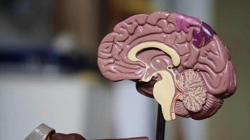
The core pathophysiology underlying the Parkinsonism syndrome, regardless of its specific etiology, centers on a profound disruption of the dopaminergic system within the basal ganglia. In idiopathic Parkinson’s Disease, this disruption is caused by the progressive degeneration and death of dopamine-producing neurons located in the substantia nigra pars compacta (SNpc). These neurons project via the nigrostriatal pathway to the striatum, where dopamine acts as a key neurotransmitter regulating the balance between the direct and indirect pathways of the basal ganglia motor loop. The basal ganglia function as a critical modulator of motor control, selectively facilitating desired movements (via the direct pathway) and inhibiting unwanted movements (via the indirect pathway).
The widespread loss of dopamine—which must exceed 60 to 80 percent of the SNpc neurons before motor symptoms become clinically apparent—fundamentally shifts the delicate balance of the basal ganglia circuitry. Dopamine normally stimulates the direct pathway (via D1 receptors) and inhibits the indirect pathway (via D2 receptors). The resulting dopamine depletion leads to under-activation of the direct pathway and over-activation of the indirect pathway. This pathological imbalance results in excessive inhibitory output from the basal ganglia’s output nuclei (the globus pallidus interna and substantia nigra pars reticulata) to the thalamus. Consequently, the thalamus is inhibited, leading to reduced excitatory input to the motor cortex, which clinically translates into the characteristic slowness, poverty, and difficulty initiating movement, known collectively as bradykinesia.
While dopamine deficiency is the primary neurochemical hallmark, the specific cellular pathology helps differentiate various forms of Parkinsonism. In idiopathic PD, the defining neuropathological feature is the presence of Lewy bodies—intracellular inclusions composed primarily of misfolded alpha-synuclein protein—found in the surviving neurons of the SNpc and other brain regions. In contrast, atypical Parkinsonian syndromes involve distinct proteinopathies. For instance, Progressive Supranuclear Palsy (PSP) and Corticobasal Degeneration (CBD) are primarily tauopathies (characterized by tau protein aggregation), while Multiple System Atrophy (MSA) involves glial cytoplasmic inclusions of alpha-synuclein. This difference in the specific misfolded protein and its anatomical distribution explains why atypical forms often present with non-dopaminergic symptoms—such as ophthalmoplegia in PSP or severe cerebellar signs in MSA—and respond poorly to Levodopa therapy, which only addresses the dopamine deficit.
Etiologies and Classification of Parkinsonism
Parkinsonism is broadly classified into three major groups based on the underlying cause: Primary (Idiopathic), Secondary (Acquired), and Atypical (Parkinson-Plus Syndromes). Idiopathic Parkinson’s Disease (PD) constitutes the vast majority of cases and remains a diagnosis of exclusion, defined by the characteristic unilateral onset, excellent and sustained response to Levodopa, and the presence of resting tremor. PD is often associated with non-motor features that can precede the motor symptoms by many years, including anosmia (loss of smell), constipation, and REM sleep behavior disorder. The progressive nature of PD stems from ongoing neurodegeneration, though the precise trigger for the alpha-synucleinopathy remains unknown.
Secondary Parkinsonism refers to cases where the syndrome is caused by an identifiable external factor or specific disease process. The most common cause within this category is Drug-Induced Parkinsonism (DIP), often resulting from medications that block dopamine receptors, such as antipsychotics or certain antiemetics. Other causes include Vascular Parkinsonism (resulting from multiple small strokes in the basal ganglia or related pathways), infectious causes (e.g., post-encephalitic Parkinsonism), and exposure to neurotoxins (e.g., MPTP). The hallmark of secondary Parkinsonism is often a symmetrical onset of symptoms, absence of classic resting tremor, and a generally poor or absent response to dopaminergic replacement therapy, contrasting sharply with the response seen in PD.
The third category, Atypical Parkinsonism Syndromes (or Parkinson-Plus syndromes), describes a group of primary neurodegenerative diseases that present with Parkinsonian motor features along with distinctive additional neurological signs that are not typical of PD. These syndromes tend to progress more rapidly, respond poorly to Levodopa, and have a more severe prognosis. Key examples include Multiple System Atrophy (MSA), which is characterized by early and severe autonomic dysfunction (e.g., orthostatic hypotension); Progressive Supranuclear Palsy (PSP), defined by early, prominent postural instability and vertical gaze palsy; and Corticobasal Degeneration (CBD), which often features severe asymmetrical apraxia and cortical sensory loss. The correct identification of these syndromes is critical because their management focuses more on supportive care and addressing the non-motor, non-dopaminergic features of the illness.
Drug-Induced Parkinsonism (DIP)
Drug-Induced Parkinsonism (DIP) is a critically important subtype of secondary Parkinsonism, representing the second most common cause of Parkinsonian symptoms after idiopathic PD, especially among the elderly. DIP is overwhelmingly caused by pharmaceutical agents that interfere with dopaminergic neurotransmission, most notably the dopamine receptor antagonists. The primary culprits are typically antipsychotic medications (neuroleptics), particularly the first-generation or typical antipsychotics (e.g., haloperidol, chlorpromazine), which exert their therapeutic effect by blocking D2 dopamine receptors in the mesolimbic pathway, but also inadvertently block D2 receptors in the nigrostriatal pathway, mimicking the neurochemical deficit of PD. Certain antiemetics (e.g., metoclopramide) and calcium channel blockers (e.g., flunarizine, cinnarizine) can also precipitate or exacerbate Parkinsonian symptoms.
The clinical presentation of DIP often differs subtly from classical PD. Symptoms typically have a rapid, symmetrical onset, and the resting tremor, if present, is usually less pronounced than the bradykinesia and rigidity. Furthermore, DIP often lacks the non-motor features characteristic of PD, such as anosmia or REM sleep behavior disorder. A crucial diagnostic clue is the temporal relationship between the initiation of the offending medication and the onset of motor symptoms, usually appearing within days to months of starting the drug or increasing its dose. Since the pathophysiology involves receptor blockade rather than neuronal death, DIP is generally reversible upon cessation or dose reduction of the causative agent, although recovery can take weeks or, in some cases, many months, particularly in older patients whose dopaminergic reserve is naturally diminished.
Management of DIP requires careful clinical judgment. The first line of action is the discontinuation or substitution of the offending drug, if medically feasible. In psychiatric populations, this often means switching from a typical antipsychotic to an atypical antipsychotic (such as quetiapine or clozapine) that has a lower affinity for D2 receptors or a more favorable D2/5HT2A receptor binding profile, thereby reducing the risk of motor side effects. While Levodopa is generally ineffective and not recommended for DIP—as the underlying neurons are intact and the receptors are merely blocked—anticholinergic agents may sometimes be used to manage severe tremor or rigidity, though their use is limited by potential cognitive side effects, particularly in the elderly. The successful resolution of symptoms following drug withdrawal confirms the diagnosis, underscoring the importance of a comprehensive medication history in all patients presenting with Parkinsonism.
Atypical Parkinsonism Syndromes (Parkinson-Plus Syndromes)
Atypical Parkinsonism Syndromes, often referred to as Parkinson-Plus syndromes, represent a group of distinct neurodegenerative disorders that combine Parkinsonian features with additional, non-dopaminergic signs indicative of widespread system degeneration beyond the substantia nigra. These conditions are characterized by rapid progression, early onset of severe disability, and a notoriously poor or absent therapeutic response to Levodopa. Key syndromes in this category include Progressive Supranuclear Palsy (PSP), Multiple System Atrophy (MSA), Corticobasal Degeneration (CBD), and Dementia with Lewy Bodies (DLB). Recognizing the distinguishing features of these syndromes is critical because their prognosis is significantly worse than that of PD, and their management strategies differ substantially.
Progressive Supranuclear Palsy (PSP), a primary tauopathy, is perhaps the most distinctive of the atypical syndromes. While patients exhibit bradykinesia and rigidity, the distinguishing features include early and severe postural instability leading to frequent, often backward, falls, and the characteristic supranuclear vertical gaze palsy (difficulty looking up or down). Axial rigidity, particularly neck hyperextension, is also common. The pathology affects the brainstem and subcortical structures, leading to deficits that Levodopa cannot correct. Multiple System Atrophy (MSA) is defined by the combination of Parkinsonism and severe autonomic failure (e.g., neurogenic orthostatic hypotension, urinary incontinence, erectile dysfunction) or cerebellar dysfunction (ataxia). MSA is further subclassified into MSA-P (predominantly Parkinsonian) and MSA-C (predominantly Cerebellar), both involving alpha-synuclein pathology in glial cells.
Corticobasal Degeneration (CBD) is characterized by profound asymmetrical cortical dysfunction alongside Parkinsonian motor features. Clinical hallmarks often include limb apraxia (inability to perform learned movements), alien limb phenomenon (a feeling that the limb is acting independently), and severe asymmetrical rigidity and dystonia. The pathology involves tau protein accumulation in cortical and subcortical regions. Finally, Dementia with Lewy Bodies (DLB) is characterized by Parkinsonism that occurs concurrently with or shortly after the onset of fluctuating cognition, recurrent visual hallucinations, and severe REM sleep behavior disorder. DLB shares the alpha-synuclein pathology of PD but is distinguished by the widespread cortical distribution of Lewy bodies, leading to the pronounced cognitive and psychiatric features that define the disorder. The presence of these “plus” features acts as crucial flags for clinicians differentiating these syndromes from PD.
Differential Diagnosis
The diagnostic process for Parkinsonism is primarily clinical, relying on the careful history and neurological examination to distinguish idiopathic PD from secondary and atypical syndromes. The initial differentiation hinges on identifying features that argue against a typical PD diagnosis. These “red flags” include rapid symptom progression, early onset of falls or cognitive impairment, symmetrical onset of motor symptoms, lack of a prominent resting tremor, and the presence of severe early autonomic dysfunction or pyramidal tract signs. A key diagnostic tool is the therapeutic trial of Levodopa: a robust and sustained improvement in motor function strongly favors idiopathic PD, whereas minimal or transient response points toward an atypical or secondary etiology.
Beyond clinical observation, neuroimaging plays an increasingly supportive role. While conventional MRI scans are often normal in early PD, they can be instrumental in identifying structural causes of secondary Parkinsonism, such as hydrocephalus (Normal Pressure Hydrocephalus) or strategically placed vascular lesions (Vascular Parkinsonism). Furthermore, specific imaging patterns can suggest atypical syndromes; for instance, midbrain atrophy (“hummingbird sign”) is characteristic of PSP, and putaminal atrophy is often seen in MSA. The use of Dopamine Transporter (DaT) scans (Ioflupane I 123 injection SPECT) has become a powerful adjunctive diagnostic tool. DaT scans visualize the density of dopamine transporters in the striatum; an abnormal scan showing reduced binding confirms the presence of dopaminergic deficit (as seen in PD, MSA, PSP, and CBD), effectively ruling out conditions like Essential Tremor or Drug-Induced Parkinsonism, where the dopaminergic neurons are functionally intact.
The differential diagnosis must also systematically exclude other movement disorders that can mimic aspects of Parkinsonism. Essential Tremor (ET), the most common tremor disorder, is often confused with PD but is distinguishable by its action or intention-based nature, its usual absence at rest, and its responsiveness to alcohol or beta-blockers, rather than dopaminergic drugs. Psychogenic Parkinsonism, though rare, must also be considered, characterized by inconsistent symptoms and non-organic signs. Ultimately, the comprehensive differential diagnosis involves a layered approach: confirming the presence of the Parkinsonian syndrome, determining whether it is Levodopa-responsive, utilizing imaging to rule out structural causes, and assessing for the presence of “plus” features that indicate an atypical neurodegenerative disorder.
Management and Treatment Approaches
The management of Parkinsonism is highly tailored to its specific etiology, aiming to improve motor function and enhance the patient’s quality of life. For idiopathic Parkinson’s Disease, treatment is primarily pharmacological and focuses on restoring dopaminergic function. The gold standard therapy remains Levodopa (combined with carbidopa or benserazide), a dopamine precursor that effectively crosses the blood-brain barrier and is converted into dopamine. Levodopa provides the most potent symptomatic relief, especially for bradykinesia and rigidity, and its efficacy is a key diagnostic factor. However, chronic use often leads to motor complications such as dyskinesia (involuntary writhing movements) and “wearing off” periods, necessitating careful dose titration and scheduling.
Other pharmacological agents are used alongside or prior to Levodopa, particularly in younger patients, to delay the onset of motor complications. These include Dopamine Agonists (e.g., pramipexole, ropinirole), which directly stimulate dopamine receptors but carry a higher risk of impulse control disorders and hallucinations. MAO-B Inhibitors (e.g., rasagiline, selegiline) are used to prevent the breakdown of endogenous dopamine, offering mild symptomatic relief. Amantadine may be used to reduce dyskinesia. When motor fluctuations become severe and medically intractable, advanced therapies such as Deep Brain Stimulation (DBS) or Levodopa intestinal gel infusion may be considered, targeting the subthalamic nucleus or globus pallidus interna to modulate the overactive basal ganglia output.
In contrast, the management of Atypical and Secondary Parkinsonism is significantly more challenging due to the limited effectiveness of dopaminergic drugs. Patients with Atypical Syndromes (PSP, MSA, CBD) typically derive minimal or no benefit from Levodopa, meaning treatment must focus intensely on managing the non-motor symptoms and providing extensive supportive care. This includes medication for autonomic symptoms (in MSA), palliative therapies for rigidity and dystonia, and crucially, non-pharmacological interventions. For Drug-Induced Parkinsonism, the primary intervention is the careful withdrawal of the causative agent. Non-pharmacological interventions are essential across all forms of Parkinsonism, including tailored physical therapy to address gait and balance, occupational therapy to maintain independence in daily tasks, and speech therapy for dysphagia and hypophonia, ensuring a multidisciplinary approach to comprehensive care.
Prognosis and Quality of Life Considerations
The prognosis for an individual diagnosed with Parkinsonism is highly dependent upon the underlying etiology. Patients with idiopathic Parkinson’s Disease generally experience a slow, progressive decline over many years, with motor symptoms often remaining manageable with dopaminergic therapy for a decade or more. The primary determinants of long-term quality of life in PD are the development of motor complications (dyskinesia and fluctuations) and the emergence of severe non-motor symptoms, particularly dementia, depression, and psychosis, which often become more disabling than the motor symptoms late in the disease course. Mortality is often linked to complications such as aspiration pneumonia or injuries resulting from falls caused by postural instability.
Conversely, patients diagnosed with Atypical Parkinsonism Syndromes face a significantly more severe and rapid prognosis. Conditions like Progressive Supranuclear Palsy (PSP) and Multiple System Atrophy (MSA) progress quickly, leading to profound disability within five to eight years of diagnosis. The lack of effective Levodopa response means symptomatic control is minimal, and life expectancy is notably reduced compared to PD. For these patients, quality of life is severely impacted by early and frequent falls, severe swallowing difficulties (dysphagia), and autonomic crises (in MSA). Therefore, management shifts rapidly toward palliative care, focusing on preventing complications like aspiration and providing comprehensive multidisciplinary support to maintain dignity and comfort.
Addressing non-motor symptoms is paramount for improving the overall quality of life across all forms of Parkinsonism. Non-motor features—including anxiety, depression, apathy, sleep disturbances (insomnia, RBSD), and pain—are common and often untreated, yet they contribute substantially to disability and caregiver burden. Comprehensive management involves screening for these symptoms regularly and utilizing appropriate pharmacological and psychological interventions. Furthermore, patient education, caregiver support, and involvement in support groups are crucial components of long-term care, helping individuals and families cope with the chronic, progressive nature of the syndrome and maximizing functional independence for as long as possible.