Photogenic Epilepsy: Understanding Visual Seizure Triggers
Photogenic epilepsy, sometimes referred to as photosensitive epilepsy, constitutes a distinct and specialized form of reflex epilepsy. This neurological disorder is fundamentally characterized by the reliable initiation of epileptic seizures following exposure to specific visual stimuli or aberrations. The defining feature is the direct causal link between the visual input and the resulting cerebral hyperexcitability, meaning that seizures are not spontaneous but are instead preempted by particular environmental conditions involving light and pattern. This condition underscores the intricate relationship between sensory processing in the visual cortex and the mechanisms governing seizure threshold within the brain. Understanding photogenic epilepsy requires a detailed examination of the types of triggers, the underlying physiological mechanisms, and effective strategies for stimulus avoidance and management to minimize the frequency and severity of epileptic events.
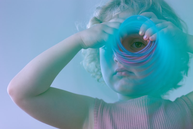
The classic presentation involves a sensitivity to flickering light, but the spectrum of effective visual triggers is broad and highly individualized, ranging from the flicker rate of older cathode ray tube televisions to specific geometric patterns, or even rapid transitions between light and shadow. The diagnosis of photogenic epilepsy demands the documentation of this reflexive relationship, typically achieved through specialized electroencephalographic testing involving controlled photic stimulation. Management of the condition often relies heavily on patient education regarding environmental risk factors and the meticulous application of avoidance strategies, supplemented by pharmacological intervention when necessary to elevate the overall seizure threshold and enhance the patient’s quality of life, which can otherwise be significantly compromised by the pervasive nature of potential visual triggers in modern society.
Classification and Terminology
Photogenic epilepsy is formally categorized within the larger group of reflex epilepsies, which are defined by their dependence on specific external or internal triggers. While some forms of epilepsy are purely idiopathic or structural, reflex epilepsies provide a clear path of causation: stimulus leads to seizure. Within the framework established by the International League Against Epilepsy (ILAE), photosensitivity often overlaps with certain syndromes, most notably Juvenile Myoclonic Epilepsy (JME), though it can occur in isolation or in conjunction with other generalized or focal epilepsy types. This connection suggests a shared genetic or physiological predisposition to generalized cortical excitability, which is then uniquely expressed or amplified by visual input, differentiating it from other reflex epilepsies triggered by activities like reading or sudden startling.
The terminology surrounding the condition requires precision. While “photogenic epilepsy” strictly refers to seizures generated by light, the broader term photosensitive epilepsy is often used synonymously in clinical practice and encompasses the sensitivity to various visual inputs, including not just flicker but also specific patterns and contrast levels. It is crucial to distinguish this condition from non-epileptic events triggered by light, such as migraine with aura or simple eye strain, which do not involve the characteristic paroxysmal electrical discharge observed in the epileptic brain. The identification of a genuine photogenic response confirms a primary neurological vulnerability that transcends mere visual discomfort, firmly placing the condition within the domain of core neurological disorders requiring specialized treatment protocols.
Historically, the phenomenon gained significant public and clinical attention following incidents such as the 1997 “Pokémon Shock” in Japan, where specific flashing light sequences in an animated television program induced seizures in hundreds of children. This event served as a stark demonstration of the potency of certain visual frequencies and confirmed the need for regulatory guidelines regarding broadcast standards and media content creation. This historical context solidified the recognition of flicker frequency as a critical factor, with frequencies typically ranging between 15 and 25 Hertz (Hz) being the most epileptogenic, although the specific bandwidth varies significantly depending on the individual patient’s cerebral excitability profile.
Etiology and Pathophysiology
The underlying etiology of photogenic epilepsy is complex, typically involving a blend of genetic predisposition and specific cortical network hyperexcitability. While the exact genes involved are not always identified, many cases show familial clustering, suggesting an inherited component that lowers the threshold for visual-induced seizures. Pathophysiologically, the primary mechanism involves the abnormal hypersensitivity of the visual processing areas, predominantly located in the occipital lobes. When the visual input—such as a specific contrast, pattern, or flicker rate—reaches this highly excitable cortex, it triggers a cascade of synchronous neuronal firing that spreads rapidly across the brain, leading to a generalized epileptic discharge.
The physiological signature of photogenic epilepsy is the photoparoxysmal response (PPR), which is evident during EEG testing. The PPR manifests as generalized spike-and-wave discharges that are time-locked precisely to the visual stimulation. This response is critical for diagnosis and involves a rapid, uncontrolled synchronization of neuronal activity, often propagating from the occipital region to the frontal lobes via thalamocortical networks. Research suggests that this widespread synchronized firing is facilitated by underlying dysfunctions in inhibitory neurotransmission, particularly within the GABAergic system. A deficit or impairment in GABA-mediated inhibition allows excitatory signals generated by the visual stimulus to overwhelm the normal regulatory mechanisms, thus leading to the uncontrolled spread of electrical activity.
Specific characteristics of the stimulus contribute significantly to its epileptogenicity. The key factors that maximize the risk of triggering a PPR include high contrast (e.g., black and white stripes), high luminance (brightness), and specific flicker frequencies. Studies indicate that patterns with strong spatial properties, such as high-density geometric patterns, can be just as potent as temporal flicker. Furthermore, the size of the visual field stimulated is relevant; the risk of seizure increases dramatically when the stimulus covers a large portion of the visual field, maximizing the cortical area involved in the initial hyperexcitability. This knowledge of critical parameters allows clinicians to advise patients on specific viewing modifications necessary for safety.
Clinical Manifestations and Seizure Types
The clinical presentation of photogenic epilepsy is diverse, dictated both by the specific visual trigger and the manner in which the epileptic discharge propagates through the brain. The most common and impactful seizure type resulting from photic stimulation is the generalized tonic-clonic seizure (GTC), which involves loss of consciousness, stiffening of the body (tonic phase), and rhythmic jerking (clonic phase). However, patients frequently experience less severe seizure types initially, such as myoclonic jerks (sudden, brief muscle twitches, often involving the arms or shoulders) or absence seizures (brief periods of staring and unresponsiveness). These milder forms often precede a full GTC seizure if the stimulus is not immediately removed.
The range of potent visual triggers is extensive and requires careful identification by the patient and clinician. Common environmental triggers include:
- Television and Computer Screens: Especially older CRT monitors or rapidly flashing sequences in video games and animated content.
- Strobe Lights and Disco Lights: Found in entertainment venues, where the flicker rate is often intentionally optimized to be within the epileptogenic range.
- Natural Light Patterns: Sunlight flickering through trees while driving, or reflections off water (the “sun-flicker phenomenon”).
- Specific Patterns: High-contrast, repeating geometric designs, such as those found in certain textiles or architecture.
The time between exposure to the trigger and the onset of the seizure (latency) is typically very short, often only seconds, highlighting the direct and rapid nature of the reflex arc involved in the pathogenesis.
Patients often develop highly adaptive, albeit sometimes socially limiting, behaviors to mitigate seizure risk. They may instinctively shield their eyes, look away from flickering sources, or cover one eye to reduce the total area of cortical stimulation. In some complex cases, patients may experience visual symptoms immediately preceding the seizure onset, such as flashing lights or distortions, which serve as a warning aura. Recognizing these subtle pre-seizure manifestations is vital, as it allows the patient a brief window to intervene by immediately closing their eyes, removing the stimulus, or moving into a safer environment before the full-blown convulsive event takes hold.
Diagnostic Criteria and Testing
The diagnosis of photogenic epilepsy relies critically on confirming the relationship between the visual stimulus and the epileptic discharge, which is achieved primarily through a specialized Electroencephalogram (EEG) with photic stimulation. Standard diagnostic criteria require the demonstration of the Photoparoxysmal Response (PPR) during controlled testing procedures. The goal of the EEG is to safely reproduce the condition under medical supervision to precisely characterize the patient’s sensitivity profile.
During the photic stimulation phase of the EEG, a high-intensity stroboscopic light is placed near the patient’s eyes, and the frequency of the flashes is varied systematically, typically starting at low frequencies (1–3 Hz) and increasing incrementally up to 60 Hz. The testing focuses particularly on the most epileptogenic range (15–25 Hz). The PPR is confirmed if the characteristic spike-and-wave or polyspike-and-wave discharges are recorded on the EEG traces, appearing synchronously with the light flashes. Important diagnostic parameters recorded include:
- The specific frequency range that elicits the PPR.
- The field of sensitivity (e.g., full visual field vs. monocular sensitivity).
- Whether the PPR persists after the light stimulation stops (a more severe indication).
The sensitivity profile derived from the EEG guides both pharmacological choices and lifestyle recommendations, offering concrete data on the patient’s neurological vulnerability.
Differential diagnosis is essential to rule out other conditions that might mimic light sensitivity or visual-induced symptoms. These include visual migraines, certain types of non-epileptic attacks (psychogenic seizures), or focal seizures originating in the occipital lobe that may produce visual phenomena but are not strictly reflexively triggered by external light. Furthermore, patients with photosensitivity due to systemic issues, such as certain metabolic disorders or drug toxicity, must be excluded. The consistent, reproducible nature of the PPR on EEG is the definitive factor that distinguishes true photogenic epilepsy from these other conditions, ensuring that treatment is appropriately focused on managing cortical hyperexcitability.
Treatment Modalities
Treatment for photogenic epilepsy is multifaceted, prioritizing stimulus avoidance and utilizing anti-epileptic medications (AEDs) to raise the seizure threshold. The most effective non-pharmacological strategy is rigorous environmental and behavioral modification. Patients must be educated on identifying and systematically avoiding their specific triggers. This includes maintaining a safe viewing distance from television screens, reducing screen brightness and contrast, and ensuring adequate ambient lighting to minimize the perception of flicker.
Specific visual aids are often recommended to mitigate risk. For instance, wearing glasses with tinted lenses, particularly those with a blue or Z1 filter, can reduce the perceived intensity and contrast of flickering light, thereby decreasing the likelihood of triggering a PPR. Furthermore, employing techniques such as monocular viewing (covering one eye) when exposed to a high-risk stimulus can reduce the size of the cortical area being stimulated, offering a rapid, protective measure. Careful modification of high-risk activities, such as reducing time spent playing video games or using specific digital devices, is often mandatory to maintain seizure freedom.
Pharmacological treatment often involves broad-spectrum AEDs known to be effective against generalized epilepsies. Medications such as Valproate (sodium valproate) and Levetiracetam are commonly prescribed because they effectively stabilize neuronal membranes and enhance GABAergic inhibition, thereby raising the general seizure threshold and specifically dampening the occipital cortex’s response to visual input. In many cases, effective management is achieved through a combination of low-dose AEDs and strict adherence to avoidance strategies. The goal of therapy is not only to achieve seizure freedom but also to minimize the constraints placed on the patient’s daily life, allowing for greater social integration and educational participation without undue risk of a visually induced event.
Prognosis and Quality of Life
The prognosis for individuals diagnosed with photogenic epilepsy is generally favorable, particularly because the condition is highly predictable and manageable once the specific triggers are identified. Unlike spontaneous epilepsies, the patient holds a significant degree of control over the seizure risk through diligent stimulus avoidance. While photogenic epilepsy often persists from childhood or adolescence into adulthood, effective medication combined with robust behavioral strategies can lead to long periods of seizure freedom, allowing many individuals to lead fulfilling and largely unrestricted lives, provided they remain vigilant about their environment.
However, the condition can impose significant challenges on quality of life, primarily due to the necessary restrictions and the pervasive nature of visual triggers in modern society. Patients may experience heightened anxiety regarding exposure to unexpected light sources, leading to avoidance of public spaces like concerts, cinemas, or certain workplaces. Children and adolescents may face difficulties in educational settings or social interactions involving popular media, requiring accommodations such as specialized protective screens for classroom computers or modifications to physical education activities. Addressing the psychological impact, including anxiety and potential depression resulting from social isolation or perceived stigma, is an integral part of comprehensive care.
In some cases, especially those beginning in early childhood, the photosensitivity may naturally decrease or remit entirely with age, though this outcome is not guaranteed. Ongoing neurological monitoring, including periodic EEG assessments, is necessary to track the patient’s sensitivity profile and adjust treatment as needed. For the majority of patients, photogenic epilepsy remains a lifelong trait that requires continuous awareness. The long-term prognosis is intrinsically linked to the patient’s ability to maintain discipline in trigger avoidance and adherence to pharmacological regimens, enabling them to effectively navigate a visually stimulating world while minimizing their neurological risk.