Pituitary Function: The Master Gland of Your Mental Health
- Introduction to Pituitarism
- The Critical Role of the Pituitary Gland
- Hypopituitarism: Deficiency States
- Hyperpituitarism: States of Excess
- Etiology and Pathogenesis of Pituitary Disorders
- Clinical Manifestations and Symptomology
- Diagnostic Procedures and Assessment
- Management and Therapeutic Strategies
- Prognosis and Long-Term Monitoring
Introduction to Pituitarism
Pituitarism refers broadly to any condition characterized by the disordered functioning of the pituitary gland. This critical endocrine organ, often termed the “master gland,” regulates numerous vital processes throughout the body by controlling the function of other endocrine glands, including the adrenal glands, thyroid gland, ovaries, and testes. A disruption in its operation leads to systemic hormonal imbalances, profoundly impacting metabolism, growth, reproduction, and overall homeostasis. The presentation of pituitarism is highly variable, depending entirely on which specific hormones are affected and whether the gland is producing too little or too much of them.
The spectrum of pituitarism is typically categorized into two main forms: hypopituitarism, which signifies an underactive state resulting in hormonal deficiencies, and hyperpituitarism, which denotes an overactive state leading to the excessive secretion of one or more pituitary hormones. While the root causes can range from benign tumors (adenomas) and trauma to genetic defects and inflammatory conditions, the ultimate consequence is a failure in the finely tuned feedback loops that govern endocrine function. Recognizing this duality is essential, as deficiency states require replacement therapy, whereas excess states often necessitate intervention to reduce hormone production or tumor burden.
Because the pituitary gland influences virtually every major physiological system, the clinical consequences of pituitarism are widespread and often non-specific in their early stages, making timely and accurate diagnosis challenging. Symptoms can mimic those of many other common chronic diseases, leading to significant diagnostic delays. Given the critical nature of certain pituitary hormones, such as adrenocorticotropic hormone (ACTH), which governs cortisol production essential for stress response, untreated pituitarism can potentially lead to life-threatening endocrine crises. Therefore, an understanding of the complex pathophysiology and the necessity of prompt, specialized endocrine management is paramount for ensuring patient survival and maintaining long-term quality of life.
The Critical Role of the Pituitary Gland
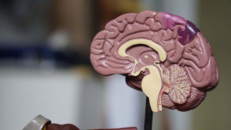
The pituitary gland is a small, pea-sized structure located at the base of the brain within the sella turcica. Structurally and functionally, it is divided into two distinct parts: the anterior pituitary (adenohypophysis) and the posterior pituitary (neurohypophysis). The anterior lobe is responsible for synthesizing and secreting six major tropic hormones: Growth Hormone (GH), Thyroid-Stimulating Hormone (TSH), Adrenocorticotropic Hormone (ACTH), Follicle-Stimulating Hormone (FSH), Luteinizing Hormone (LH), and Prolactin (PRL). These hormones are released under the direct control of releasing and inhibiting hormones produced by the adjacent hypothalamus, which connects to the pituitary via a specialized portal vascular system.
The posterior pituitary, conversely, does not synthesize hormones but rather stores and releases two hormones produced in the hypothalamus: Antidiuretic Hormone (ADH), crucial for water balance and osmoregulation, and Oxytocin, involved in uterine contractions and milk ejection. This intricate anatomical relationship between the hypothalamus and the pituitary gland means that disorders affecting the pituitary often also involve hypothalamic dysfunction, further complicating the clinical picture and diagnosis. Disruption at any point in this complex axis—whether due to compression, infiltration, or vascular compromise—can lead to profound endocrine failure.
The regulatory function of the pituitary gland operates through sophisticated negative feedback loops. For example, the release of ACTH stimulates the adrenal glands to produce cortisol. As cortisol levels rise in the bloodstream, they signal back to the hypothalamus and pituitary to reduce the output of CRH and ACTH, thereby maintaining equilibrium. Pituitarism occurs when these feedback mechanisms are broken, either because the pituitary cells themselves are damaged or hyper-functional, or because the target organs are failing to respond correctly, although the latter is generally classified as a primary endocrine disorder rather than pituitarism itself. Understanding these axes—the HPA (hypothalamic-pituitary-adrenal), HPT (thyroid), and HPG (gonadal)—is crucial for interpreting diagnostic test results.
Hypopituitarism: Deficiency States
Hypopituitarism is defined as the deficient secretion of one or more pituitary hormones. This condition can range from an isolated deficiency of a single hormone to panhypopituitarism, which involves the complete or near-complete cessation of hormone production by the anterior pituitary gland. Common causes include large pituitary tumors (macroadenomas) that compress and destroy normal pituitary tissue, cranial radiation therapy, pituitary surgery, or vascular events such as Sheehan’s syndrome (postpartum pituitary necrosis) or hemorrhagic infarction of an adenoma (pituitary apoplexy). The order in which hormones are lost often follows a typical pattern, with GH and gonadotropins (FSH/LH) often being the first to show deficiencies.
The clinical features of hypopituitarism are directly tied to the specific hormones lacking. Deficiency of ACTH leads to secondary adrenal insufficiency, presenting with severe fatigue, weight loss, hypotension, and risk of adrenal crisis, which is a medical emergency requiring immediate glucocorticoid replacement. A lack of TSH results in secondary hypothyroidism, characterized by cold intolerance, sluggishness, and constipation. Deficiencies in gonadotropins cause hypogonadism, manifesting as amenorrhea, infertility, loss of libido, and potentially osteoporosis in both sexes due to reduced sex steroid levels. Furthermore, GH deficiency in adults leads to reduced muscle mass, increased central adiposity, and diminished quality of life.
Diagnosis requires not only the measurement of basal hormone levels but also dynamic testing, such as stimulation tests, to assess the reserve capacity of the pituitary gland. For instance, the insulin tolerance test (ITT) or the glucagon stimulation test are used to evaluate the release of GH and ACTH. Management relies heavily on precise and lifelong hormone replacement therapy. Patients must receive physiological doses of glucocorticoids, thyroid hormone, and often sex steroids. Crucially, the administration of cortisol replacement must precede or accompany thyroid hormone replacement, as initiating T4 therapy alone in an ACTH-deficient patient can precipitate an adrenal crisis by increasing the metabolic clearance of residual cortisol.
Hyperpituitarism: States of Excess
Hyperpituitarism typically results from the excessive, autonomous secretion of hormones, most commonly due to the presence of a functional pituitary adenoma. These tumors are classified based on the hormone they predominantly secrete. The most frequently occurring hypersecretory tumor is the prolactinoma, which secretes Prolactin (PRL). Other significant functional tumors include somatotroph adenomas (GH-secreting) and corticotroph adenomas (ACTH-secreting). Unlike deficiencies which affect multiple axes, hyperpituitarism often presents initially as a specific endocrine syndrome related to the single hormone in excess.
Two of the most clinically striking forms of hyperpituitarism are Acromegaly and Cushing’s Disease. Acromegaly results from chronic GH excess in adults (or Gigantism if onset is before epiphyseal plate fusion). Symptoms include characteristic facial changes, enlargement of the hands and feet, joint pain, hypertension, and significantly increased risk of cardiovascular disease and diabetes mellitus. Cushing’s Disease, caused by an ACTH-secreting tumor, leads to adrenal hyperplasia and excessive cortisol production. Clinical signs include central obesity, facial plethora (moon face), proximal muscle weakness, easy bruising, and psychiatric disturbances, all indicative of hypercortisolism.
Prolactinomas cause symptoms related both to the excess Prolactin and the mass effect of the tumor itself. High PRL levels commonly cause galactorrhea (inappropriate milk production) and hypogonadism through suppression of the HPG axis, leading to menstrual irregularities and infertility in women, and erectile dysfunction and loss of libido in men. Unlike GH- and ACTH-secreting tumors, which often require surgery, prolactinomas are uniquely sensitive to medical management using dopamine agonists (e.g., cabergoline or bromocriptine), which can effectively shrink the tumor and normalize Prolactin levels in the majority of patients, often avoiding the need for invasive procedures.
Etiology and Pathogenesis of Pituitary Disorders
The overwhelming majority of pituitary disorders, particularly those leading to hyperpituitarism, are caused by pituitary adenomas, which are benign monoclonal tumors arising from one of the hormone-secreting cell lines of the anterior pituitary. These tumors are categorized as microadenomas (less than 10 mm in diameter) or macroadenomas (10 mm or larger). Functional adenomas cause disease primarily through hormone excess, while non-functional adenomas—and large functional adenomas—can cause symptoms via mass effect, compressing the surrounding normal gland tissue and leading to secondary hypopituitarism, or impinging on the optic chiasm causing visual deficits.
While adenomas dominate the etiology, non-tumorous causes are responsible for a significant proportion of hypopituitarism cases. These include infiltrative diseases, such as sarcoidosis, histiocytosis X (Langerhans cell histiocytosis), and hemochromatosis, which deposit abnormal materials within the gland, progressively destroying normal tissue. Infections, though rare today, such as tuberculosis or fungal infections, can also cause severe inflammation (hypophysitis). Furthermore, traumatic brain injury (TBI) and subarachnoid hemorrhage are increasingly recognized causes of delayed-onset hypopituitarism, particularly affecting GH and gonadotropin axes, due to damage to the pituitary stalk or the gland itself.
Genetic predisposition plays a role in a small percentage of pituitarism cases. For example, the multiple endocrine neoplasia type 1 (MEN1) syndrome, an autosomal dominant disorder, involves germline mutations in the MEN1 tumor suppressor gene and predisposes individuals to developing pituitary adenomas (often prolactinomas), along with tumors of the parathyroid and pancreas. Additionally, genetic mutations affecting specific transcription factors, such as PROP1, are linked to congenital forms of combined pituitary hormone deficiency, resulting in severe childhood-onset hypopituitarism affecting multiple hormone lines.
Clinical Manifestations and Symptomology
The clinical presentation of pituitarism can be broadly divided into symptoms arising from local mass effect and those resulting from systemic hormonal derangements. Mass effect symptoms typically manifest when a tumor grows large enough (macroadenoma) to impinge upon adjacent structures. The most common and serious local symptom is visual disturbance, specifically bitemporal hemianopsia—loss of peripheral vision in both eyes—caused by compression of the optic chiasm situated directly superior to the pituitary fossa. Other local symptoms include chronic headaches, often severe and resistant to standard analgesics, and, in rare cases, cranial nerve palsies affecting eye movement.
Systemic symptoms related to hormone deficiencies are often insidious and chronic. Patients with ACTH or TSH deficiency frequently report profound, unexplained fatigue, weakness, and significant changes in body weight or temperature regulation. Gonadotropin deficiency leads to sexual dysfunction, infertility, and in premenopausal women, secondary amenorrhea. In children, GH deficiency results in dwarfism or growth failure, whereas in adults, it contributes to adverse body composition changes (increased visceral fat) and reduced exercise capacity. Because these symptoms overlap with depression, chronic fatigue syndrome, and menopause, a high index of suspicion is required to identify the underlying endocrine cause.
Conversely, symptoms resulting from hormone excess are often more distinctive but equally widespread. In Acromegaly (GH excess), patients exhibit progressive coarse facial features, prognathism (protrusion of the jaw), macroglossia, and soft tissue swelling, often leading to carpal tunnel syndrome and debilitating arthritis. Chronic excess cortisol in Cushing’s Disease results in pathognomonic physical signs, including the redistribution of fat (buffalo hump, truncal obesity), easy skin bruising, purple striae, and significant psychological symptoms ranging from irritability to severe depression and psychosis. The duration and severity of the excess determine the irreversible nature of these systemic complications, highlighting the urgency of treatment.
Diagnostic Procedures and Assessment
The diagnostic workup for suspected pituitarism is meticulous and requires a combination of biochemical assays and radiological imaging. Initial screening involves measuring basal levels of peripheral hormones (e.g., free T4, cortisol, IGF-1, sex steroids) and the corresponding pituitary tropic hormones (TSH, ACTH, FSH, LH, Prolactin). However, basal measurements alone are often insufficient, especially for diagnosing subtle deficiencies or excesses, because of the pulsatile nature of hormone release and diurnal variation (e.g., cortisol).
Therefore, dynamic endocrine testing is crucial. For diagnosing GH deficiency, the Insulin Tolerance Test (ITT) remains the gold standard, though less risky alternatives like the glucagon stimulation test are often used. For ACTH deficiency, the low-dose ACTH stimulation test is used to assess adrenal reserve, while the ITT assesses the pituitary reserve. Hypersecretory states are confirmed using suppression tests; for example, Cushing’s disease is diagnosed using the low-dose overnight or 48-hour Dexamethasone Suppression Test, and Acromegaly is confirmed by demonstrating failure of GH suppression following an oral glucose tolerance test (OGTT).
Once biochemical evidence of pituitarism is established, Magnetic Resonance Imaging (MRI) of the pituitary and hypothalamus is mandatory. The MRI, often performed with gadolinium enhancement, provides high-resolution anatomical detail necessary to identify pituitary adenomas, cysts, or destructive lesions. It is essential for differentiating between microadenomas and macroadenomas and assessing their proximity to critical structures, such as the optic chiasm and the cavernous sinuses, which dictates the surgical approach. In cases where ACTH-dependent Cushing’s Syndrome is confirmed, inferior petrosal sinus sampling (IPSS) may be required to definitively localize the source of ACTH production to the pituitary gland versus an ectopic source.
Management and Therapeutic Strategies
The management of pituitarism is complex, requiring a multidisciplinary approach involving endocrinologists, neurosurgeons, and radiation oncologists. The primary goals are twofold: first, to normalize hormone levels, and second, to remove or reduce the mass effect of any underlying tumor. For hypopituitarism, the treatment is lifelong hormone replacement therapy, prioritizing life-sustaining hormones such as glucocorticoids (hydrocortisone or prednisone) and thyroid hormone (levothyroxine). Sex steroids (estrogen, progesterone, testosterone) are replaced to restore fertility and bone health, and GH replacement is considered for adults with documented GH deficiency to improve body composition and quality of life.
For hyperpituitarism caused by functional adenomas, treatment depends on the specific tumor type. Transsphenoidal surgery, performed through the nasal cavity, is the preferred initial treatment for most GH- and ACTH-secreting tumors, offering the best chance for cure and immediate decompression of the optic chiasm. For prolactinomas, medical therapy with dopamine agonists is the first-line treatment due to its high efficacy in shrinking tumors and normalizing Prolactin levels. Pharmacological agents, such as somatostatin analogs (e.g., octreotide, lanreotide), are also crucial in managing GH-secreting tumors, often used as adjunctive therapy if surgery is unsuccessful or for patients who are not surgical candidates.
When surgery fails to achieve remission or if the tumor recurs, radiation therapy is utilized. Techniques such as conventional fractionated radiotherapy or highly focused stereotactic radiosurgery (Gamma Knife or CyberKnife) deliver precise radiation doses to the residual tumor tissue while minimizing damage to surrounding healthy brain tissue and the remaining normal pituitary gland. However, radiation carries the risk of inducing delayed-onset hypopituitarism, necessitating continued close monitoring for new hormone deficiencies months or years post-treatment. Effective management requires constant titration of medications and regular follow-up to maintain optimal hormonal balance and prevent complications.
Prognosis and Long-Term Monitoring
The prognosis for individuals with pituitarism varies significantly based on the underlying etiology, the extent of pituitary damage, and the success of definitive treatment for any tumors. Patients with isolated, mild hormone deficiencies that are adequately replaced can maintain a near-normal life expectancy and quality of life. Conversely, those presenting with acute conditions like pituitary apoplexy or those whose hormone excess conditions (Acromegaly, Cushing’s) have led to severe secondary complications (cardiovascular disease, diabetes) face a more guarded prognosis, emphasizing the importance of early diagnosis and intervention.
Long-term care for all patients with pituitarism necessitates rigorous, lifelong endocrine follow-up. This includes regular biochemical screening to ensure replacement hormone dosages are optimized and, for patients treated for tumors, monitoring for recurrence of hormone excess or the development of new hormone deficiencies, especially after radiation therapy. Monitoring is particularly critical for patients on glucocorticoid replacement, who must be educated on stress dosing—increasing their corticosteroid intake during illness, trauma, or surgery—to prevent potentially fatal adrenal crisis. Furthermore, surveillance imaging (MRI) is typically performed annually for several years post-treatment.
Finally, managing pituitarism extends beyond hormonal balance to addressing psychological and quality of life issues. Patients often struggle with chronic fatigue, cognitive changes, and body image issues resulting from the disorder or its treatment. Therefore, comprehensive care includes assessing for comorbid conditions like depression and anxiety and providing appropriate psychological support. Successful management requires a collaborative effort between the patient and the healthcare team to ensure consistent adherence to complex medication regimens and lifestyle adjustments necessary to mitigate the chronic effects of this complex endocrine disorder.