Pneumoencephalography: Medicine’s Dark Diagnostic Past
- Introduction and Definition
- Historical Context and Development (1918)
- Mechanism of Action: Contrast and Visualization
- The Procedure of Pneumoencephalography
- Clinical Applications and Diagnostic Utility
- Limitations, Risks, and Patient Experience
- The Rise of Modern Neuroimaging and Obsolescence
- Legacy and Contribution to Neuroscience
Introduction and Definition
Pneumoencephalography (PEG) was a critical, albeit invasive, diagnostic method employed in neurology and neurosurgery from its inception in 1918 until its decline in the mid-1980s. Defined as a radiological technique, its primary purpose was the detailed visualization and analysis of the cerebral ventricles and the subarachnoid space within the brain. This was achieved by introducing a gaseous contrast medium—typically filtered air—into the cerebrospinal fluid (CSF) system. The resulting radiographic image, known specifically as a pneumoencephalogram, provided the only available method for decades to examine these critical internal structures in living patients.
The mechanism relies on fundamental principles of radiology concerning tissue density. Standard X-rays pass easily through soft tissues like the brain parenchyma, resulting in minimal contrast and poor visualization of internal structures. However, air possesses a vastly lower density compared to both brain tissue and cerebrospinal fluid. When air is administered into the CSF system via a lumbar puncture, it displaces the fluid and fills the ventricles and cisterns. On the resulting X-ray film, this low-density air appears as a distinct, dark shadow against the surrounding higher-density structures, thus creating the necessary contrast for anatomical study.
Despite being highly effective for its time, the procedure was notoriously challenging and associated with significant patient discomfort and potential morbidity. Its eventual obsolescence was brought about by the monumental advancements in non-invasive radiological technologies. The rapid development and clinical adoption of Computed Tomography (CT) scanning in the 1970s, followed by the superior soft-tissue resolution offered by Magnetic Resonance Imaging (MRI) in the 1980s, entirely eliminated the medical necessity for this invasive test, marking a profound shift toward safer neurodiagnostic practices.
Historical Context and Development (1918)
The development of pneumoencephalography is irrevocably linked to the American neurosurgeon Walter Dandy, who introduced the technique in 1918. Prior to Dandy’s innovation, neurologists relied almost exclusively on clinical manifestations and post-mortem examination to diagnose intracranial diseases. The inability to visualize space-occupying lesions, hydrocephalus, or cortical atrophy in a living patient severely limited the effectiveness of neurosurgery, which was often exploratory and fraught with risk. Dandy’s genius lay in recognizing that the introduction of air could serve as a reliable, albeit artificial, contrast agent within the closed system of the central nervous system.
Dandy initially experimented with ventriculography, a related but even more invasive technique where air was injected directly into the lateral ventricles via burr holes drilled into the skull. Pneumoencephalography represented a critical refinement, as it utilized the existing pathway of the spinal column through a lumbar puncture, allowing the injected air to ascend into the cranial vault and fill the ventricular system. This method, while still highly invasive, offered a marginally safer and more accessible route for visualization, contributing to its rapid acceptance as the standard diagnostic tool in neurosurgery across the globe during the ensuing decades.
The early 20th century was defined by a desperate search for methods to localize brain pathology before surgery. PEG provided this crucial localization data, fundamentally transforming the practice of neurosurgery. By allowing surgeons to map the precise location and extent of mass effects—inferred by the distortion of the ventricular system—surgical planning became significantly more precise. This diagnostic capability propelled advancements in the understanding and treatment of neurological disorders, establishing PEG as the technological backbone of neurodiagnosis for over half a century.
Mechanism of Action: Contrast and Visualization
The effectiveness of pneumoencephalography is rooted in the principle of differential X-ray absorption. Soft tissues, such as the brain parenchyma, offer little variation in density, making them nearly transparent to conventional X-ray beams. Cerebrospinal fluid (CSF) similarly provides inadequate contrast. The introduction of gas, possessing an exceptionally low specific gravity and density, creates a stark, high-contrast interface against the denser surrounding tissues and fluid, enabling visualization through standard radiography.
The procedure is contingent upon the successful exchange of CSF for the gaseous medium. After the gas (typically filtered air, though oxygen was sometimes used to hasten absorption) is injected via the lumbar puncture, it begins its upward migration. This process is assisted by gravity and the differential pressures within the CNS. The technical challenge lay in ensuring that the gas adequately filled the deep-seated structures, including the lateral, third, and fourth ventricles, as well as the intricate network of basal cisterns and subarachnoid spaces surrounding the brain.
Once the gas filled these cavities, the resulting pneumoencephalogram displayed the delicate outlines of the ventricular system as dark, sharply defined spaces. Interpreting these images required profound neuroanatomical expertise. Pathological conditions were diagnosed indirectly: a tumor, though invisible itself, would manifest by causing a measurable compression, displacement, or distortion of the normally symmetrical ventricular outline. Similarly, atrophy was evidenced by the symmetrical enlargement of the ventricles and widening of the cortical sulci, illustrating the loss of surrounding brain tissue.
The Procedure of Pneumoencephalography
The procedure for pneumoencephalography was complex, demanding meticulous technique and often local anesthesia, meaning the patient remained conscious throughout the potentially agonizing process. The patient was typically seated upright and secured in a specialized chair or rotating frame, which allowed the medical team to precisely control the angle of the patient’s head and body, crucial for directing the gas flow within the cranium.
The first critical step involved performing a standard lumbar puncture in the lower spine. Following the insertion of the needle, small, measured amounts of CSF were carefully withdrawn. Crucially, an equal volume of gas was immediately injected to replace the withdrawn fluid, thus maintaining intracranial pressure as closely as possible and minimizing the risk of severe pressure imbalance. This fluid-gas exchange was performed incrementally, often involving dozens of separate withdrawal and injection cycles until a sufficient volume of gas (ranging typically from 30 to 50 mL) had been introduced.
Radiographic imaging was performed almost continuously as the exchange progressed and immediately thereafter. The patient was rapidly maneuvered into numerous positions—including various degrees of flexion, extension, and rotation of the neck and head—to ensure the gas traversed the foramina and filled all four ventricular chambers. Because the gas was constantly being absorbed by the surrounding tissues, the imaging sequence had to be executed swiftly. A complete diagnostic study involved capturing a significant number of X-ray views from multiple angles (anteroposterior, lateral, oblique) to create a comprehensive visualization of the internal cerebral structures.
Clinical Applications and Diagnostic Utility
For several decades, pneumoencephalography served as the definitive diagnostic tool for a wide array of neurosurgical and neurological conditions. Its primary utility lay in its capacity to visualize the effects of intracranial pathology on the ventricular system. The procedure was paramount in diagnosing various forms of hydrocephalus, allowing clinicians to distinguish between obstructive (non-communicating) types, where gas flow was blocked, and non-obstructive (communicating) types, where the gas distributed throughout the system but the flow of CSF was impaired elsewhere.
Furthermore, PEG was invaluable in identifying cortical and subcortical atrophy, particularly in cases of degenerative neurological diseases. In patients suffering from dementia or other conditions leading to significant tissue loss, the pneumoencephalogram would reveal characteristic findings such as marked dilation of the cerebral ventricles and pronounced widening of the cortical sulci, providing objective, measurable evidence of generalized or localized brain shrinkage that could not be achieved through other means.
Most critically, PEG was used to localize tumors and other space-occupying lesions. Since the mass itself was often radiographically occult, diagnosis relied on observing the secondary effects—the mass effect—on the visible, air-filled structures. A tumor in a specific hemisphere would cause a midline shift or distinct indentation on the adjacent ventricle, providing the necessary spatial coordinates for neurosurgical intervention. This ability to localize lesions transformed neurosurgery from a speculative field into a targeted medical discipline.
Limitations, Risks, and Patient Experience
Despite its diagnostic power, pneumoencephalography was burdened by severe limitations, chiefly concerning patient safety and comfort. The procedure was notoriously painful, primarily due to the intense meningeal irritation caused by the introduction of air into the subarachnoid space and the significant, though temporary, fluctuations in intracranial pressure during the CSF-gas exchange. Patients routinely suffered severe, debilitating headaches, nausea, protracted vomiting, and vertigo for days following the examination, frequently requiring intensive post-procedural care and strong analgesics.
The procedure carried serious, potentially fatal, risks. The greatest danger was the possibility of brain herniation, particularly in patients presenting with pre-existing, poorly localized masses that had already caused elevated intracranial pressure. The withdrawal of CSF could critically destabilize the pressure gradient, leading to the rapid downward displacement of brain tissue through the tentorium or foramen magnum. This extreme risk meant that PEG was reserved only for cases where the diagnostic information was absolutely essential for life-saving neurosurgical intervention.
Additionally, the diagnostic quality of the pneumoencephalogram was highly dependent on technical execution. If the gas failed to adequately fill the target areas or if the patient moved during the rapid series of X-ray exposures, the resulting image could be non-diagnostic or misleading. The procedure also involved high doses of ionizing radiation due to the necessity of taking multiple images from various angles, further contributing to the imperative for developing safer diagnostic alternatives.
The Rise of Modern Neuroimaging and Obsolescence
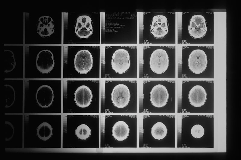
The reign of pneumoencephalography came to an abrupt end with the introduction and refinement of revolutionary cross-sectional imaging technologies. The pivotal turning point occurred in the early 1970s with the clinical availability of Computed Tomography (CT) scanning. CT utilized advanced computer processing to reconstruct detailed cross-sectional images of the brain from numerous X-ray projections. Crucially, CT could directly visualize soft tissue structures and intrinsic pathologies, such as tumors, edema, and hemorrhage, without any need for invasive CSF manipulation or the injection of contrast agents into the subarachnoid space.
The transition away from PEG accelerated dramatically with the widespread adoption of Magnetic Resonance Imaging (MRI) throughout the 1980s. MRI offered unparalleled clarity and superior differentiation of soft tissues, providing exquisite detail of the brain parenchyma, white matter tracts, and subtle pathological changes that were entirely invisible on a pneumoencephalogram. Furthermore, MRI achieved this level of detail without using ionizing radiation, eliminating a major health concern associated with earlier radiological techniques.
The combination of superior image quality, non-invasiveness, and patient safety provided by CT and MRI rendered pneumoencephalography medically obsolete almost instantaneously. By the late 1980s, PEG was virtually eliminated from standard clinical practice in developed countries, marking the end of an era defined by reliance on invasive physical manipulation of the CNS for diagnostic purposes. This rapid abandonment exemplifies how technological breakthroughs can fundamentally redefine the landscape of medical diagnosis and drastically improve patient outcomes.
Legacy and Contribution to Neuroscience
Despite its archaic nature and associated patient suffering, the legacy of pneumoencephalography remains critically important to the history of neuroscience. It was the first method to provide a systematic, reproducible, and verifiable means of visualizing internal cerebral structures in a living human. The vast amount of data collected via pneumoencephalograms over fifty years provided the foundational anatomical and pathological knowledge necessary for understanding the spatial relationships between the ventricular system and surrounding cerebral structures.
The diagnostic insights gained through PEG were fundamental to establishing the clinical concepts of mass effect, hydrocephalus classification, and the radiological interpretation of cerebral atrophy. This hard-won knowledge base provided the initial framework that guided the development and interpretation of subsequent neuroimaging technologies. For example, the recognition of specific ventricular distortions caused by tumors, first mapped by PEG, became a crucial benchmark used by early CT scan interpreters.
Ultimately, the inherent limitations and extreme risks of pneumoencephalography served as a powerful catalyst for innovation. The urgent medical need to replace this painful and hazardous procedure drove intense research into alternative, less invasive imaging modalities. In this sense, PEG not only paved the way by providing the first visualizations but also created the necessary technological vacuum that was eventually filled by the revolutionary advancements of CT and MRI, ensuring a safer and more precise future for neurodiagnosis.