Polioencephalitis: How Inflammation Shapes Mental Health
- The Core Definition of Polioencephalitis
- Etiology and Pathogenesis
- Historical Discovery and Context
- Clinical Manifestations and Cognitive Impact
- A Practical Case Study: Post-Infectious Cognitive Decline
- Significance in Neuropsychology and Treatment Implications
- Connections to Related Neurological Disorders
The Core Definition of Polioencephalitis
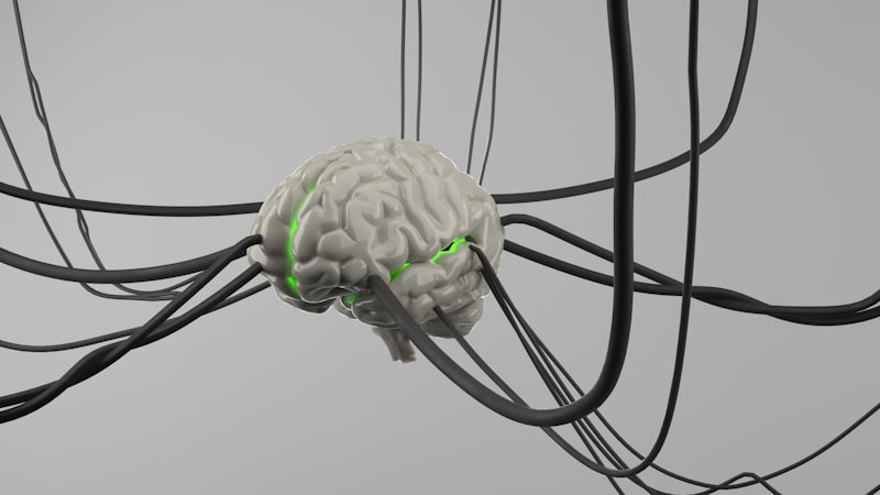
Polioencephalitis is defined fundamentally as the inflammation of the gray matter of the central nervous system, specifically within the brain, resulting from an infectious illness. This term, derived from the Greek “polios” (gray), “enkephalos” (brain), and “-itis” (inflammation), specifies the anatomical location of the primary pathology, differentiating it from generalized forms of encephalitis which may affect both gray and white matter indiscriminately. The core mechanism involves the body’s immune response to a pathogen—be it viral, bacterial, fungal, or parasitic—which breaches the protective barriers of the brain, leading to localized immune activity, cellular damage, and subsequent swelling. This swelling, or edema, is particularly dangerous because the brain is encased within the rigid skull, meaning any expansion drastically increases intracranial pressure, compromising blood flow and neuronal function.
The key distinguishing principle of polioencephalitis lies in its targeting of the gray matter, which is primarily composed of neuronal cell bodies, dendrites, unmyelinated axons, glial cells, and capillaries. Since the gray matter is responsible for processing information—including memory, sensation, and motor control—damage to these regions quickly manifests as severe cognitive, behavioral, and motor deficits. Unlike conditions primarily affecting the white matter (myelinated axons), polioencephalitis disrupts the core processing units themselves. The initial trigger is always an infectious agent, which either directly invades and destroys the neurons (as seen in some viral infections) or initiates a robust inflammatory cascade that leads to secondary damage through excitotoxicity and oxidative stress. Understanding this specific mechanism is vital for predicting the psychological and neurological outcomes experienced by the patient.
When clinicians state, “The patient is suffering from polioencephalitis as a result of a milder case of MRSA,” they are confirming that a systemic infection, such as Methicillin-resistant Staphylococcus aureus (MRSA), has progressed to cause inflammation localized within the brain’s processing centers. While the primary infection might be treatable or resolving, the resulting central nervous system inflammation requires specialized neurological intervention due to the high risk of permanent damage to cognitive functions. The severity of the psychological sequelae often correlates directly with the extent and location of the gray matter destroyed during the acute inflammatory phase, impacting areas such as the cerebral cortex, basal ganglia, and brainstem nuclei.
Etiology and Pathogenesis
The origins of polioencephalitis are diverse, encompassing a wide range of infectious agents. Historically, the most famous causative agent was the Poliovirus, which specifically targets motor neurons in the anterior horn of the spinal cord (a gray matter structure), leading to paralytic poliomyelitis—a form of spinal polioencephalitis. However, modern cases are frequently linked to viral infections like Herpes Simplex Virus, West Nile Virus, and certain enteroviruses, as well as bacterial infections that cause meningitis or systemic sepsis, such as the aforementioned MRSA or pneumococcus. The pathogenesis often involves a breakdown of the blood-brain barrier (BBB), a highly selective semipermeable membrane that normally protects the central nervous system from circulating pathogens and toxins.
Once the infectious agent successfully crosses the BBB, it enters the parenchymal tissue. In viral cases, the virus often exhibits neurotropism, meaning it has an affinity for neuronal cells, directly replicating within them and causing cell death. In bacterial or severe systemic sepsis cases, the mechanism is often indirect; the overwhelming systemic inflammation triggers the release of pro-inflammatory cytokines and chemokines. These powerful signaling molecules, while intended to fight the infection, cause collateral damage to the delicate neuronal environment. This inflammatory storm leads to microglial activation and astrogliosis, where the brain’s resident immune cells become overactive, contributing to the swelling and destruction of the gray matter architecture.
The consequence of this localized damage is profound, particularly because the gray matter contains the majority of the brain’s synapses—the points of communication between neurons. When these areas are inflamed and damaged, the intricate network responsible for complex psychological functions—such as executive planning, emotional regulation, and procedural memory—is severely compromised. The specific pattern of gray matter involvement is highly predictive of the ensuing psychological symptoms. For instance, involvement of the temporal lobes is strongly associated with memory impairment and language difficulties, while damage to the frontal lobes often results in pronounced personality changes and impaired judgment.
Historical Discovery and Context
The concept of specific inflammation within the central nervous system has roots stretching back to early neurological observations, but the differentiation of gray matter involvement became crucial in the late 19th and early 20th centuries. The most significant historical context surrounds the study of poliomyelitis, which defined a clear association between a specific infectious agent (the Poliovirus) and localized damage to gray matter structures (the anterior horn cells). This provided an undeniable template for understanding how pathogens could selectively target functional parts of the nervous system, leading to distinct clinical syndromes.
Further historical refinement came with the study of viral encephalitis outbreaks. Researchers began to categorize encephalitic conditions not just by the causative virus, but by the pattern of brain involvement observed post-mortem or through early imaging techniques. This rigorous pathological observation allowed clinicians to recognize that certain infections consistently produced a “polioencephalitic” pattern, focusing on the cortex, deep nuclei (like the thalamus and basal ganglia), or brainstem, rather than a diffuse spread across all tissues. This development was critical for the emerging field of neurology, as it helped establish the principle of clinicopathological correlation—linking specific brain lesions to specific observed symptoms.
This historical progression helped solidify the understanding that polioencephalitis, regardless of the primary infectious agent, demands rapid medical recognition due to the vulnerability of the gray matter structures. The insights gained from studying historical epidemics, where cognitive and motor functions were permanently altered by inflammatory destruction of neuronal bodies, underscored the urgent need for antiviral or antimicrobial treatments coupled with aggressive anti-inflammatory strategies to mitigate long-term psychological and neurological disability.
Clinical Manifestations and Cognitive Impact
The symptoms of polioencephalitis are typically acute and severe, reflecting the rapid disruption of core brain function. Initial psychological manifestations often include an abrupt onset of confusion, delirium, and profound disorientation. Patients may struggle with attention and concentration, exhibiting fluctuating levels of consciousness. Unlike subtle neurological conditions, polioencephalitis often presents as a medical emergency because the inflammatory process quickly compromises the patient’s ability to interact coherently with their environment or maintain vital autonomic functions if the brainstem is affected.
Beyond the acute phase, the psychological sequelae are often the most devastating long-term consequence, requiring extensive rehabilitation from neuropsychology specialists. Survivors frequently exhibit significant cognitive impairment, particularly in domains mediated by the damaged gray matter regions. Common deficits include executive dysfunction (difficulty with planning, initiating tasks, and problem-solving), anterograde amnesia (inability to form new memories, especially if the hippocampus or related temporal lobe structures are involved), and personality changes. These behavioral alterations can range from apathy and emotional blunting to heightened irritability and impulsivity, severely impacting the patient’s quality of life and social reintegration.
Motor symptoms are also prevalent, corresponding to damage in the motor cortex or basal ganglia. These can include involuntary movements (dystonia, chorea), muscle weakness, or paralysis. However, it is the combination of motor and cognitive deficits that defines the clinical picture, highlighting the interconnected nature of the gray matter structures. A patient might exhibit severe working memory deficits alongside significant gait instability, demonstrating the widespread destruction caused by the acute inflammatory episode that characterizes polioencephalitis.
A Practical Case Study: Post-Infectious Cognitive Decline
Consider a practical scenario involving a 55-year-old male who was successfully treated for a severe systemic MRSA infection contracted during a hospital stay following surgery. While the MRSA was eradicated from his bloodstream, he began displaying subtle but worrying neurological symptoms: persistent high fever, severe headache, and moments of incoherent speech. Subsequent MRI confirmed areas of edema and hyperintensity localized predominantly within the gray matter of the bilateral temporal lobes and basal ganglia, confirming a diagnosis of bacterial polioencephalitis secondary to the MRSA septicemia.
The application of the polioencephalitis principle in this case explains the subsequent psychological decline. The damage to the temporal lobes, rich in gray matter and housing structures critical for memory (hippocampus), resulted in severe amnesia. The basal ganglia damage, associated with procedural memory and motor control, led to involuntary tremors and difficulty initiating complex movements. The “how-to” of the psychological principle is broken down step-by-step:
- Infectious Trigger: The systemic MRSA infection triggers a massive inflammatory response.
- BBB Breach: Inflammatory mediators or the bacteria itself penetrate the Blood-Brain Barrier.
- Gray Matter Specificity: The resulting inflammation primarily targets the metabolically active neuronal cell bodies (gray matter) in regions like the temporal lobes.
- Functional Deficit: The destruction of gray matter in the temporal lobe results directly in the patient’s inability to recall recent events or learn new information (amnesia).
- Behavioral Outcome: Due to memory loss and confusion stemming from the damaged processing centers, the patient exhibits profound psychological distress, requiring extensive cognitive rehabilitation focusing on compensatory strategies.
This case illustrates that the psychological consequences are not merely side effects of a physical illness, but direct, structural results of inflammation localized to the brain’s core processing units.
Significance in Neuropsychology and Treatment Implications
Polioencephalitis holds significant importance for the fields of neurology and neuropsychology because it provides clear evidence of how selective damage to gray matter impacts specific cognitive functions. By studying the precise lesion location in polioencephalitis survivors, researchers can further refine the mapping of brain function, reinforcing the principle of localization of function. For example, if a specific viral agent consistently damages the insula (gray matter structure involved in interoception and emotion), the resulting psychological symptoms (e.g., altered emotional processing) provide invaluable data for understanding the neural substrates of consciousness and emotion.
In clinical practice, recognizing polioencephalitis quickly impacts treatment profoundly. The immediate goal is twofold: treating the underlying infection (with powerful antivirals or antibiotics) and aggressively managing the resulting brain inflammation (typically with high-dose corticosteroids or other immunomodulatory agents). Rapid intervention is crucial because neuronal death in the gray matter is often irreversible; every hour counts in reducing the inflammatory cascade that dictates the extent of permanent psychological disability.
Furthermore, the neuropsychological rehabilitation pathway for polioencephalitis survivors is highly specialized. Therapies focus on maximizing recovery and teaching compensatory strategies for lost functions, such as severe memory or executive control deficits. Therapists work to rebuild cognitive capacity by leveraging undamaged brain regions (plasticity) and employing external aids (diaries, structured routines) to manage the permanent psychological consequences resulting from the initial gray matter destruction.
Connections to Related Neurological Disorders
Polioencephalitis exists within a broader category of neuroinfectious and neuroinflammatory diseases. Its most immediate relationship is with generalized encephalitis, differing only in the specificity of the tissue involved. While polioencephalitis focuses on gray matter, other conditions, such as leukoencephalopathy, specifically involve the white matter, often leading to different clinical symptoms, such as demyelination and disruptions in signal transmission speed rather than processing capacity.
Another important connection is to toxic or metabolic encephalopathies that mimic infectious polioencephalitis. For instance, Wernicke-Korsakoff Syndrome, caused by severe thiamine deficiency (often related to chronic alcoholism), specifically damages gray matter structures around the third and fourth ventricles, leading to a profound amnestic syndrome. This condition, sometimes referred to as Wernicke’s polioencephalitis, highlights that similar patterns of gray matter damage can arise from non-infectious causes, though the resulting psychological deficits (e.g., severe memory loss, confabulation) bear striking resemblance to those seen in post-infectious polioencephalitis cases.
Ultimately, polioencephalitis serves as a key concept within the subfield of clinical neuroscience and neuroinfectious diseases. It underscores the fragility of the brain’s processing centers and the immediate psychological risk posed by systemic infections that breach the central nervous system defenses. Studying these specific gray matter inflammations provides neurologists and psychologists alike with essential data for understanding the relationship between pathology, cognitive function, and long-term psychological outcome.