Posterior Directionality: Mapping the Brains Spatial Logic
- Etymology and Foundational Definition
- Anatomical Context in Vertebrates (Quadrupeds)
- Application in Human Anatomy (Bipedal Context)
- The Posterior in Neuroscience: Brain Structures
- Clinical and Surgical Significance of Posterior Structures
- Related Directional Terminology
- Developmental Biology and Posterior Specification
- Summary and Conceptual Importance
Etymology and Foundational Definition
The term posterior serves as a critical directional adjective within the lexicon of anatomy, zoology, and neuroscience, defining a positional relationship relative to the front or head of an organism or structure. Derived from the Latin root post, meaning “after” or “behind,” the term fundamentally indicates a position toward the rear end of the body, or simply the back surface of a structure. Its precise interpretation is inherently dependent upon the axis of the organism being described; while universally signifying the opposite of anterior (the front or head end), the physical orientation changes dramatically between creatures exhibiting horizontal body plans, such as most quadrupeds, and those exhibiting vertical, erect body plans, such as humans. Understanding the distinction between these applications is paramount for accurate biological communication, especially when discussing comparative anatomy or surgical approaches.
In its most generalized anatomical usage, posterior describes the caudal aspect—that which is closest to the tail or hind region. This clear, unambiguous definition establishes the basic spatial coordinate system utilized in analyzing morphology and physiological processes across the animal kingdom. The term is inherently relational; a structure is only posterior when compared to another structure located more toward the anterior pole. For example, the hips are posterior to the ribs. This foundational concept ensures that descriptions of organ placement, limb attachment, and neurological pathways maintain rigorous consistency, allowing scientists globally to interpret spatial relationships within complex biological systems without ambiguity.
Anatomical Context in Vertebrates (Quadrupeds)
In the vast majority of four-legged vertebrates, or quadrupeds, the body axis is primarily oriented horizontally. In this context, posterior is synonymous with the caudal direction, strictly referring to the tail end of the animal. For instance, when describing the anatomy of a dog, alligator, or mouse, the posterior portion of the torso refers to the pelvic region and the subsequent tail structure. This clarity is essential when discussing features such as the hind limbs, which are often referred to as the posterior extremities. The original illustrative example, concerning the skinning of an alligator starting from its posterior, highlights this specific application, where the term clearly denotes the caudal, tail-ward end of the animal.
The distinction between dorsal and posterior is often maintained in quadrupeds, even though the back surface is superior (above the ventral surface). Dorsal specifically refers to the upper surface or back, whereas posterior refers to the direction along the long axis toward the tail. For example, the vertebrae are located dorsally, but the tail is located posteriorly. This meticulous segregation of terms prevents confusion regarding the three-dimensional placement of organs and skeletal components. Failure to distinguish these directional terms can lead to significant errors in surgical planning or comparative anatomical studies, emphasizing the necessity of adhering to established anatomical standards based on the organism’s standard anatomical position.
Furthermore, the orientation of limbs requires careful application of the posterior designation. Within the limbs themselves, the directional terms proximal and distal are more frequently used, describing positions relative to the point of attachment to the trunk. However, when describing the muscle groups or fascial layers of the entire limb structure relative to the body, the posterior aspect of the hind limb contains muscle masses responsible for extension and propulsion, contrasting functionally with the anterior aspect. The consistent use of posterior helps map evolutionary changes and functional adaptations across various species, particularly those related to locomotion and defense mechanisms.
Application in Human Anatomy (Bipedal Context)
The shift to bipedal posture in humans introduces a significant complication to the application of anatomical directional terms. Because the human trunk is oriented vertically, the horizontal axis that defines posterior in quadrupeds (caudal direction) now bends. Consequently, posterior often becomes synonymous with dorsal, referring to the back surface of the body, as opposed to the front surface, or anterior (ventral). For example, the shoulder blades are located on the posterior aspect of the thoracic cavity. This shift requires careful contextualization, as the term posterior in human neuroanatomy still refers to the caudal direction of the central nervous system, which remains mostly horizontal within the skull, while the trunk follows the vertical axis.
When discussing structures within the human head, posterior refers to structures positioned toward the occiput (the back of the head). In the trunk, the term typically describes anything closer to the spine and the back musculature. For instance, the posterior chain of muscles includes the hamstrings, gluteals, and erector spinae, all located along the rear surface of the body. Conversely, the posterior pituitary gland is located behind the anterior pituitary gland, illustrating a relative positional description within a compact organ system rather than a broad body coordinate. This nuanced application requires specialized knowledge to avoid misinterpreting anatomical descriptions, emphasizing that human anatomy employs posterior both in terms of surface location (dorsal) and relative depth/position within confined spaces.
The application of posterior is crucial in describing anatomical planes. The coronal plane, which divides the body into anterior and posterior sections, is fundamental to medical imaging and surgical planning. Furthermore, describing the surfaces of internal organs often relies heavily on this terminology. The spleen, for example, has a posterior surface that abuts the diaphragm and ribs. Precise identification of these posterior surfaces is not only academically necessary but clinically vital, guiding the interpretation of radiological images where views are often described as posteroanterior (PA) or anteroposterior (AP), depending on the direction of the X-ray beam or imaging sensor relative to the patient’s body.
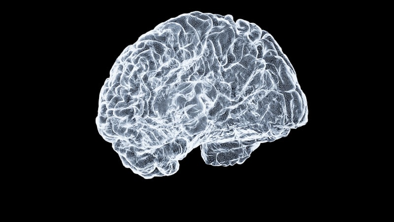
The Posterior in Neuroscience: Brain Structures
In neuroanatomy, the term posterior is used extensively to delineate regions of the central nervous system, particularly the brain and spinal cord. Given that the main axis of the brain is largely horizontal, posterior usually aligns with the caudal direction, pointing toward the spinal cord, or the rear of the cerebrum. Key structures located in this region include the posterior fossa, a significant cavity in the skull that houses the cerebellum and brainstem, and the occipital lobe, which constitutes the most posterior portion of the cerebral cortex and is primarily responsible for processing visual information. Damage to this highly specialized posterior region often results in visual field deficits or cortical blindness.
The functionality of posterior brain regions is often associated with integration and output processing, contrasting with the anterior regions often tied to planning and motor initiation. For instance, the posterior parietal cortex (PPC) plays a fundamental role in spatial awareness, attention, and integrating sensory input to guide movement. Lesions here can result in complex syndromes like hemineglect, where the patient fails to acknowledge stimuli on the side opposite the lesion. Furthermore, major vascular supply systems, such as the posterior cerebral artery (PCA), provide blood flow to these critical caudal structures; occlusion of the PCA is a common cause of stroke affecting visual and memory functions, underscoring the functional importance concentrated in the posterior brain.
Within the spinal cord, posterior (or dorsal) roots carry sensory information (afferent signals) into the central nervous system, a key anatomical fact underpinning neurological diagnosis. The posterior columns of the white matter transmit vital information regarding proprioception, vibration, and fine touch, enabling the brain to interpret body position in space. The integrity of these posterior pathways is routinely assessed during neurological examinations. Thus, in the context of the nervous system, posterior describes not only location but frequently correlates with specific sensory and integrative functional roles, making it an indispensable descriptor for clinicians and researchers alike.
Clinical and Surgical Significance of Posterior Structures
The designation of posterior holds immense practical value in clinical medicine, guiding surgical approaches, diagnostic imaging interpretation, and therapeutic interventions. Surgeons frequently plan procedures based on posterior approaches to minimize damage to vital anterior structures and facilitate access to deep-seated anatomy. For example, spinal fusion surgery often utilizes a posterior approach (posteriolateral or midline posterior) to access the vertebral bodies and intervertebral discs, providing a direct route to the affected segment while avoiding the abdominal cavity. Similarly, certain hip replacement techniques favor a posterior approach, though this must be weighed against the potential for higher dislocation risk compared to anterior methods.
Diagnostic imaging relies heavily on identifying posterior anatomical landmarks. In chest X-rays, the description of a lesion as being in the “posterior segment of the right upper lobe” provides the precise localization necessary for targeted biopsy or radiation therapy. Radiologists use terms like posterior displacement or posterior compression to describe pathological changes, such as a herniated intervertebral disc compressing the posterior spinal nerves, which is the underlying cause of conditions like sciatica. The clarity afforded by the posterior descriptor directly influences treatment protocols and patient outcomes.
Furthermore, in ophthalmology, the posterior segment of the eye, which includes the vitreous humor, retina, and optic nerve, is frequently the site of severe pathology, such as diabetic retinopathy or macular degeneration. Accessing and treating this region requires specialized instrumentation and techniques, often referred to as posterior segment surgery. The careful delineation of anterior versus posterior compartments in clinical contexts ensures that interventions are localized correctly, whether discussing the chambers of the eye, the ligaments of the knee (e.g., posterior cruciate ligament), or the valves of the heart (e.g., posterior leaflet of the mitral valve).
Related Directional Terminology
To fully appreciate the meaning and utility of posterior, it must be understood within the matrix of related anatomical directional terms that together establish the three-dimensional coordinate system of the body. These terms are often used in conjunction with posterior to provide exceptionally precise descriptions of location or movement. Understanding this relational framework is fundamental to all biological sciences.
The primary complementary term is anterior, which denotes the front or head end. Other critical related terms include those defining the dorsal/ventral and cranial/caudal axes. The following list outlines the most frequently paired terms used alongside posterior:
- Anterior: Opposite of posterior; toward the front or head end.
- Dorsal: Toward the back surface (often synonymous with posterior in human anatomy).
- Ventral: Toward the belly surface (opposite of dorsal).
- Caudal: Toward the tail or inferior end (often synonymous with posterior in quadrupeds).
- Cranial (or Superior): Toward the head (opposite of caudal/inferior).
- Medial: Toward the midline of the body.
- Lateral: Away from the midline of the body.
When describing complex locations, these terms are often combined, creating compound descriptors such as posterosuperior (behind and above) or posterolateral (behind and to the side). These combinations allow for granular precision, such as describing the exact entry point of a nerve or the precise location of a metastatic lesion within an organ. The rigorous application of these standardized terms ensures that anatomical descriptions are universally understandable, regardless of the reader’s native language or specific field of study.
Developmental Biology and Posterior Specification
The definition of the posterior pole is not merely descriptive in mature organisms but is established through highly conserved and critical processes during early embryonic development. The formation of the anterior-posterior (A-P) axis is one of the very first patterning events following fertilization, determining which end of the developing embryo will form the head and which will form the tail/rear. This process, known as axis specification, involves complex molecular signaling cascades that establish concentration gradients across the embryonic tissue.
Key signaling molecules, such as members of the Wnt signaling pathway and various growth factors, are often expressed at high concentrations at the prospective posterior end of the embryo, gradually diffusing toward the anterior end. Cells interpret their position along this gradient to determine their ultimate fate. Cells exposed to high levels of posterior signals are induced to differentiate into caudal structures, while those exposed to lower levels or antagonistic anterior signals differentiate into rostral (head) structures. This foundational patterning event dictates the entire subsequent organization of the body plan, including the placement of the nervous system and the segmentation of the trunk.
The Hox gene cluster plays a crucial role in maintaining and interpreting this posterior identity. These genes are master regulatory genes that are expressed colinearly along the A-P axis, meaning the genes located physically toward one end of the chromosome are expressed in the corresponding posterior regions of the body. They determine the segment identity—for example, ensuring that ribs grow only from the thoracic vertebrae and not from the lumbar (more posterior) vertebrae. Disruptions in the precise establishment or maintenance of the posterior axis during embryogenesis can result in severe congenital defects, including caudal regression syndrome or improperly formed spinal segments, highlighting the absolute necessity of accurate A-P specification.
Summary and Conceptual Importance
The term posterior transcends a simple definition of “behind”; it functions as a cornerstone of biological communication, providing the necessary spatial anchor for describing structures across disparate fields, from comparative zoology to human neurosurgery. Its utility is defined by its consistency as the opposite of anterior, yet its precise application must always be contextualized based on the organism’s standard anatomical position, differentiating between caudal orientation in quadrupeds and dorsal orientation in bipedal humans.
The integration of posterior terminology is essential for achieving diagnostic clarity and surgical precision. Whether identifying the location of a brain tumor within the posterior fossa, planning a surgical approach to the spine, or analyzing the early signals that specify the caudal end of an embryo, the term provides an unambiguous reference point. The ability to articulate complex spatial relationships using terms like posterior is what elevates biological description from generalized narrative to rigorous scientific analysis, ensuring that discoveries and clinical data are accurately and reliably communicated across the scientific community.
Ultimately, the concept of the posterior axis is not merely an arbitrary linguistic convention but a reflection of the fundamental, evolutionarily conserved body plan shared across the animal kingdom. Mastery of this term, along with its related directional complements, is the foundation for any serious study of anatomy, physiology, and developmental biology, allowing for the comprehensive mapping and understanding of life’s intricate structures.