Projection Neurons: The Brain’s Long-Distance Messengers
Scientifically Reviewed · Dr. Marwa Abd-Alazim · September 30, 2026
Review Criteria & Clinical Standards
This content undergoes rigorous scientific peer-review and medical editorial standards at Arab Psychology Network to ensure clinical accuracy, validity, and compliance with evidence-based guidelines from leading psychological and healthcare authorities (APA / WHO).
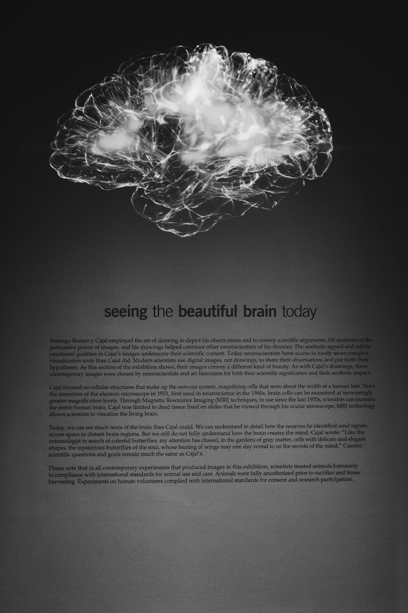
Introduction to Projection Neurons and Their Role in Neural Communication
In the complex architecture of the central nervous system, projection neurons serve as the primary conduits for long-distance communication. Unlike interneurons, which facilitate local circuitry and internal processing within a specific brain region, projection neurons possess specialized anatomical features that allow them to transmit information across vast distances. These neurons are the structural backbone of the brain’s macro-scale networks, linking disparate anatomical regions to form cohesive functional systems. Their primary role is to integrate processed information from one area and convey it to another, ensuring that the brain operates as a unified entity rather than a collection of isolated modules.
The significance of projection neurons cannot be overstated, as they are fundamental to every aspect of human experience, from the basic perception of environmental stimuli to the execution of complex motor sequences and the regulation of deep-seated emotional states. By acting as the “highways” of the brain, these cells enable the rapid exchange of electrical and chemical signals necessary for survival. Their unique ability to span multiple centimeters—or even meters in the case of spinal projections—distinguishes them as a critical focus in the field of neurobiology and psychology. Understanding their mechanics is essential for comprehending how the mind perceives and interacts with the world.
The study of these neurons has revealed a high degree of specialization and diversity. Depending on their location and the specific pathways they serve, projection neurons may utilize different neurotransmitters and exhibit varying firing patterns. This article provides a comprehensive overview of their structure, their role in major physiological pathways, their involvement in significant neurological and psychiatric disorders, and the cutting-edge research that is currently shaping our understanding of neural connectivity. By examining these cells, researchers gain insights into the very nature of brain connectivity and the catastrophic effects that occur when these connections are severed or dysfunctional.
To understand the breadth of their influence, it is helpful to categorize their primary functions:
- Information Integration: Synthesizing inputs from local interneurons and other projection neurons.
- Long-Distance Signaling: Facilitating communication between non-adjacent brain regions.
- Signal Amplification: Ensuring that neural messages maintain their integrity over long axonal distances.
- Pathway Formation: Constructing the white matter tracts that define the brain’s structural connectome.
Structural Morphology and Cellular Components
The unique functional capabilities of projection neurons are directly derived from their specialized morphology. Like most neurons, they are composed of a cell body (soma), an axon, and a complex dendritic tree. However, the proportions and specializations of these components are tailored for long-range transmission. The soma serves as the metabolic and genetic center of the cell, housing the nucleus and the machinery required for protein synthesis. Because projection neurons must maintain incredibly long axons, their metabolic demands are significantly higher than those of local circuit neurons, requiring robust transport systems to move nutrients and organelles along the length of the cell.
The defining feature of a projection neuron is its axon, which can extend significantly beyond the immediate vicinity of the cell body. These axons are often wrapped in a myelin sheath, a fatty insulating layer produced by oligodendrocytes in the central nervous system. Myelination is crucial because it allows for saltatory conduction, where electrical impulses jump between gaps in the myelin called nodes of Ranvier, drastically increasing the speed and efficiency of signal transmission. Without this structural adaptation, the time required for a signal to travel from the motor cortex to the base of the spinal cord would be too slow to support coordinated movement.
On the receiving end, the dendrites of projection neurons are often highly branched and covered in dendritic spines. These structures serve as the primary sites for synaptic input, allowing a single projection neuron to receive and integrate thousands of signals from diverse sources. This integration is vital because the decision to fire an action potential—which will then travel down the long axon—is based on the summation of these various excitatory and inhibitory inputs. Consequently, the projection neuron acts not just as a cable, but as a sophisticated processor that determines which information is important enough to be broadcast to other brain regions.
Key structural differences between projection neurons and interneurons include:
- Axonal Length: Projection neurons have long axons that exit their region of origin, while interneurons have short axons contained within a single region.
- Myelination: Projection axons are frequently myelinated for speed; interneurons are often unmyelinated.
- Target Connectivity: Projection neurons target distant nuclei or cortical areas; interneurons target neighboring cells.
- Volume of Cytoplasm: Projection neurons typically have a larger total volume due to the massive surface area of their extended axons.
Functional Integration in Sensory Processing Pathways
In the context of sensory pathways, projection neurons are the essential links that carry information from the periphery to the higher-order processing centers of the brain. When a sensory organ—such as the eye, ear, or skin—detects a stimulus, the information is converted into electrical signals. Projection neurons then take these signals and transmit them through a series of relay stations. A classic example is the thalamocortical pathway, where projection neurons in the thalamus receive sensory data and “project” it to the primary sensory cortex. This transmission is not a simple relay; it involves filtering and modulation, ensuring that the most relevant sensory information reaches conscious awareness.
The efficiency of these pathways is what allows for the near-instantaneous perception of our environment. For instance, in the visual system, retinal ganglion cells act as projection neurons that carry visual information from the retina, through the optic nerve, to the lateral geniculate nucleus of the thalamus. From there, another set of projection neurons carries the data to the visual cortex. Each stage of this journey relies on the structural integrity of the projection neurons involved. If these neurons are damaged or if their myelination is compromised, the result is a significant delay or total loss of sensory perception, illustrating their vital role in our interface with the external world.
Furthermore, projection neurons in sensory pathways are involved in the phenomenon of sensory gating. This process allows the brain to ignore redundant or irrelevant stimuli, such as the feeling of clothes against the skin, while remaining highly sensitive to novel or dangerous stimuli. By modulating the firing rates of projection neurons, the brain can effectively “turn up” or “turn down” the volume of specific sensory inputs. This sophisticated control mechanism is a hallmark of healthy neural function and highlights the active role projection neurons play in shaping our subjective experience of reality.
Motor Pathways and the Execution of Voluntary Movement
The motor pathways of the nervous system rely heavily on projection neurons to translate mental intent into physical action. This process begins in the motor cortex, where “upper motor neurons”—a specific class of projection neurons—originate. These cells send their axons down through the brainstem and into the spinal cord, forming major tracts such as the corticospinal tract. These axons are among the longest in the human body, spanning the entire distance from the top of the head to the lower back. Their primary function is to carry the commands for voluntary movement to the “lower motor neurons” located in the spinal cord, which then stimulate the muscles to contract.
Precision and timing are the most critical factors in motor control, and projection neurons are optimized for both. The high degree of myelination on motor axons ensures that signals travel at speeds exceeding 100 meters per second. This rapid communication is what allows for the complex coordination required for tasks like playing a musical instrument, typing, or maintaining balance while walking. Any disruption in the flow of information along these projection neurons leads to motor deficits, ranging from mild incoordination to total paralysis, emphasizing that these neurons are the physical manifestation of the brain’s “will” over the body.
Beyond simple muscle contraction, projection neurons in the motor system are also involved in feedback loops with the cerebellum and basal ganglia. These loops allow for the constant refinement of movement. For example, as you reach for an object, projection neurons are constantly sending information about the position of your limb back to the brain, while other projection neurons send corrective signals down to the muscles. This continuous, high-speed dialogue is made possible by the robust connectivity of projection neurons, which integrate sensory feedback with motor output to ensure smooth, accurate, and efficient movement patterns.
Regulation of Emotion and Limbic Circuitry
In addition to sensory and motor functions, projection neurons are fundamental to the emotion pathways that govern our psychological state and behavioral responses. The limbic system, which includes structures like the amygdala, hippocampus, and prefrontal cortex, is interconnected by a dense network of projection neurons. These neurons facilitate the communication between the “emotional” centers of the brain and the “rational” centers. For example, when a threat is perceived, projection neurons from the amygdala send signals to the hypothalamus to trigger the stress response, while simultaneously communicating with the prefrontal cortex to evaluate the situation.
This interplay is essential for emotional regulation and the formation of complex behaviors. The projection neurons that link the prefrontal cortex to the amygdala are particularly important, as they provide top-down control over emotional impulses. In a healthy individual, these neurons allow the brain to suppress inappropriate emotional outbursts or to modulate fear responses based on logic. When the connectivity provided by these projection neurons is weakened or imbalanced, it can lead to various mood disorders and behavioral issues, as the brain’s ability to integrate emotional information with cognitive context becomes impaired.
The reward system of the brain also depends on projection neurons, specifically those that utilize dopamine as a neurotransmitter. Neurons originating in the ventral tegmental area project to the nucleus accumbens and the prefrontal cortex, creating the neural basis for motivation, pleasure, and reinforcement learning. These long-range projections ensure that a rewarding stimulus—such as food or social interaction—is translated into a widespread neural signal that encourages the repetition of the beneficial behavior. The complexity of these emotional circuits demonstrates that projection neurons are not just structural elements, but are the primary mediators of our internal psychological landscape.
Pathophysiology in Neurodevelopmental and Psychiatric Disorders
Given their central role in brain connectivity, it is unsurprising that projection neurons are implicated in a wide array of neurological disorders and psychiatric conditions. In neurodevelopmental disorders such as autism spectrum disorder (ASD), research suggests that the “wiring” of projection neurons may be atypical. Specifically, there may be an overabundance of local connections (interneurons) and a deficit in long-range connections (projection neurons). This imbalance can lead to difficulties in integrating information across different brain regions, which may explain the sensory sensitivities and social communication challenges observed in individuals with ASD.
In the case of schizophrenia, the dysfunction of projection neurons is often linked to impaired communication between the prefrontal cortex and other areas, such as the thalamus and the hippocampus. This “disconnectivity” is thought to contribute to the cognitive fragments and hallucinations associated with the disorder. When projection neurons fail to synchronize their firing or when their axonal integrity is compromised, the brain’s ability to maintain a coherent stream of thought and a clear distinction between internal and external stimuli is significantly diminished. Current theories suggest that the misfiring or improper development of these cells is a core component of the disease’s pathology.
Furthermore, the synaptic pruning process—where the brain eliminates excess connections during adolescence—is a critical period for projection neurons. In several psychiatric conditions, it is believed that this process goes awry, either pruning too many or too few projection axons. This leads to a brain network that is either inefficient or overly noisy. The study of projection neurons in these contexts provides a biological framework for understanding symptoms that were once viewed purely through a behavioral lens, offering hope for more targeted physiological interventions in the future.
Neurodegenerative Implications: Parkinson’s and Alzheimer’s Diseases
The vulnerability of projection neurons is a hallmark of many neurodegenerative diseases, most notably Parkinson’s disease and Alzheimer’s disease. In Parkinson’s, the pathology specifically targets the dopaminergic projection neurons of the substantia nigra. These neurons project to the striatum and are essential for the initiation and smoothing of movement. As these projection neurons die off, the long-distance communication required for motor control is lost, resulting in the characteristic tremors, rigidity, and bradykinesia (slowness of movement) seen in patients. The loss of the physical “bridge” provided by these axons is the direct cause of the motor symptoms.
In Alzheimer’s disease, the destruction of projection neurons occurs on a more global scale but often begins in areas critical for memory, such as the entorhinal cortex and the hippocampus. These neurons are responsible for projecting information into the cerebral cortex for long-term storage and retrieval. The accumulation of amyloid-beta plaques and tau tangles within and around these projection neurons disrupts their transport systems and eventually leads to cell death. As these long-range connections are severed, the brain’s various regions become isolated from one another, leading to the profound cognitive decline and memory loss that define the condition.
The specific susceptibility of projection neurons to neurodegeneration may be due to their high metabolic demands and the sheer length of their axons. Maintaining a cellular structure that spans long distances requires immense energy and efficient axonal transport. When cellular processes like mitochondrial function or protein clearance fail, these long-distance cells are often the first to suffer. Understanding why projection neurons are particularly at risk is a major focus of current medical research, as preserving these connections is key to slowing or preventing the progression of neurodegenerative disorders.
Advancements in Genetic Research and Molecular Identification
Recent years have seen a surge in research focused on the genetic underpinnings of projection neuron development and function. Scientists have identified specific gene expression profiles that distinguish projection neurons from other cell types and even different subtypes of projection neurons from each other. These genes govern everything from the direction of axonal growth during embryonic development to the specific neurotransmitters the neuron will use in adulthood. By mapping these genetic markers, researchers can now identify which specific populations of projection neurons are affected in different diseases, allowing for a level of precision that was previously impossible.
One of the most exciting areas of genetic research involves the study of transcription factors that determine the “identity” of a projection neuron. For instance, certain genes act as molecular switches that tell a developing neuron to send its axon to the spinal cord rather than to another area of the cortex. Disruptions in these genetic instructions can lead to “miswiring,” where neurons project to the wrong targets. This research is vital for understanding the etiology of various congenital brain abnormalities and provides a potential pathway for regenerative medicine, where scientists might one day “reprogram” cells to replace lost projection neurons.
In addition to developmental genetics, researchers are investigating how epigenetic changes—modifications to gene expression caused by environmental factors—affect the health of projection neurons over a lifetime. Factors such as chronic stress, toxin exposure, and aging can alter the way genes in these neurons are expressed, potentially making them more or less resilient to disease. This holistic view of neurogenetics is bridging the gap between our DNA and our actual brain connectivity, providing a more comprehensive understanding of how individual differences in projection neuron function contribute to personality, intelligence, and disease risk.
Technological Progress in Neuroimaging and Connectomics
The ability to study projection neurons in the living human brain has been revolutionized by advancements in imaging techniques. Traditional MRI provides a look at brain structure, but newer methods like Diffusion Tensor Imaging (DTI) allow researchers to visualize the actual pathways of white matter—the bundles of myelinated axons belonging to projection neurons. DTI measures the movement of water molecules along axons, allowing scientists to map the brain’s connectome in high detail. This has proven invaluable for identifying subtle changes in connectivity that occur in disorders like traumatic brain injury or multiple sclerosis.
Furthermore, functional imaging techniques, such as fMRI, allow researchers to observe the activity of projection neurons in real-time. By looking at which brain regions “fire” together during a task, scientists can infer the strength and efficiency of the projection neurons connecting them. This has led to the discovery of resting-state networks, which are groups of brain regions that remain in constant communication via projection neurons even when a person is at rest. These networks are essential for maintaining a sense of self and for the “background” processing that occurs during daydreaming or reflection.
In the laboratory, techniques such as optogenetics and two-photon microscopy allow for the manipulation and observation of individual projection neurons with incredible precision. Optogenetics involves using light to turn specific projection neurons on or off, allowing researchers to see exactly how a particular long-range connection influences behavior. These tools have confirmed that projection neurons are not just passive transmitters but are active participants in neural computation. The combination of macro-scale human imaging and micro-scale laboratory techniques is providing a multi-level understanding of how projection neurons sustain the complexity of the human mind.
Future Directions in Therapeutic Interventions
The ultimate goal of studying projection neurons is the development of new and more effective treatments for neurological and psychiatric conditions. Because many of these disorders are essentially “connectivity diseases,” the focus is shifting toward therapies that can repair or bypass damaged projection pathways. One promising area is Deep Brain Stimulation (DBS), which uses implanted electrodes to modulate the activity of projection neurons in specific circuits. This has already shown remarkable success in treating the motor symptoms of Parkinson’s disease and is currently being explored for treatment-resistant depression and obsessive-compulsive disorder.
Another frontier is the use of stem cell therapy and nerve growth factors to encourage the regeneration of projection axons. In cases of spinal cord injury or stroke, the primary challenge is the brain’s limited ability to regrow long-distance connections. Researchers are investigating ways to create a “scaffold” that guides regrowing axons to their original targets, effectively rebuilding the lost bridges of the nervous system. While still in the experimental stages, the prospect of restoring lost functions by repairing projection neurons is one of the most ambitious and potentially transformative goals in modern medicine.
Finally, the rise of pharmacogenomics—tailoring drug treatments to an individual’s genetic profile—holds great promise for disorders involving projection neurons. By understanding the specific molecular defects in a patient’s projection neurons, doctors may be able to prescribe medications that specifically address those issues, rather than using broad-spectrum drugs with numerous side effects. As our understanding of projection neurons continues to grow, so too does our ability to intervene in the complex machinery of the brain, offering hope for a future where the devastating effects of brain connectivity disorders can be mitigated or even reversed.
References
Bing, J., & Li, X. (2020). Projection neurons: Structure, function, and role in neurological disorders. Frontiers in Neuroscience, 14, 596. https://doi.org/10.3389/fnins.2020.00596
Khan, S. Y., & Sohal, V. S. (2020). Projection neurons: A review of their role in neurological disorders. Frontiers in Neuroscience, 14, 595. https://doi.org/10.3389/fnins.2020.00595
Pang, T. Y., & Ching, S. (2020). Recent advances in projection neuron research. Neuroscience & Biobehavioral Reviews, 109, 306–319. https://doi.org/10.1016/j.neubiorev.2020.04.008