Cellular Mitosis: The Blueprint of Biological Growth
The Core Definition of Prophase
Prophase is the pivotal initial stage of mitosis, a fundamental process of cell division occurring in all eukaryotic cells. This phase marks the crucial transition from the interphase growth and DNA replication to the organized distribution of genetic material into two daughter cells. Essentially, prophase acts as the meticulous preparation stage where the cell undertakes significant internal reorganizations to ensure the accurate and equitable segregation of its genetic blueprint, encapsulated within its chromosomes. Without the precise execution of events during prophase, the subsequent stages of cell division would be compromised, leading to genetic abnormalities that can have severe consequences for the organism.
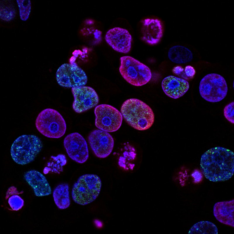
During prophase, several key cellular events are initiated or completed that are indispensable for the successful progression of mitosis. The most visually striking event is the dramatic condensation of chromosomes, transforming the diffuse chromatin network into compact, rod-like structures that are discernible under a light microscope. Simultaneously, the nuclear envelope, which encloses the genetic material, begins to disintegrate, paving the way for the chromosomes to interact with the newly forming mitotic spindle. Concurrently, the mitotic spindle apparatus, a dynamic structure composed primarily of microtubules, starts to assemble, originating from structures called centrosomes that migrate to opposite poles of the cell.
The culmination of these preparatory steps in prophase is to ready the duplicated chromosomes for their eventual separation. Each chromosome at this stage consists of two identical copies, known as sister chromatids, joined at a constricted region called the centromere. The condensation protects these delicate DNA molecules from damage and tangling during their movement, while the formation of the mitotic spindle provides the necessary machinery for their precise alignment and segregation. The breakdown of the nuclear envelope ensures that the spindle microtubules can access and attach to the centromeres of the chromosomes. This meticulous orchestration during prophase is foundational, guaranteeing that each daughter cell receives a complete and identical set of chromosomes, thereby preserving the genetic fidelity across generations of cells.
Historical Context of Mitotic Discovery
The journey to understanding cell division, and by extension, the intricate processes of prophase, began with the advent of microscopy and the pioneering observations of early cell biologists. In the 17th century, Antonie van Leeuwenhoek’s groundbreaking work with improved microscopes revealed the existence of cells and microorganisms, opening up an entirely new world of biological inquiry. However, the internal dynamics of cell division remained largely unknown for centuries, primarily due to the limitations of optical technology and staining techniques. Early observations hinted at nuclear changes during cell proliferation, but a comprehensive, systematic description of the stages of cell division was yet to emerge, leaving a significant gap in the understanding of how organisms grow and reproduce at the cellular level.
The pivotal breakthroughs in elucidating mitosis, including the distinct stage of prophase, are largely attributed to the German anatomist and cell biologist Walther Flemming in the late 19th century. Using newly developed aniline dyes to stain chromosomes and vastly improved microscopes, Flemming meticulously observed and documented the intricate movements of thread-like structures within the nucleus of salamander larval cells. In his seminal 1882 work, “Zellsubstanz, Kern und Zelltheilung” (Cell-Substance, Nucleus, and Cell-Division), he not only coined the term “mitosis” (from the Greek “mitos” meaning thread, referring to the chromosomal structures) but also provided the first detailed description of its successive stages: prophase, metaphase, anaphase, and telophase. His observations of chromosome condensation and the formation of the spindle apparatus during the initial phase laid the groundwork for our modern understanding of prophase.
Flemming’s rigorous and systematic approach revolutionized cell biology, shifting the focus from static cellular structures to dynamic cellular processes. His discovery of the consistent pattern of chromosome behavior during mitosis provided compelling evidence for the continuity of genetic material and its faithful transmission from parent to daughter cells. The recognition of prophase as the initial stage, where chromosomes become visible and organized, was a critical step in deciphering the entire cell cycle. This historical foundation paved the way for subsequent research that elucidated the molecular mechanisms controlling prophase, its checkpoints, and its profound implications for genetics, development, and disease, continuing to be a cornerstone of biological study today.
Detailed Cellular Events During Mitotic Prophase
Mitotic prophase is a period of intense cellular activity, marked by a series of precisely orchestrated events that prepare the cell for chromosome segregation. The most visually apparent transformation is the dramatic condensation of chromosomes. During the preceding interphase, the cell’s genetic material, or chromatin, exists in a decondensed, diffuse state, making it accessible for DNA replication and gene expression. As prophase begins, the long, thin chromatin fibers progressively coil and supercoil, compacting into discrete, highly organized structures. Each replicated chromosome now consists of two identical sister chromatids, joined tightly at their centromeres. This extensive condensation is crucial for preventing entanglement and ensuring that the chromosomes can be efficiently and accurately moved during the later stages of mitosis.
Concurrently with chromosome condensation, the cell begins to assemble the mitotic spindle, a dynamic structure essential for chromosome movement. In animal cells, this process starts as the two centrosomes, which duplicated during interphase, begin to migrate to opposite poles of the cell. Each centrosome acts as a microtubule-organizing center, nucleating the growth of microtubules. As the centrosomes move apart, they establish a bipolar spindle, with microtubules radiating outwards, forming structures known as asters. These microtubules will later connect to the chromosomes to facilitate their separation. The precise positioning of the centrosomes and the controlled assembly of the spindle fibers during prophase are critical for establishing the axis of cell division and ensuring that chromosomes are evenly distributed.
Another defining event of prophase is the breakdown of the nuclear envelope. In most eukaryotic cells, this membrane barrier encapsulates the nucleus during interphase, physically separating the genetic material from the cytoplasm. As prophase progresses, the nuclear envelope, a double membrane studded with nuclear pores, disassembles into numerous small vesicles. This breakdown is mediated by the phosphorylation of nuclear lamins, proteins that provide structural support to the nuclear envelope, and the integral nuclear pore proteins. The dissolution of this barrier is a prerequisite for the mitotic spindle microtubules to gain access to the condensed chromosomes and attach to their kinetochores, specialized protein complexes located at the centromeres. Furthermore, the nucleolus, a prominent structure within the nucleus involved in ribosome synthesis, typically disappears during prophase, signifying a pause in ribosomal production as the cell dedicates its resources to division.
Practical Significance and Biological Applications
The precise execution of prophase is fundamentally important for all forms of life that rely on cell division for their existence and propagation. In multicellular organisms, mitosis, initiated by prophase, is the driving force behind growth and development. From a single fertilized egg, an entire complex organism develops through countless rounds of cell division, with each new cell originating from a parent cell undergoing mitosis. Prophase ensures that as cells multiply to form tissues, organs, and organ systems, every new cell receives a complete and accurate copy of the genetic information. This fidelity is also critical for tissue repair and maintenance throughout an organism’s life, replacing old or damaged cells (e.g., skin cells, blood cells) with genetically identical new ones, thereby preserving tissue integrity and function.
Beyond growth and repair, understanding prophase and its regulatory mechanisms carries profound implications for human health and disease. Errors in the intricate processes of chromosome condensation, spindle formation, or nuclear envelope breakdown during prophase can lead to aneuploidy—an abnormal number of chromosomes—which is a hallmark of many human cancers and developmental disorders. For instance, if chromosomes fail to condense properly, they may become tangled, leading to breaks or incorrect segregation. Similarly, defects in spindle assembly or function can result in daughter cells receiving too many or too few chromosomes. Research into the molecular players that govern prophase, such as specific kinases and phosphatases, provides crucial insights into the origins of these diseases and offers potential targets for therapeutic interventions aimed at controlling uncontrolled cell proliferation, as seen in cancer.
The study of prophase extends its utility into various fields of biological and medical research. In developmental biology, understanding how prophase is regulated during embryonic development helps unravel the mechanisms that drive cell differentiation and tissue patterning. In genetics, the accurate segregation of chromosomes during mitosis, orchestrated by prophase, underpins the inheritance of traits and the stability of the genome. Furthermore, the detailed visualization of condensed chromosomes during prophase and metaphase is a cornerstone of cytogenetics, allowing for the detection of chromosomal abnormalities through techniques like karyotyping, which are vital for diagnosing genetic disorders. Thus, prophase is not merely an academic concept but a fundamental biological process with wide-ranging practical applications in medicine, biotechnology, and our overall understanding of life.
Connections to the Broader Cell Cycle and Meiosis
Prophase is an integral component of the broader cell cycle, which describes the series of events that take place in a cell leading to its division and duplication. It immediately follows the interphase, a period of cell growth and DNA replication, specifically the G2 phase where the cell prepares for division. The transition from G2 to prophase is tightly regulated by cell cycle checkpoints that ensure the cell is ready to divide, with all DNA replicated and no damage present. Once prophase is complete, the cell transitions into metaphase, where condensed chromosomes align at the cell’s equatorial plate. This is followed by anaphase, characterized by the separation of sister chromatids, and finally telophase, where new nuclear envelopes form around the separated chromosome sets. The entire process culminates in cytokinesis, the physical division of the cytoplasm, yielding two genetically identical daughter cells.
While the events described for prophase pertain to mitosis, a process that produces somatic cells, a distinct and more complex form of prophase exists in meiosis, the specialized cell division that produces gametes (sperm and egg cells). Meiosis involves two successive divisions, and its first stage, Prophase I, is notably prolonged and elaborate. Unlike mitotic prophase where sister chromatids condense independently, Prophase I of meiosis is further subdivided into five substages: leptotene, where chromosomes begin to condense; zygotene, where homologous chromosomes pair up to form bivalents via a protein structure called the synaptonemal complex; pachytene, during which crossing over (genetic recombination) occurs between non-sister chromatids; diplotene, where homologous chromosomes begin to separate but remain joined at chiasmata (sites of crossing over); and finally, diakinesis, where chromosomes condense further and the nuclear envelope breaks down. This intricate Prophase I is essential for generating genetic diversity, a key feature of sexual reproduction.
The fundamental principles underlying chromosome condensation, spindle formation, and nuclear envelope dynamics are shared between mitotic prophase and prophase I of meiosis, highlighting their common evolutionary origins and the universal requirements for accurate chromosome handling. However, the unique events of homologous pairing and crossing over in meiotic prophase I underscore the distinct outcomes of these two types of cell division: genetic identity in mitosis versus genetic diversity in meiosis. Thus, prophase, in its various forms, stands as a cornerstone in cell biology and genetics, representing the initial, crucial steps that dictate the fate of genetic information during cellular reproduction and, ultimately, the continuity and evolution of life itself. Its study continues to illuminate fundamental biological mechanisms and offers pathways for understanding and treating a wide array of diseases.