Pyramidotomy: The Dark History of Motor Control Surgery
- Introduction to Pyramidotomy: Definition and Context
- Neuroanatomical Basis: The Pyramidal Tract
- Historical Development and Early Indications
- Surgical Methodology and Technique
- Clinical Applications and Expected Outcomes
- Complications and Iatrogenic Effects
- Decline in Practice and Ethical Review
- Modern Neuromodulatory Alternatives
Introduction to Pyramidotomy: Definition and Context
Pyramidotomy is defined historically as a radical neurosurgical procedure involving the deliberate and precise surgical cutting or transection of the pyramidal tract, which is the principal efferent pathway responsible for voluntary motor control. This intervention, performed primarily during the mid-20th century, was classified as an ablative functional neurosurgery, designed explicitly to interrupt the descending motor signals originating from the cerebral cortex. The procedure targeted the motor pathways, often at the level of the brainstem, specifically the medullary pyramids, from which the operation derives its name. The fundamental purpose of this destructive surgery was not to cure the underlying disease but rather to alleviate severe, debilitating motor symptoms, particularly those characterized by uncontrollable involuntary movements or extreme, refractory spasticity, by intentionally inducing a partial or complete motor deficit.
The core rationale underpinning pyramidotomy was rooted in the understanding that certain severe movement disorders resulted from pathological hyperactivity or dysregulation within the motor system, leading to signals that could not be suppressed pharmacologically or managed through conservative physical therapy. By creating a controlled lesion in the primary motor output pathway, surgeons aimed to isolate the affected musculature from the aberrant central commands, thereby trading a disabling involuntary movement for a more manageable, albeit permanent, state of paralysis or paresis. This decision was only made in cases where the patient’s quality of life was severely compromised by the uncontrolled movements, necessitating a drastic intervention to restore a measure of functional rest and reduce pain associated with chronic spasm.
It is crucial to contextualize pyramidotomy within the history of neurological surgery, particularly the pioneering era of functional neurosurgery that flourished before the widespread availability of advanced pharmacology and non-ablative neuromodulation techniques. Alongside procedures like pallidotomy and thalamotomy, pyramidotomy represented the surgical frontier in managing catastrophic neurological symptoms. While ethically challenging by modern standards due to its irreversible nature, at the time of its development, it offered one of the few avenues for relief for patients suffering from conditions deemed otherwise intractable, highlighting the desperate need for effective treatments for severe motor pathology.
Neuroanatomical Basis: The Pyramidal Tract
A comprehensive understanding of pyramidotomy requires detailed knowledge of the neuroanatomical structure targeted: the pyramidal tract, also known as the corticospinal tract. This tract represents the longest and most crucial efferent pathway in the human central nervous system, originating primarily from the motor, premotor, and somatosensory cortices of the brain. Its fibers descend through the cerebral hemispheres, converging dramatically in the internal capsule before traversing the midbrain (cerebral peduncles) and the pons. It is the integrity of these fibers that dictates the ability to execute skilled, voluntary, and fractionated movements, particularly those involving the distal musculature of the hands and feet.
The procedure specifically targets the fibers as they pass through the medulla oblongata, the lowest part of the brainstem, where they form the recognizable, paired structures known as the medullary pyramids on the ventral surface. This anatomical localization is critical because, just caudal to the pyramids, approximately 85% of the fibers decussate (cross over) to form the lateral corticospinal tract, which descends contralaterally (on the opposite side of the body) in the spinal cord. The remaining uncrossed fibers form the anterior corticospinal tract. Therefore, a successful pyramidotomy at this specific brainstem level results in a highly predictable, severe neurological deficit on the contralateral side of the body, affecting the motor control of the limbs and trunk.
The primary challenge and inherent danger of performing a pyramidotomy stems from the dense packing of vital structures within the brainstem. The medullary pyramids lie in close proximity to numerous critical cranial nerve nuclei (controlling functions like breathing, swallowing, and eye movement), as well as essential ascending and descending tracts (such as the medial lemniscus for sensory information). Therefore, the success of the procedure hinged entirely upon the surgeon’s ability to create a lesion that was perfectly confined to the pyramidal fibers while sparing adjacent, non-motor nuclei. Any deviation could result in catastrophic iatrogenic effects far exceeding the intended motor deficit, leading to profound sensory loss, autonomic dysfunction, or even mortality.
Historical Development and Early Indications
The concept of surgically interrupting the motor pathway to treat severe neurological symptoms gained traction in the early to middle parts of the 20th century, coinciding with a period of intense experimentation in functional neurosurgery. The realization that severe spasticity and certain hyperkinetic disorders were related to an imbalance or excess of motor drive led researchers to postulate that a controlled lesion could reset the pathological state. Early attempts were often experimental and highly invasive, but they established the principle that severing the descending motor tract could effectively abolish the uncontrollable movements that defied all existing medical therapies.
The primary clinical indications for pyramidotomy were conditions characterized by excruciatingly severe, refractory motor dysfunction. These included extreme spasticity, frequently observed in advanced cerebral palsy, severe post-stroke syndromes, or certain forms of spinal cord injury, where muscle rigidity and painful contractures rendered the patient immobile or caused chronic pain. In some specific cases, pyramidotomy was also explored as a treatment for highly debilitating forms of dystonia or choreoathetosis, where the involuntary writhing movements were so violent and constant that they led to exhaustion, self-injury, and total loss of functional independence.
The surgical community viewed pyramidotomy as a procedure of last resort. The strict selection criteria demanded that the patient had failed extensive trials of pharmacological management, physical therapy, and less invasive neurosurgical options (if available). The ethical justification rested on a utilitarian calculation: the induced motor deficit (weakness) was considered an acceptable and necessary sacrifice if it resulted in the complete abolition of a far more disabling and painful hyperkinesia. This required meticulous preoperative evaluation to ensure that the patient and family fully understood the certainty of resulting motor weakness alongside the potential reduction in involuntary symptoms.
Surgical Methodology and Technique
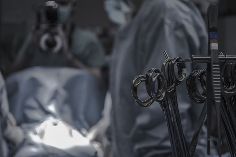
Performing a pyramidotomy was technically demanding and required exceptional surgical skill and anatomical precision, especially considering the constraints of mid-century neurosurgical technology. The procedure generally necessitated a complex craniotomy, often involving a posterior fossa approach, to gain access to the ventral surface of the brainstem (medulla oblongata). This access route itself carried significant risk due to the necessity of maneuvering around the cerebellum and major blood vessels supplying the posterior circulation of the brain. The surgeon had to navigate deep into the surgical field to reach the paired medullary pyramids.
Once the target was visualized, the core of the procedure involved creating a highly localized, controlled lesion. Early techniques relied on manual incision using fine surgical knives or needles, aiming for a precise transection of the white matter fibers composing the pyramid. As technology progressed, techniques shifted toward using stereotactic guidance, allowing for more accurate targeting, and employing radiofrequency ablation (thermal coagulation) to create the lesion. Radiofrequency allowed for a more controlled destruction of the tissue, minimizing the mechanical trauma associated with physical cutting, although the fundamental goal remained the same: irreversible interruption of the motor pathway.
To mitigate the high risk of collateral damage, intraoperative neurophysiological monitoring became increasingly important, particularly in later applications of the procedure. Surgeons utilized techniques like somatosensory evoked potentials (SSEPs) or motor evoked potentials (MEPs) to map the functional boundaries of the pyramidal tract and to ensure that the lesion did not encroach upon adjacent structures, particularly the sensory pathways or the nuclei of vital cranial nerves (such as the vagus or hypoglossal nerve). Despite these precautions, the proximity of critical structures meant that the margin for error in pyramidotomy remained extremely narrow, contributing significantly to its overall morbidity rate.
Clinical Applications and Expected Outcomes
The successful clinical outcome of a pyramidotomy was measured primarily by the degree of reduction in the target pathological movement. In cases of severe spasticity, the desired effect was a transition from rigid, high-tone muscle spasms to a more flaccid or reduced-tone state, which would ideally allow for passive movement, improved hygiene, and reduced chronic pain. For hyperkinetic disorders, success was defined by the cessation or dramatic attenuation of the involuntary movements that were rendering the patient non-functional. The immediate and unavoidable consequence, however, was contralateral hemiparesis (weakness) or hemiplegia (paralysis) in the limbs controlled by the severed tract.
In the short term, the procedure often resulted in a period of spinal shock followed by flaccid paralysis on the side opposite the lesion. Over time, as the spinal cord adjusted and reflexes returned, this flaccidity often converted into a more chronic state of spasticity, though theoretically less severe than the preoperative condition, or settled into a state of permanent weakness. The effectiveness of pyramidotomy was highly variable. For some patients with intractable spasticity, the reduction in tonic muscle activity was significant enough to improve nursing care and comfort, thus justifying the loss of voluntary motor function, which may have already been heavily compromised by the underlying disease.
Long-term assessment revealed that while the procedure could effectively abolish involuntary movements in some instances, the resulting motor deficit was profoundly disabling. The permanent loss of fine motor control, especially in the hand, meant that even if the patient was relieved of spasm, they faced severe functional limitations. Furthermore, the induced motor deficit did not always remain static; adaptation and reorganization within the central nervous system could sometimes lead to the development of new, maladaptive movement patterns, suggesting that simple transection did not fully address the complex etiology of the underlying neurological disorder.
Complications and Iatrogenic Effects
Pyramidotomy carried an exceptionally high risk profile, both immediately related to the surgery and as long-term functional deficits. Since the target area is deep within the brainstem, one of the most critical immediate risks was the potential for surgical trauma, hemorrhage, or edema within this compact structure, which could easily lead to life-threatening complications such as respiratory failure, profound autonomic instability, and death. Damage to surrounding structures, even minor errors in lesion placement, frequently resulted in debilitating non-motor deficits.
Specific iatrogenic effects related to damage adjacent to the pyramidal tract included injury to the cranial nerve nuclei, particularly the lower motor cranial nerves. Damage to the hypoglossal nerve (CN XII) could cause tongue weakness and difficulty speaking (dysarthria), while damage to the vagus nerve (CN X) or glossopharyngeal nerve (CN IX) could impair swallowing (dysphagia) and affect vocalization, severely impacting the patient’s ability to communicate and sustain nutrition. These complications, stemming from the proximity of the target to the neural centers controlling vital functions, were often more distressing than the motor deficit itself.
The most predictable and intended complication was the permanent loss of motor function, ranging from profound hemiplegia to severe paresis. While this weakness was considered the necessary trade-off for symptom relief, in reality, the degree of voluntary function loss often complicated rehabilitation and reduced the overall functional gain. In cases where the lesion was incomplete or poorly targeted, the patient might suffer the debilitating motor weakness without achieving the desired reduction in spasticity, leaving them with two significant, compounding disabilities. This highly unfavorable risk-benefit profile ultimately contributed significantly to the procedure’s eventual obsolescence.
Decline in Practice and Ethical Review
The widespread application of pyramidotomy began to decline rapidly from the 1970s onwards, a period marked by parallel advances in neurology and neurosurgery. The primary factor driving this decline was the advent of effective pharmacological agents capable of modulating muscle tone and reducing spasticity without requiring destructive brain surgery. Drugs targeting neurotransmitters and muscle relaxation offered a reversible, less invasive alternative to manage symptoms that previously required radical intervention, dramatically shifting the risk-benefit analysis away from ablative procedures.
Furthermore, as neurological imaging improved and surgical techniques became more refined, the medical community increasingly recognized the ethical complexities inherent in performing highly destructive, irreversible procedures for non-life-threatening, chronic conditions. Pyramidotomy involves deliberately destroying healthy tissue (even if functionally misguided) to manage symptoms, a concept that became ethically difficult to justify as less invasive, reversible options emerged. The procedure came under scrutiny regarding informed consent, particularly concerning the high probability of permanent, severe functional deficit in exchange for only potential symptom relief.
The shift in neurosurgical philosophy moved decisively away from large, open ablative lesions towards highly targeted, minimally invasive, and ideally reversible interventions. The high morbidity associated with brainstem access, coupled with the permanent nature of the induced deficit, rendered pyramidotomy medically unsustainable as neuroscientists gained a deeper, more nuanced understanding of motor circuit pathology. The procedure transitioned from being a therapeutic option to becoming primarily a historical case study in the evolution of functional neurosurgery and the ethics of ablative brain interventions.
Modern Neuromodulatory Alternatives
Contemporary management of severe, refractory movement disorders and spasticity has entirely supplanted pyramidotomy through the development of sophisticated neuromodulatory and targeted surgical techniques. The most significant advancement is Deep Brain Stimulation (DBS), a reversible technique that involves implanting electrodes into specific deep brain nuclei (such as the subthalamic nucleus or globus pallidus interna). DBS does not destroy brain tissue but rather delivers electrical impulses that modulate pathological circuit activity, effectively managing symptoms of Parkinson’s disease, essential tremor, and certain dystonias with high efficacy and reversibility.
For severe spasticity, the modern approach often favors targeted peripheral interventions or intrathecal drug delivery. The use of precisely administered Botulinum toxin (Botox) injections can temporarily paralyze specific hyperactive muscles, offering localized and reversible relief. Additionally, the implantation of intrathecal pumps delivering drugs like baclofen directly into the spinal fluid provides highly concentrated relief to the spinal cord and peripheral nerves, bypassing systemic side effects and avoiding the need for brain surgery altogether. These methods offer superior safety profiles and maintain the integrity of the motor pathways.
While highly precise, non-reversible stereotactic radiofrequency lesions (e.g., pallidotomy or thalamotomy) are still performed in rare, highly selected cases where DBS is contraindicated or unavailable, the critical distinction is their immense precision and target location, usually avoiding the densely packed, vital structures of the brainstem. Pyramidotomy, targeting the critical medullary pyramid, represents a technique that is now entirely obsolete. The evolution from destructive transection to fine-tuned electrical or pharmacological modulation underscores a fundamental paradigm shift in neurology: treating motor dysregulation as a circuit problem requiring correction, rather than a pathway requiring permanent severance.