Neuroimaging: Mapping the Architecture of the Human Mind
Core Definition and Scope of Radiology
Radiology is a highly specialized medical discipline utilizing sophisticated imaging technologies to visualize the internal structures and functions of the human body. At its core, it serves two primary functions: the diagnosis of disease or injury through diagnostic imaging techniques, and the treatment of various conditions, particularly cancers, using precisely controlled radioactive substances or image-guided interventions. The field is fundamentally interdisciplinary, requiring a deep understanding of physics, anatomy, physiology, and pathology to accurately interpret complex visual data and translate those findings into actionable clinical recommendations for referring physicians.
The fundamental mechanism underlying modern radiology involves the interaction of different forms of energy—such as electromagnetic waves, sound waves, or magnetic fields—with biological tissues. By measuring how these energies are absorbed, scattered, or reflected, high-resolution images can be reconstructed, revealing vital information about organ density, blood flow, and structural integrity. This capacity for non-invasive internal inspection has revolutionized medicine, moving diagnostics away from reliance on exploratory surgery and toward rapid, accurate, and often outpatient procedures. Furthermore, the practice demands meticulous attention to detail, as subtle visual cues on an image can differentiate benign conditions from life-threatening pathologies, making the radiologist a critical gatekeeper in the patient care pathway.
The scope of this specialty extends far beyond traditional film interpretation. It encompasses modalities ranging from simple projectional X-rays to complex functional studies like Positron Emission Tomography (PET) scans and Magnetic Resonance Imaging (MRI). Moreover, radiologists often collaborate closely with oncologists, surgeons, and internists, not only providing the initial diagnosis but also guiding biopsies, monitoring treatment efficacy, and performing therapeutic procedures themselves. This dual role—diagnostic interpreter and clinical interventionist—defines the modern practice of radiology, emphasizing both intellectual rigor in interpretation and technical skill in procedural execution.
Historical Foundations of Medical Imaging
The history of radiology is relatively recent, dating back only to the late 19th century, yet its impact was immediate and transformative. The pivotal moment occurred in 1895 when German physicist Wilhelm Conrad Röntgen discovered a new type of electromagnetic radiation that could penetrate opaque materials, including human flesh, and leave an image on photographic film. He famously called these unknown waves “X-rays.” The first human radiograph was of his wife’s hand, clearly showing the bones and a ring, demonstrating the unprecedented ability to see inside a living body without incision. This discovery earned Röntgen the first Nobel Prize in Physics in 1901 and laid the entire foundation for the modern field of diagnostic imaging.
Following Röntgen’s discovery, the medical community rapidly adopted the new technology. Within months, X-rays were being used in military hospitals to locate bullets and shrapnel, showcasing their immediate practical utility in trauma care. However, the early years were also characterized by a profound lack of understanding regarding the biological risks associated with this new energy source. Early practitioners, often viewing the process as harmless, suffered severe burns, hair loss, and eventually cancer due to cumulative exposure to high levels of ionizing radiation. This tragic context spurred subsequent research into radiation safety, dosimetry, and shielding, which remain critical components of radiological practice today, codified by principles like ALARA (As Low As Reasonably Achievable).
The mid-20th century witnessed a dramatic expansion of radiological techniques, driven by advancements in electronics and computer science. The introduction of technologies such as fluoroscopy (real-time X-ray imaging) allowed for dynamic studies of organ function, while the development of ultrasound in the 1940s and 1950s provided a non-ionizing alternative, particularly useful in obstetrics and cardiology. The true revolution, however, came with the invention of Computed Tomography (CT) scanning in the 1970s by Godfrey Hounsfield and Allan Cormack, which transformed two-dimensional projection images into detailed, cross-sectional anatomy, paving the way for the complex multi-modality approach that defines contemporary radiology.
Diagnostic Modalities: Non-Ionizing Techniques
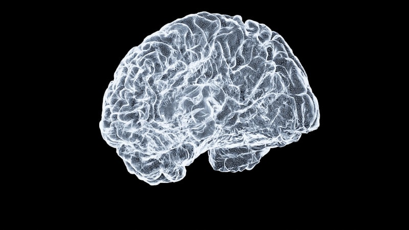
Non-ionizing diagnostic modalities represent a crucial segment of diagnostic imaging, providing detailed anatomical and physiological information without the use of potentially harmful ionizing radiation. The two primary techniques in this category are Magnetic Resonance Imaging (MRI) and Ultrasound. MRI utilizes powerful magnetic fields and radio waves to excite hydrogen nuclei (protons) within the body’s tissues. As these protons return to their resting state, they emit energy signals that are detected by sophisticated scanners. Because the density and environment of water molecules vary dramatically between different tissue types—such as fat, muscle, and cerebrospinal fluid—MRI excels at providing exceptional soft-tissue contrast, making it indispensable for evaluating the brain, spinal cord, joints, and internal organs where subtle differences in tissue composition are paramount to diagnosis.
Ultrasound, or sonography, operates on entirely different physical principles, relying on high-frequency sound waves. A transducer emits these waves into the body and then records the echoes that bounce back when the waves encounter boundaries between tissues of different densities, such as fluid, soft tissue, and bone. These returning echoes are processed into a real-time, dynamic image. Ultrasound is highly valued for its portability, low cost, and ability to assess moving structures, particularly blood flow (via Doppler ultrasound), making it the preferred method for assessing the abdomen, monitoring fetal development during pregnancy, and guiding minimally invasive procedures like targeted biopsies or fluid drainage. The absence of radiation makes it exceptionally safe for repeated use, even in vulnerable populations.
While MRI and Ultrasound offer distinct advantages, they also present specific limitations. MRI requires the patient to remain perfectly still inside a noisy, confined space for extended periods, and it is contraindicated for patients with certain metal implants or pacemakers. Ultrasound image quality is highly dependent on the skill of the operator and can be severely degraded by air or bone, making it challenging to image structures obscured by lung tissue or deep within the pelvic cavity. Radiologists must therefore judiciously select the appropriate non-ionizing modality based on the clinical question, the patient’s physical condition, and the specific anatomy under investigation, often integrating these findings with information obtained from ionizing techniques for a complete diagnostic picture.
Diagnostic Modalities: Ionizing Techniques
Ionizing diagnostic modalities, while requiring careful management of radiation exposure, remain the cornerstone of much of modern medical practice due to their speed, accessibility, and ability to visualize dense structures. These techniques include projectional X-rays, Computed Tomography (CT), and Nuclear Medicine studies. Conventional radiography (X-ray) involves passing a small, controlled beam of radiation through the patient to create a shadow image on a detector plate. This technique is unmatched for its utility in assessing bone fractures, diagnosing pneumonia or other lung pathologies, and identifying foreign bodies, providing a rapid initial assessment in emergency settings.
Computed Tomography (CT) scanning represents a significant advancement over traditional X-rays. Instead of a single projection, the CT scanner rotates an X-ray source and multiple detectors 360 degrees around the patient. Sophisticated computer algorithms then process the thousands of individual measurements taken from different angles to reconstruct detailed cross-sectional, or “slice,” images of the body. CT is indispensable for evaluating acute trauma, diagnosing strokes, staging cancer, and assessing complex abdominal pathologies. Its speed and high resolution have made it the modality of choice for immediate evaluation in emergency rooms, often requiring the use of intravenous contrast agents to enhance the visualization of blood vessels and specific organ perfusion.
Nuclear Medicine, often grouped with radiology but distinct in its methodology, uses small amounts of radioactive material, called radiopharmaceuticals or tracers, which are injected into the patient. These tracers are designed to accumulate in specific organs or tumors based on their physiological function. Specialized cameras (like SPECT or PET scanners) then detect the radiation emitted from within the patient’s body, creating functional maps rather than purely anatomical images. This technique is highly effective for detecting early-stage cancers, assessing cardiac function, and evaluating thyroid disorders, demonstrating the physiological activity of cells long before structural changes become visible on traditional CT or MRI scans. The safe and effective use of all ionizing modalities requires strict adherence to radiation protection protocols to maximize diagnostic yield while minimizing patient risk.
Interventional Radiology: A Practical Example
Interventional radiology (IR) exemplifies the evolution of the field from purely diagnostic interpretation to therapeutic action, representing a unique blend of surgical skill and imaging expertise. IR physicians use real-time diagnostic imaging—such as fluoroscopy, CT, or ultrasound—to guide miniature instruments, like catheters, wires, and needles, through the body’s vessels or organs to treat diseases minimally invasively. This approach often replaces major surgery, resulting in shorter hospital stays, reduced pain, and faster recovery times for the patient. The practical example of an IR procedure often cited is image-guided angioplasty and stenting to treat peripheral artery disease (PAD).
The “how-to” of this procedure begins when a patient presents with symptoms of PAD, typically leg pain caused by blockages in the arteries. First, the IR physician performs an angiogram, injecting contrast dye into the blood vessels and using fluoroscopy (real-time X-ray) to map the precise location and severity of the arterial narrowing. Once the target lesion is identified, the radiologist accesses the femoral artery through a tiny incision, often less than 2 millimeters, and threads a guidewire and a catheter into the vascular system, meticulously navigating to the diseased segment under continuous visual guidance. This highly technical maneuvering ensures that no healthy tissue is unnecessarily disturbed.
Next, a balloon catheter is inflated at the site of the blockage to compress the plaque against the artery wall and restore blood flow—a process known as angioplasty. To maintain the artery’s patency and prevent future collapse, a mesh tube called a stent is often deployed, permanently holding the artery open. Crucially, the radiologist monitors the entire process on screen, confirming placement and immediate therapeutic success before withdrawing the instruments. This image-guided approach minimizes tissue trauma, avoids the risks associated with general anesthesia required for open surgery, and demonstrates the powerful integration of high-resolution diagnostic imaging with sophisticated therapeutic techniques, which is the hallmark of modern interventional radiology.
Significance, Impact, and Safety Considerations
The significance of radiology in modern healthcare cannot be overstated; it acts as the “eyes” of clinical medicine, enabling physicians to accurately diagnose hundreds of conditions without resorting to invasive procedures. Before the widespread adoption of advanced imaging, physicians often relied on physical examination and laboratory results alone, or had to perform exploratory surgeries to confirm suspicions about internal pathologies. Today, CT and MRI scans provide definitive, high-resolution evidence that guides treatment plans, monitors disease progression, and dramatically improves patient outcomes across specialties from oncology to neuroscience. This proactive diagnostic capability has made early detection of diseases like cancer or aneurysm rupture routine, fundamentally lowering morbidity and mortality rates globally.
The application of radiological principles is pervasive across the entire healthcare spectrum. In emergency medicine, rapid imaging protocols (e.g., trauma CT scans) save lives by quickly identifying internal bleeding or organ damage. In cancer care, imaging is used for staging the disease, planning the precise delivery of radiation therapy, and evaluating the patient’s response to chemotherapy. Furthermore, the growth of interventional radiology has provided minimally invasive alternatives for complex procedures such as treating liver tumors via embolization, draining abscesses, or managing acute bleeding, effectively bridging the gap between diagnosis and treatment and improving the efficiency of hospital systems by reducing recovery times.
Despite its profound benefits, the field maintains a critical focus on patient safety, particularly concerning the use of ionizing radiation. Regulatory bodies and professional organizations rigorously enforce safety standards, ensuring that equipment is properly calibrated and that practitioners adhere to dose-reduction strategies. The key ethical and practical consideration is the risk-benefit analysis: the diagnostic or therapeutic information gained must always outweigh the small associated risk of radiation exposure. Continuous technological improvements, such as iterative reconstruction algorithms in CT and low-dose fluoroscopy techniques, are constantly being developed and implemented to ensure that the powerful diagnostic capabilities of radiology are delivered with maximum safety and precision.
Subspecialties and Related Disciplines
Radiology is not a monolithic field; it is highly fragmented into numerous subspecialties, each requiring extensive fellowship training and deep expertise in specific anatomical regions or disease processes. These subspecialties ensure focused knowledge and the highest level of diagnostic accuracy for complex cases. Key areas include Neuroradiology (specializing in the brain, spine, and head/neck), Musculoskeletal Radiology (focusing on joints, bone, and soft tissue injuries), Pediatric Radiology (dealing exclusively with imaging children, requiring specialized protocols for dose reduction and sedation), and Women’s Imaging (encompassing mammography and reproductive tract imaging). This specialization allows radiologists to serve as dedicated consultants for clinical teams, interpreting the most challenging and nuanced images within their respective domains.
The relationship between radiology and other medical disciplines is highly symbiotic. Radiology is inextricably linked with Oncology, where diagnostic images are essential for cancer detection and radiation oncology relies on image guidance to target radiation beams precisely to tumors while sparing healthy surrounding tissue. It also shares significant overlap with Cardiology (in the form of cardiac MRI and CT angiography), Gastroenterology (for imaging the digestive tract), and Pathology, particularly when performing image-guided biopsies that provide tissue samples necessary for definitive diagnosis. The radiologist serves as a central hub of information, synthesizing data from multiple sources to provide comprehensive reports that drive multidisciplinary clinical decision-making.
Ultimately, radiology is categorized broadly as a discipline within Clinical Medicine, but its foundational reliance on physics and technology places it at the intersection of science and patient care. Its relationship with Biomedical Engineering is particularly strong, as advancements in imaging hardware, software processing, and artificial intelligence (AI) continually push the boundaries of what can be visualized and treated. For example, AI algorithms are now being developed to assist radiologists in rapidly identifying subtle findings, such as pulmonary nodules on chest CT scans, enhancing both the speed and accuracy of diagnostic imaging interpretation and solidifying its role as one of the most technologically advanced and vital medical fields.