Neurotransmission: How Cells Build Your Reality
Scientifically Reviewed · Dr. Marwa Abd-Alazim · September 14, 2026
Review Criteria & Clinical Standards
This content undergoes rigorous scientific peer-review and medical editorial standards at Arab Psychology Network to ensure clinical accuracy, validity, and compliance with evidence-based guidelines from leading psychological and healthcare authorities (APA / WHO).
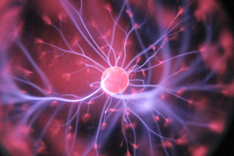
Introduction to Receptor Sites
Receptor sites represent the fundamental machinery by which cells perceive and interact with their environment, serving as highly specialized molecular interfaces for intercellular communication. These sites, which are typically composed of complex protein or lipid structures, are strategically positioned either on the exterior surface of the cell membrane or deep within the cytoplasm or nucleus. Their indispensable function is to recognize, bind to, and respond to specific incoming signals, collectively known as ligands. These ligands can be diverse in nature, ranging from endogenous signaling molecules such as hormones, neurotransmitters, and growth factors, to exogenous stimuli like pharmacological agents or toxins. The integrity and precise functioning of receptor sites are crucial for maintaining homeostasis, coordinating developmental processes, and enabling adaptive responses to environmental changes, making them central to virtually all physiological and psychological processes.
The interaction between a receptor site and its specific ligand initiates a process called signal transduction, a cascade of molecular events that translates the external signal into a meaningful internal cellular action. This mechanism ensures that external cues, whether chemical, electrical, or mechanical, are effectively relayed across the cellular boundary. For example, when a neurotransmitter binds to a receptor on a postsynaptic neuron, it might trigger the opening of ion channels, altering the membrane potential and propagating an electrical impulse. Conversely, the binding of a hormone to an internal receptor might directly influence gene expression, leading to the synthesis of new proteins. Understanding these initial binding events is paramount in fields ranging from endocrinology and neurobiology to pharmacology and clinical medicine, as receptor sites are often the primary targets for therapeutic intervention.
The sophisticated design of receptor sites allows for exceptional specificity and high affinity toward their target ligands, ensuring that the cell responds only to appropriate signals while ignoring the vast array of other molecules present in the extracellular matrix. This specificity is governed by the complementary three-dimensional structure of the receptor’s binding pocket and the ligand’s shape and chemical properties, a concept often likened to a lock-and-key mechanism. Furthermore, the functional output of receptor activation is finely tuned; the same ligand acting on different subtypes of receptors can elicit vastly different cellular responses. Therefore, the study of receptor sites is not merely about identification but encompasses the detailed examination of their molecular architecture, dynamics, regulation, and the downstream signaling pathways they govern, which together dictate the ultimate fate and behavior of the cell.
Detailed Definition and Molecular Architecture
A receptor site is formally defined as a macromolecular component, generally a protein, located either on the cell surface or within the cell, which is capable of recognizing and binding to a specific endogenous or exogenous chemical signal (the ligand) with high affinity. This binding event must subsequently result in a measurable functional change within the cell, thereby mediating the transmission of information. Structurally, receptor proteins are highly complex, often featuring multiple domains: the extracellular domain, responsible for ligand binding; one or more transmembrane domains, anchoring the receptor within the lipid bilayer; and the intracellular domain, which interacts with and activates internal cellular effectors or signaling molecules. The precise arrangement of these domains dictates the receptor’s specific function and its mechanism of signal relay.
The chemical nature of receptor sites varies depending on their location and function. The vast majority of membrane-bound receptors are complex integral proteins, sometimes existing as oligomers composed of multiple subunits. These proteins possess intricate tertiary and quaternary structures, forming a specialized binding pocket that precisely accommodates the ligand. For example, G protein-coupled receptors (GPCRs), one of the largest families of cell surface receptors, are characterized by a structure featuring seven transmembrane alpha-helices. The binding site for the ligand is often nestled within the transmembrane bundle or formed by parts of the extracellular loops. This structural complexity allows for subtle conformational changes upon ligand binding, which are essential for coupling the external signal to the internal signaling machinery.
In contrast to membrane receptors, intracellular receptors—such as those for steroid hormones (e.g., cortisol, estrogen) or thyroid hormones—are typically soluble proteins found in the cytoplasm or nucleus. Because their ligands are small, lipophilic molecules capable of diffusing directly across the cell membrane, these receptors do not require external binding domains. Instead, they feature a ligand-binding domain, a DNA-binding domain, and a transcriptional activation domain. Upon binding the ligand, the receptor undergoes a conformational change that allows it to translocate to the nucleus (if initially cytoplasmic) and bind directly to specific regulatory sequences on the DNA (Hormone Response Elements or HREs), thereby modulating gene transcription. This mechanism highlights the versatility of receptor sites in controlling cellular activities, ranging from rapid membrane depolarization to slow, sustained changes in protein synthesis.
The molecular specificity of a receptor site is determined by non-covalent interactions that occur between the ligand and the amino acid residues lining the binding pocket. These interactions include hydrogen bonds, ionic interactions, van der Waals forces, and hydrophobic interactions. The cumulative strength and geometric arrangement of these forces define the receptor’s affinity (how tightly the ligand binds) and its specificity (which ligands it will accept). Even minor changes in the molecular structure of the ligand or a single point mutation in the receptor protein can drastically alter these binding properties, explaining why small variations in drug structure can lead to profound differences in pharmacological efficacy and side-effect profiles.
Classification Based on Location and Mechanism
Receptor sites are broadly classified into four major superfamilies based on their structure, location, and the mechanism by which they transduce the external signal into a cellular response. This functional classification includes ligand-gated ion channels (Ionotropic Receptors), G protein-coupled receptors (Metabotropic Receptors), enzyme-linked receptors, and intracellular receptors. Understanding these classifications is essential for comprehending the speed and nature of cellular communication, as they dictate whether the response is immediate and transient (like synaptic transmission) or slow and sustained (like hormonal regulation).
Ligand-gated ion channels, or ionotropic receptors, are complex transmembrane proteins that function as both a receptor and an ion channel. When the appropriate ligand binds to the receptor site, it causes a rapid conformational shift that opens the central pore of the channel, allowing specific ions (such as Na+, K+, Cl-, or Ca2+) to flow across the membrane down their electrochemical gradient. This rapid influx or efflux of ions instantly changes the membrane potential of the cell, leading to excitation or inhibition. Examples include the Nicotinic Acetylcholine Receptor (nAChR) and the GABA-A receptor. Because the effect is direct and involves the rapid movement of electrical charge, ionotropic receptors are primarily responsible for the millisecond-scale speed required for synaptic transmission in the nervous system.
G protein-coupled receptors (GPCRs), often termed metabotropic receptors, represent the largest and most diverse family of cell surface receptors, mediating responses to a vast range of ligands including biogenic amines, peptides, and lipids. Unlike ionotropic receptors, GPCRs do not contain an intrinsic ion channel. Instead, their mechanism involves activation of an intermediary signaling molecule, the heterotrimeric G protein, located on the inner surface of the cell membrane. Upon ligand binding, the GPCR undergoes a conformational change, catalyzing the exchange of GDP for GTP on the G protein, which subsequently dissociates into active subunits. These active G protein subunits then modulate the activity of various effector enzymes or ion channels, leading to the generation of intracellular second messengers.
Enzyme-linked receptors are transmembrane proteins that possess, or are tightly associated with, intrinsic enzyme activity. This class typically includes receptors for growth factors, cytokines, and certain hormones, playing critical roles in cell growth, proliferation, differentiation, and survival. The most common type is the receptor tyrosine kinase (RTK). Upon ligand binding, RTKs typically dimerize, resulting in the activation of their cytoplasmic tyrosine kinase domains. These domains then phosphorylate specific tyrosine residues both on themselves (autophosphorylation) and on various downstream signaling proteins. This phosphorylation cascade acts as a molecular switch, activating intricate pathways such as the MAPK pathway, ultimately influencing gene expression and cellular behavior over minutes to hours.
Finally, Intracellular Receptors, as discussed previously, are located within the cytoplasm or nucleus and are distinct because their ligands must be lipid-soluble to traverse the cell membrane. These receptors primarily function as ligand-activated transcription factors. Their mechanism is slower than that of membrane receptors, involving the modification of gene expression and subsequent protein synthesis, but the resulting cellular changes are often long-lasting and profound. This category is crucial for endocrine signaling involving steroid hormones, providing a mechanism for delayed, yet sustained, control over fundamental cellular physiology.
The Principles of Ligand Binding: Specificity and Affinity
The functional efficacy of any receptor site hinges on two critical parameters: specificity and affinity. Specificity refers to the ability of a receptor to discriminate between its natural ligand and other molecules, ensuring that the cellular response is appropriate for the signal received. This high degree of molecular recognition is achieved through the precise complementary fit between the ligand and the receptor’s binding pocket, a concept first articulated by Paul Ehrlich. The binding pocket’s unique arrangement of amino acid residues ensures that only molecules possessing the correct size, shape, and distribution of charged and hydrophobic regions can form stable, favorable interactions.
The classical explanation for receptor-ligand interaction is the lock-and-key hypothesis, proposed by Emil Fischer and later applied to pharmacological receptors by Ehrlich. This model posits that the ligand (the key) and the receptor (the lock) possess rigid, preformed structures that are perfectly complementary, allowing for a precise and instantaneous fit. While elegant in its simplicity, this model fails to account for the dynamic nature of biological macromolecules. A more refined and currently accepted model is the induced-fit hypothesis. According to this model, the binding site is flexible; the initial interaction between the ligand and the receptor induces subtle, yet crucial, conformational changes in the receptor protein. These changes optimize the fit, maximizing stabilizing non-covalent interactions and, crucially, translating the binding energy into the functional conformational shift required to initiate signal transduction.
Affinity measures the strength of the reversible interaction between the ligand and the receptor, typically quantified by the dissociation constant (Kd). A low Kd value indicates high affinity, meaning the ligand binds strongly and remains associated with the receptor for a longer period. Affinity is a crucial determinant of the dose-response relationship; receptors with high affinity require lower concentrations of the ligand to achieve a significant level of occupancy and activation. Pharmacologists exploit this principle by designing drugs (ligands) that possess extremely high affinity for specific receptor subtypes, thereby minimizing the required therapeutic dose and reducing potential off-target effects.
The interaction between ligands and receptor sites also introduces the concepts of agonism and antagonism. An agonist is a ligand that, upon binding, activates the receptor and elicits a maximal biological response, mimicking the action of the endogenous signal. Conversely, an antagonist is a ligand that binds to the receptor site but fails to induce the necessary conformational change for activation, effectively blocking the site and preventing the endogenous agonist from binding and exerting its effect. Furthermore, some ligands are classified as partial agonists (producing a submaximal response) or inverse agonists (stabilizing the receptor in an inactive conformation, thereby reducing basal activity), highlighting the intricate spectrum of functional outcomes stemming from the receptor-ligand interaction.
Signal Transduction: From Receptor Activation to Cellular Response
Signal transduction is the complex process by which a receptor site converts the binding of an external ligand into a functional intracellular event. For membrane-bound receptors, this process must bridge the physical and chemical discontinuity between the extracellular space and the cytoplasm. The mechanism begins with the initial conformational change induced by ligand binding, which serves as the primary activation step. This change is then amplified and diversified through intricate downstream pathways involving intermediary proteins, enzymes, and small, diffusible molecules known as second messengers.
In the case of GPCRs, the activated receptor acts as a guanine nucleotide exchange factor (GEF), prompting the associated G protein to release GDP and bind GTP. The activated G protein subunits then diffuse along the inner membrane surface to interact with effector enzymes, such as adenylyl cyclase or phospholipase C. Activation of adenylyl cyclase results in the synthesis of cyclic AMP (cAMP) from ATP, which acts as a crucial second messenger, primarily by activating protein kinase A (PKA). PKA, in turn, phosphorylates numerous target proteins—including ion channels, metabolic enzymes, and transcription factors—leading to diverse cellular responses like muscle contraction, glycogen breakdown, or altered gene expression.
Alternatively, activation of phospholipase C (PLC) by certain G proteins cleaves the membrane lipid phosphatidylinositol 4,5-bisphosphate (PIP2) into two distinct second messengers: Inositol 1,4,5-trisphosphate (IP3) and Diacylglycerol (DAG). IP3 is soluble and rapidly diffuses through the cytosol, binding to receptors on the endoplasmic reticulum and triggering the release of stored calcium ions (Ca2+), a ubiquitous and potent signaling molecule. DAG remains tethered to the membrane, where it activates protein kinase C (PKC). The simultaneous rise in intracellular Ca2+ concentration and the activation of PKC synergistically regulate various cellular functions, including exocytosis, cell proliferation, and cytoskeletal reorganization.
For enzyme-linked receptors, the transduction mechanism often relies on a direct phosphorylation cascade. Following dimerization and autophosphorylation of the receptor itself, docking sites are created for specific signaling proteins containing Src homology 2 (SH2) domains. These proteins, once recruited, become phosphorylated and activated, initiating linear or branching signaling pathways. A prominent example is the activation of the Ras/MAPK pathway, crucial for transmitting signals from growth factor receptors (like the Epidermal Growth Factor Receptor, EGFR) to the nucleus, regulating cell division and differentiation. The sheer complexity of these intersecting pathways allows a limited number of external signals to produce an enormous variety of highly specific and context-dependent cellular outcomes.
Regulation and Desensitization of Receptor Function
To prevent overstimulation, protect cellular integrity, and maintain sensitivity to fluctuating levels of external signals, receptor sites are subject to sophisticated regulatory mechanisms that modulate their activity, density, and responsiveness. These dynamic processes ensure that the magnitude and duration of the cellular response are tightly controlled. The primary regulatory mechanisms include desensitization, down-regulation, and up-regulation, which collectively govern receptor homeostasis.
Desensitization, often referred to as tachyphylaxis, is a rapid process where the receptor becomes temporarily unresponsive to continued stimulation by the agonist, even if the ligand remains bound. A key mechanism of homologous desensitization (where only the stimulated receptor is affected) involves phosphorylation of the receptor’s intracellular domain by specific kinases, such as G protein-coupled receptor kinases (GRKs). This phosphorylation promotes the binding of arresting proteins (e.g., β-arrestin), which sterically hinder the receptor from interacting with the G protein, effectively uncoupling the receptor from its signaling pathway within seconds or minutes.
Following rapid desensitization, prolonged exposure to high concentrations of an agonist often leads to down-regulation, a slower, long-term process that reduces the total number of receptor sites available on the cell surface. This process typically involves the internalization of the receptor-arrestin complex into endocytic vesicles via clathrin-coated pits. Once internalized, the receptor can either be dephosphorylated and recycled back to the cell membrane, restoring sensitivity (resensitization), or it can be targeted for degradation within lysosomes. Down-regulation serves as a protective mechanism against chronic overstimulation, often seen in addiction or chronic exposure to certain therapeutic drugs, where receptor numbers decrease, necessitating higher doses to achieve the original effect.
Conversely, up-regulation occurs when cells are chronically exposed to low levels of a ligand or, more commonly, when the receptor site is persistently blocked by an antagonist. In this scenario, the cell compensates for the lack of signal transmission by increasing the synthesis of new receptor proteins and inserting them into the plasma membrane. This mechanism increases the cell’s sensitivity to the ligand. Clinically, this phenomenon is critical, as the sudden withdrawal of a receptor-blocking drug (antagonist) can lead to a period of hypersensitivity due to the unnaturally high number of available receptors, often resulting in severe rebound effects. Both up- and down-regulation highlight the remarkable plastic nature of receptor sites in maintaining cellular equilibrium.
Clinical and Pharmacological Significance
Receptor sites are indisputably the most important class of drug targets in modern medicine, with the majority of currently marketed therapeutic agents exerting their effects by interacting with specific receptor proteins. The ability to design small molecules that selectively modulate receptor function—acting as agonists, antagonists, or modulators—allows clinicians to precisely control physiological and pathological processes. By targeting specific receptor subtypes, pharmacologists can maximize therapeutic efficacy while minimizing unwanted side effects, a cornerstone of rational drug design.
In the treatment of neurological and psychiatric disorders, receptor sites are central. For instance, many antidepressants target monoamine receptors and transporters to modulate levels of serotonin and norepinephrine in the synaptic cleft. Antipsychotic medications often function as antagonists at dopamine D2 receptors, reducing excessive dopaminergic signaling implicated in psychosis. The development of highly selective agonists for opioid receptors is crucial for pain management, while the manipulation of GABA-A receptors by benzodiazepines provides effective treatment for anxiety and seizure disorders. The selectivity required for these interventions underscores the importance of understanding the subtle structural differences between various receptor subtypes.
Receptor dysfunction is also a key mechanism underlying numerous disease states. Genetic mutations can lead to non-functional or constitutively active receptors, resulting in inherited endocrine disorders or developmental defects. Furthermore, autoimmune diseases, such as Myasthenia Gravis, involve the production of antibodies that attack and block or destroy specific receptors (in this case, the nicotinic acetylcholine receptor at the neuromuscular junction), leading to impaired muscle contraction and severe weakness. In cancer, aberrant activity of growth factor receptors, particularly receptor tyrosine kinases like EGFR and HER2, drives uncontrolled cell proliferation.
Targeting dysfunctional receptors has become a highly successful therapeutic strategy. For example, in oncology, tyrosine kinase inhibitors (TKIs) are small-molecule drugs designed to block the ATP-binding site of hyperactive RTKs, thereby halting the proliferative signaling cascade. Similarly, monoclonal antibodies are engineered to bind specifically to the extracellular domain of receptors (like Trastuzumab targeting HER2), blocking ligand access and promoting receptor degradation. The ongoing research into allosteric modulation—where a ligand binds to a site separate from the main orthosteric binding site to modify receptor activity—represents the next frontier in drug discovery, offering novel ways to fine-tune receptor function with greater precision than traditional agonists or antagonists.
Historical Development and Foundational Theories
The concept of a specific molecular target, or receptor site, predates the identification of any actual receptor molecule. The foundational theory emerged in the late 19th and early 20th centuries, primarily through the work of two pioneering scientists: Paul Ehrlich and John Newport Langley. Paul Ehrlich, a German physician and pharmacologist, hypothesized that drugs and toxins must bind to specific sites on cells to exert their effects. He famously used the phrase “corpora non agunt nisi fixata” (substances do not act unless they are bound) to express this idea. Ehrlich developed his “side-chain theory,” suggesting that certain chemical groups (side chains) on the cell surface served the dual purpose of binding nutrients and binding toxins or drugs. This concept provided the first theoretical framework for the molecular basis of drug action and the highly selective nature of pharmacological agents.
Simultaneously, British physiologist John Newport Langley provided crucial experimental evidence supporting the receptor hypothesis. Working on the effects of alkaloids on muscle contraction, Langley observed in the early 1900s that nicotine and curare had antagonistic effects on skeletal muscle, and that these effects were localized to the junction between nerve and muscle, rather than affecting the nerve or the muscle fiber itself. He concluded that there must be a “receptive substance” in the muscle membrane that mediated the chemical stimulus. Langley’s work provided the empirical basis for the location and function of the first identified receptor site, laying the groundwork for modern neuropharmacology.
Following these initial theories, the field of receptor biology languished until the mid-20th century. The advent of radioligand binding assays in the 1970s, pioneered by researchers like Alfred Gilman and Robert Lefkowitz, revolutionized the study of receptors. By using radioactively labeled ligands with high affinity, researchers could finally quantify, localize, and characterize receptor sites directly, confirming the existence of discrete binding molecules predicted decades earlier. This technological leap allowed for the precise determination of receptor affinity (Kd) and density (Bmax), transitioning receptor theory from a conceptual model into a quantifiable molecular science.
The subsequent decades saw the successful purification, sequencing, and cloning of numerous receptor proteins, revealing the molecular structure of the major superfamilies, including GPCRs and ligand-gated ion channels. The detailed understanding of the seven-transmembrane architecture of GPCRs, for example, provided a structural explanation for their functional diversity and opened avenues for targeted drug development. Today, structural biology techniques like X-ray crystallography and cryo-electron microscopy continue to provide atomic-resolution images of receptor-ligand complexes, further validating and refining the induced-fit model and driving the rational design of therapeutic molecules that precisely interact with these critical cellular gatekeepers.
Further Reading
The study of receptor sites remains one of the most dynamic and critical areas in biomedical research. The following resources provide detailed insights into the historical development, molecular mechanisms, and pharmacological applications of receptor theory.
- Ehrlich, P. (1890). Über Farbensehen und Farbstoffe. Zeitschrift für Physiologische Chemie, 14(1), 265-280.
- Ferguson, S. S., & Christopoulos, A. (2002). G protein-coupled receptor structure and function. Annual Review of Biochemistry, 71(1), 657-700.
- García‐Martínez, J., & López‐Rodríguez, M. L. (2019). Chemical and molecular recognition in drug discovery. Current Opinion in Structural Biology, 56, 76-82.
- Matsumura, Y., & Nishizuka, Y. (1986). Protein kinase C and lipid signaling for sustained cellular responses. FASEB Journal, 3(3), 2105-2124.
- Takeda, S., Kadowaki, S., & Haga, T. (2005). G protein-coupled receptor and disease. Annual Review of Pharmacology and Toxicology, 45(1), 609-631.
- Lefkowitz, R. J., & Hoffman, B. B. (2013). Receptor mechanisms. In L. L. Brunton, B. A. Chabner, & B. C. Knollmann (Eds.), Goodman & Gilman’s The Pharmacological Basis of Therapeutics (12th ed., pp. 49–78). McGraw-Hill.
- Changeux, J. P. (2012). The Nicotinic Acetylcholine Receptor: An Allosteric Protein. The Journal of Biological Chemistry, 287(48), 40213-40219.