Biological Receptors: How Your Senses Shape Reality
- Definition and Fundamental Role of Receptors
- Cellular Mechanisms: Receptor Proteins and Transduction
- Classification of Receptors by Stimulus Modality
- Sensory Receptors (Sense Organs) and the Five Senses
- Specialized Examples: Photoreceptors and Chemoreceptors
- The Process of Sensory Transduction
- Clinical Significance and Malfunctions
- Future Directions in Receptor Research
Definition and Fundamental Role of Receptors
A receptor, within the context of psychology and biological sciences, is defined fundamentally as a specialized cell, group of cells, or molecular structure that detects and responds to specific stimuli originating from the environment, both internal and external. These structures serve as the critical interface between the physical or chemical world and the nervous system, initiating the crucial process known as sensory transduction. The term is utilized across multiple scales of biological organization, referring either to macroscopic sensory organs, such as the eye or the ear, or to microscopic cellular components, specifically protein molecules embedded in cell membranes or within the cytoplasm that bind signaling molecules like neurotransmitters or hormones. The essential function is always the same: to convert a form of energy (the stimulus) into an electrochemical signal that the nervous system can interpret and process.
The core necessity for receptor function lies in maintaining homeostasis and ensuring survival. Organisms must constantly monitor their environment to detect potential threats, locate resources, and regulate internal states. Receptors possess a high degree of specialization, meaning they are typically tuned to respond optimally to only one type of stimulus energy, referred to as its adequate stimulus. For example, photoreceptors are highly sensitive to light waves but relatively insensitive to temperature changes, ensuring that the organism receives clean, useful data about its surroundings. This sensitivity differentiation ensures that the flood of environmental energy is carefully filtered and organized before it reaches the central processing centers of the brain.
In the cellular definition, the receptor acts as the initial point of information transfer. The mechanism involves the receptor protein undergoing a conformational change upon binding its specific ligand (a chemical signal) or absorbing its specific energy form (e.g., photon, pressure wave). This conformational change then triggers a cascade of intracellular events, ultimately leading to a change in the cell’s membrane potential. This shift in electrical charge is the foundation of the neural signal, demonstrating how the receptor effectively translates an external input into the universal language of the nervous system. The efficiency and accuracy of this initial step are paramount; errors or malfunctions at the receptor level can lead to significant sensory deficits or pathological conditions.
Cellular Mechanisms: Receptor Proteins and Transduction
At the molecular level, receptors are typically complex proteins responsible for binding signaling molecules or responding directly to physical forces. These molecular receptors dictate the specificity and speed of communication within the organism. They are broadly categorized based on their location and mechanism of action, with the most common being cell-surface receptors and intracellular receptors. Cell-surface receptors are crucial for rapid communication, responding to external signaling molecules that cannot easily cross the lipid bilayer, such as neurotransmitters and large peptide hormones. The interaction between the ligand and the receptor is highly specific, often described by the “lock-and-key” model, ensuring that only the correct signal initiates a response.
Two major classes of cell-surface receptors are integral to neural and hormonal signaling. Firstly, Ion Channel-Linked Receptors (or ligand-gated ion channels) are rapid responders. Upon binding the ligand, the receptor protein changes shape, opening a pore that allows specific ions (like sodium, potassium, or chloride) to rush across the membrane. This rapid ion movement instantly alters the electrical potential of the cell, leading to either excitation or inhibition of the neuron. Secondly, G-Protein Coupled Receptors (GPCRs) are slower but initiate highly amplified and complex intracellular signaling cascades. When activated by a ligand, the GPCR interacts with an associated G-protein inside the cell, which then modulates enzyme activity or opens other ion channels indirectly. This mechanism is central to processes like vision, smell, and the action of many psychoactive medications.
The core action initiated by molecular receptors is signal transduction, the process by which an external stimulus is converted into an electrical response. For sensory cells, this results in a graded electrical potential known as a receptor potential. If this potential reaches a specific threshold, it triggers an action potential in the associated afferent neuron, propagating the signal toward the central nervous system (CNS). The magnitude and duration of the stimulus are encoded by the receptor cell into the frequency and pattern of these action potentials. This encoding mechanism is critical for accurately representing the sensory world, allowing the brain to distinguish between a faint whisper and a loud shout, or a light touch versus heavy pressure.
Classification of Receptors by Stimulus Modality
Receptors are systematically classified based on the origin of the stimulus they detect, providing a useful framework for understanding the body’s comprehensive monitoring system. This classification divides receptors into three primary functional groups: Exteroceptors, Interoceptors, and Proprioceptors. This categorization is vital because the central nervous system processes information differently depending on whether the input relates to the external world, the visceral state, or the body’s spatial orientation.
Exteroceptors are specialized cells or sensory nerve endings that monitor stimuli arising from the external environment. These receptors are responsible for our perception of the outside world and are concentrated in the specialized sensory organs. Key examples include the photoreceptors in the retina which detect light, the hair cells in the cochlea which respond to sound vibrations, and the various mechanoreceptors and thermoreceptors in the skin that sense touch, pressure, pain, and temperature. The information gathered by exteroceptors is generally consciously perceived and is essential for interacting with the surrounding environment, facilitating actions like locomotion, communication, and immediate defense mechanisms.
Interoceptors, conversely, monitor stimuli arising from the internal environment, specifically within the viscera and blood vessels. These receptors are crucial for maintaining systemic homeostasis and their signals are typically processed unconsciously by the autonomic nervous system. Interoceptors include chemoreceptors that monitor blood chemistry (e.g., oxygen, carbon dioxide, pH levels), baroreceptors that track blood pressure, and osmoreceptors that sense osmotic pressure and fluid balance. When interoceptors detect dangerous deviations, they initiate reflex actions, such as altering heart rate or respiration, or they might generate conscious sensations like nausea or thirst, prompting corrective behavior.
The third major class, Proprioceptors, provides the nervous system with continuous information regarding the position and movement of the body in space. These receptors are primarily located in the skeletal muscles, tendons, joints, and the inner ear (vestibular apparatus). Proprioceptors monitor the degree of muscle stretch, the tension in tendons, and the angular position of joints. This continuous, detailed feedback is essential for coordinated movement, posture maintenance, and motor learning. Without functional proprioceptors, even simple acts like standing upright or walking without looking at one’s feet become extremely challenging, highlighting their foundational role in motor control and spatial awareness.
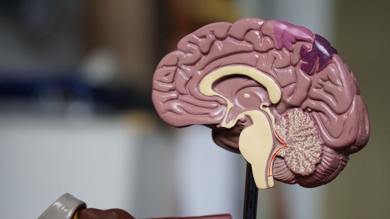
Sensory Receptors (Sense Organs) and the Five Senses
The term “receptor” frequently refers to a complete sensory organ, which represents a highly organized structure containing numerous receptor cells integrated with supportive tissues designed to maximize the detection and focusing of specific stimuli. Classical sensory organs—the eye, ear, nose, tongue, and skin—house the specialized receptor cells for the five traditional senses, allowing for complex interpretation of external reality. These organs are sophisticated biological machines that often preprocess the stimulus before the actual cellular transduction occurs. For instance, the lens system of the eye focuses light onto the photoreceptors, and the tympanic membrane and ossicles of the ear amplify sound waves before they reach the cochlear hair cells.
The auditory system provides an excellent example of receptor complexity. The ear contains mechanoreceptors—the hair cells—located within the Organ of Corti in the cochlea. These cells are specialized to respond to physical displacement caused by fluid movement initiated by sound waves. The mechanical bending of their stereocilia opens ion channels, generating the electrical signal. This arrangement allows the ear not only to detect sound but also to perform frequency analysis, as different hair cells along the cochlea are tuned to respond to different pitches, demonstrating an intricate level of organization and specialized function within a single receptor organ.
Similarly, the retina of the eye, which is arguably the most complex sensory receptor structure, contains millions of photoreceptor cells (rods and cones). These cells are arranged in layers and interact with supporting neurons to begin processing visual information immediately upon detection. The collective function of a sense organ is far greater than the sum of its individual receptor cells; the entire structure acts as a system responsible for stimulus capture, filtering, enhancement, and transfer. The accuracy of our perception of the world relies entirely on the fidelity with which these complex receptor systems execute their initial detection and signaling functions.
Specialized Examples: Photoreceptors and Chemoreceptors
Diving deeper into specific receptor types reveals the incredible diversity and evolutionary refinement of detection mechanisms. Photoreceptors, found in the retina, are a prime example of energy-converting receptor cells. These cells contain light-sensitive photopigments, such as rhodopsin in rods, which change their molecular structure when they absorb a photon. This change initiates a biochemical cascade that ultimately closes sodium channels, hyperpolarizing the cell—a unique signaling mechanism where light causes inhibition rather than excitation—thereby signaling the presence of light to the subsequent neurons. Rods are highly sensitive and crucial for low-light (scotopic) vision, while cones are responsible for color discrimination and high-acuity (photopic) vision.
Chemoreceptors specialize in detecting chemical substances, playing critical roles in fundamental biological processes, including respiration, digestion, and behavior. These receptors are central to the senses of taste (gustation) and smell (olfaction). Olfactory receptors, located in the nasal epithelium, are sensitive to volatile chemical molecules inhaled from the air. Each olfactory receptor cell typically expresses only one type of receptor protein, capable of binding to a specific range of odorants. The resulting pattern of activation across thousands of different receptor types allows the brain to distinguish a vast array of smells. Taste receptors, located in the taste buds, respond to chemicals dissolved in saliva, categorized broadly into detection of sweetness, sourness, saltiness, bitterness, and umami.
A particularly crucial specialized class are the Nociceptors, or pain receptors. Unlike other sensory receptors that generally adapt to continuous stimulation, nociceptors are uniquely designed to sensitize, meaning they become more responsive to repeated or prolonged stimuli, a phenomenon known as hyperalgesia. They respond to potentially damaging stimuli, including extreme temperatures, excessive pressure, and particularly, chemicals released by damaged tissues (e.g., prostaglandins, bradykinin). Nociceptors are essential for protective reflexes and alerting the organism to injury, demonstrating a vital specialization geared towards minimizing tissue damage and promoting recuperation.
The Process of Sensory Transduction
Sensory transduction is the essential physiological process where the energy of a stimulus is converted into the electrical energy of a neural signal. This transition is not merely a transfer but a complex conversion that requires specialized machinery to maintain fidelity. The stimulus energy, whether mechanical, electromagnetic, or chemical, must first interact with the receptor cell, causing a change in the receptor’s membrane permeability, typically by opening or closing ion channels. This alteration leads to an influx or efflux of ions, resulting in a localized change in membrane potential known as the receptor potential.
The receptor potential is a graded potential, meaning its magnitude is proportional to the intensity of the stimulus; a stronger stimulus generates a larger receptor potential. Unlike action potentials, receptor potentials do not propagate over long distances. Instead, they act locally, modulating the release of neurotransmitters onto the associated afferent neuron, or, in the case of primary sensory neurons, directly generating action potentials at the axon hillock. This graded response is critical for coding stimulus intensity: a large receptor potential drives the afferent neuron to fire action potentials more frequently, thereby informing the central nervous system that the stimulus is intense.
The sequence of events from stimulus detection to neural transmission is highly regulated and follows a predictable pathway, ensuring timely and accurate information transfer. The entire process hinges upon the initial integrity of the receptor cell and its ability to properly process the incoming energy. The fundamental steps involved in this transfer of information from the environment to the nervous system are:
- The stimulus energy is applied and interacts with the specialized receptor structure (e.g., a hair cell stereocilium is bent, or a chemical ligand binds to a protein).
- The receptor structure undergoes a conformational change, leading to the opening or closing of specific ion channels.
- Ions flow across the membrane, generating a graded receptor potential, the magnitude of which reflects stimulus intensity.
- If the receptor potential reaches the threshold for the associated afferent neuron, it triggers an action potential (a rapid, self-propagating electrical spike).
- The train of action potentials travels along the afferent pathway to specific relay centers in the brainstem and thalamus, finally reaching the sensory cortex for conscious interpretation.
Clinical Significance and Malfunctions
Given their role as the initiation point of the nervous system, receptor function is highly relevant in clinical medicine, pharmacology, and pathology. Malfunctions in receptor systems can lead to a wide range of debilitating conditions. For instance, defective molecular receptors are implicated in many inherited diseases, such as certain forms of congenital deafness resulting from non-functional hair cell channels, or various visual deficits caused by mutations in photopigment proteins. Furthermore, the concept of sensory processing disorders involves the CNS misinterpreting signals received from otherwise healthy receptors, demonstrating the complex dependency between detection and central interpretation.
Pharmacology is heavily reliant on the precise modulation of molecular receptors. A vast majority of therapeutic drugs, particularly those targeting the nervous system, function as either agonists or antagonists of neurotransmitter receptors. Agonists mimic the action of the natural ligand, binding to and activating the receptor (e.g., opioid drugs activating pain receptors), while antagonists block the receptor, preventing the natural ligand from binding and thereby inhibiting its action (e.g., beta-blockers inhibiting adrenaline receptors). The selectivity of these drugs for specific receptor subtypes is the key to designing medications that target pathological processes while minimizing unwanted side effects.
Autoimmune diseases frequently involve attacks on functional receptor systems. A classic example is Myasthenia Gravis, where the body produces antibodies that destroy or block the acetylcholine receptors at the neuromuscular junction. Since acetylcholine is the neurotransmitter required to initiate muscle contraction, the resulting loss of receptor function leads to severe, fluctuating muscle weakness. Understanding the molecular integrity and distribution of these receptors is thus crucial for diagnosing and treating diseases where the communication pathway between the nerve and the effector cell is compromised, underscoring the vital nature of receptor integrity in maintaining physiological function.
Future Directions in Receptor Research
Ongoing research into receptor biology promises significant advances in neurobiology, sensory science, and therapeutics. Modern techniques, such as high-resolution cryo-electron microscopy (Cryo-EM) and X-ray crystallography, allow researchers to determine the precise three-dimensional structure of receptor proteins, providing unprecedented insight into how ligands bind and how conformational changes initiate signaling. This structural knowledge is immediately translatable into rational drug design, enabling the creation of highly specific molecules tailored to interact with only a desired receptor subtype.
The emergence of cutting-edge technologies like optogenetics represents a revolutionary step in receptor manipulation. Optogenetics involves introducing genetically engineered light-sensitive receptors (often derived from algae) into specific populations of neurons. By shining light of a specific wavelength onto these cells, researchers can precisely turn neural activity on or off with millisecond accuracy. This technique provides unparalleled control over neural circuits and is currently being used to map the function of various receptor-mediated pathways in living animals, offering new avenues for understanding complex behaviors and sensory processing disorders.
Furthermore, a growing area of focus is the application of receptor knowledge to neuroprosthetics and sensory restoration. For individuals with sensory deficits, research aims to develop artificial systems that can bypass damaged natural receptors and directly stimulate the nervous system. Examples include advanced cochlear implants that directly stimulate auditory nerve fibers based on complex sound analysis, and emerging retinal prostheses designed to replace the function of damaged photoreceptors. The ultimate goal is to create biologically compatible interfaces that mimic the exquisite sensitivity and coding capability of natural receptor systems, ensuring that the processed and transferred information allows for meaningful and accurate perception.