Rhinolalia: Understanding Nasal Speech Resonance Disorders
- Definition and Classification of Rhinolalia
- Etiology: Causes of Abnormal Nasal Resonance
- Types of Rhinolalia: Hypernasality and Hyponasality
- Psychological and Social Impact of Resonance Disorders
- Diagnosis and Assessment Procedures
- Treatment Approaches: Medical, Surgical, and Behavioral
- Prognosis and Long-Term Management
Definition and Classification of Rhinolalia
Rhinolalia, often referred to as a resonance disorder, describes an abnormal quality of the speaking voice characterized by inappropriate nasal airflow during the production of speech sounds. This condition is fundamentally linked to the inadequate or excessive coupling of the oral and nasal cavities, a process primarily controlled by the velopharyngeal mechanism. Unlike simple articulation errors, rhinolalia reflects a physiological disruption in the balance of oral and nasal resonance, profoundly affecting the clarity and naturalness of communication. This nasal quality of the voice usually results from a structural malformation, neurological impairment, or disease of the upper vocal tract and nasal passages, necessitating a careful differential diagnosis to determine the precise underlying etiology.
In clinical speech-language pathology, rhinolalia is classified under the broader category of resonance disorders, distinguishing it from disorders of phonation (voice source) or articulation (sound shaping). The significance of accurate classification lies in the fact that treatment success hinges entirely on addressing the specific type of resonance imbalance present. A voice characterized by rhinolalia fails to properly dampen or amplify frequencies in the expected cavities, leading to sounds that listeners perceive as either excessively “stuffy” or overly “open.” This perceptual deviation is often the first indicator of underlying anatomical or physiological dysfunction, specifically concerning the soft palate (velum) and the pharyngeal walls.
The core definition highlights that rhinolalia is not merely a cosmetic issue but a significant communicative hurdle. As noted in preliminary observations, the resultant vocal quality “can be extremely irritating,” not only for the listener who struggles to process the distorted speech signal but also, crucially, for the speaker who experiences profound difficulty in producing intelligible and natural-sounding speech. This irritation can quickly translate into significant psychological distress and withdrawal from social interaction, underlining why this condition, though anatomical in origin, holds substantial relevance within the domain of applied psychology and communication sciences. Comprehensive analysis requires evaluation of both the anatomical structures and the functional psychological consequences of the chronic speech deviation.
Etiology: Causes of Abnormal Nasal Resonance
The causes of rhinolalia are diverse and typically categorized into structural, functional, and neurological origins. Structural causes involve physical anomalies or obstructions within the vocal tract, the most common and severe being velopharyngeal insufficiency (VPI), often associated with a history of cleft lip or palate. In VPI, the soft palate is too short or moves inefficiently, preventing complete closure of the velopharyngeal port during the production of non-nasal sounds. Other structural malformations include deep pharynxes, submucous clefts (where the bony structure is intact but the underlying musculature is defective), and structural damage resulting from trauma or surgical resection. These physical deficits directly prevent the necessary separation of the oral and nasal airways, leading to uncontrolled nasal airflow.
Conversely, diseases and acquired conditions frequently lead to temporary or chronic rhinolalia, particularly the closed type. Conditions such as severe chronic sinusitis, large adenoid or tonsil tissue, nasal polyps, and deviated septums obstruct the nasal passages, blocking the normal flow of air and sound through the nasal cavity. When the nasal cavity is blocked, the sounds that normally resonate there (the nasals /m/, /n/, and /ng/) sound distorted, muffled, and similar to a person speaking with a severe cold. While these conditions are often treatable medically or surgically, their persistence can establish patterns of speech production that are difficult to modify even after the physical obstruction is resolved.
Neurological factors represent a third significant etiological category. Damage to the cranial nerves responsible for innervating the velopharyngeal musculature—specifically the Vagus nerve (CN X)—can result in paresis or paralysis of the soft palate. This neurological impairment disrupts the motor planning and execution necessary for rapid and accurate velopharyngeal closure, leading to fluctuating or persistent open rhinolalia (hypernasality). Conditions such as cerebral palsy, stroke, traumatic brain injury, or degenerative neurological diseases like Amyotrophic Lateral Sclerosis (ALS) can all manifest rhinolalia as a component of dysarthria. In these instances, the underlying issue is not the structure itself, but the lack of coordinated neurological control over that structure, making treatment necessarily focused on maximizing muscle strength and coordination rather than surgical repair.
Types of Rhinolalia: Hypernasality and Hyponasality
Rhinolalia is traditionally divided into two primary, opposing types: Rhinolalia Aperta (Open Rhinolalia or Hypernasality) and Rhinolalia Clausa (Closed Rhinolalia or Hyponasality). Hypernasality occurs when there is excessive nasal resonance, meaning that acoustic energy travels into the nasal cavity during the production of oral vowels and consonants, which should normally be blocked. This is typically the result of VPI, where the velopharyngeal mechanism fails to achieve full closure. The excessive leakage of sound energy through the nose results in a thin, weak, and often distorted vocal quality, sometimes accompanied by audible nasal air emission, particularly on high-pressure consonants like /p/, /t/, and /k/. This type is commonly associated with cleft palate and neurological deficits affecting palatal movement.
In contrast, Hyponasality, or Closed Rhinolalia, involves insufficient nasal resonance. This occurs when the nasal airway is blocked, preventing the necessary acoustic energy from entering the nasal cavity during the production of the three English nasal phonemes: /m/, /n/, and /ng/. When attempting to produce these sounds, the speaker sounds like they have a severe head cold, where the nasal sounds are substituted by their oral cognates (e.g., ‘m’ sounds like ‘b’, ‘n’ sounds like ‘d’). The primary causes are physical obstructions within the nasal cavity or nasopharynx, such as enlarged adenoids (a common cause in children), severe rhinitis, or large nasal polyps. While generally less complex to treat than hypernasality, persistent hyponasality can severely impair intelligibility and vocal quality.
A third, more complex category is Mixed Rhinolalia, where both hypernasality and hyponasality coexist. This often occurs when a patient has multiple contributing factors. For example, a person with a history of VPI (causing hypernasality) may develop compensatory articulation patterns that involve posterior tongue placement, or they may concurrently suffer from chronic sinus issues or mild nasal obstruction. Determining the dominant resonance deviation and its source is critical for devising an effective treatment strategy. If a clinician only addresses the hypernasality without recognizing the complicating factor of the nasal obstruction causing hyponasality, the intervention will be incomplete and the overall speech clarity will remain compromised.
Psychological and Social Impact of Resonance Disorders
The psychological toll associated with rhinolalia is often profound, directly stemming from the challenges in achieving fluid and understandable communication. Because the vocal quality is highly distinctive and often perceived as unusual or “irritating,” speakers frequently internalize negative reactions from listeners. This leads to intense self-consciousness regarding speech production. The speaker may develop communication avoidance behaviors, choosing silence over the risk of judgment or misunderstanding. Over time, this avoidance can lead to social isolation, impacting academic performance, career opportunities, and the ability to form and maintain intimate relationships. The chronic struggle to speak clearly often results in significant communication frustration, contributing to feelings of helplessness and inadequacy.
The social consequences extend beyond mere avoidance. Individuals with persistent rhinolalia may face stigma, sometimes being inaccurately perceived as less intelligent or less capable simply based on their vocal quality. This prejudice, though often unintentional, can manifest in subtle forms of social exclusion or professional discrimination. The effort required to overcome the resonance distortion can also lead to secondary physical manifestations, such as vocal strain or hyperfunctional voice habits, further complicating the psychological landscape. The perpetual need to monitor and adjust speech makes relaxed, spontaneous conversation nearly impossible, placing a heavy cognitive load on the individual.
Psychological sequelae frequently observed in individuals with chronic resonance disorders include heightened anxiety, particularly social anxiety disorder, and clinical depression. The constant preoccupation with speech can morph into speech-related phobias (phonophobia), where the fear of speaking in public or even in small groups becomes debilitating. Therefore, successful management of rhinolalia requires more than just surgical or behavioral speech correction; it mandates robust psychological support. Counseling or cognitive behavioral therapy (CBT) can be essential tools for addressing the internalized stigma, improving self-esteem, and teaching coping mechanisms for managing communicative stress, ultimately promoting greater confidence and participation in social life.
Diagnosis and Assessment Procedures
Accurate diagnosis of rhinolalia requires a comprehensive, multidisciplinary approach involving otolaryngologists (ENT specialists), speech-language pathologists (SLPs), and sometimes plastic surgeons or orthodontists. The initial assessment relies heavily on auditory-perceptual evaluation conducted by the SLP. During this phase, the clinician judges the type and severity of rhinolalia (hypernasal, hyponasal, or mixed), the presence of nasal air emission, and the impact on articulation. Standardized speech samples, involving both sustained vowels and high-pressure consonant phrases, are utilized to characterize the resonance deviation under varying speech loads. Key to the initial assessment is determining if the disorder is primarily structural, neurological, or functional.
Instrumental assessment provides objective data to supplement the perceptual judgment. Nasometry is a primary tool, utilizing a Nasometer device to measure the acoustic energy ratio emitted simultaneously through the oral and nasal cavities during speech. This generates a numerical score (the nasalence score) that quantifies the degree of hypernasality, providing a baseline for tracking treatment progress. Aerodynamic assessments measure the airflow and air pressure differences between the oral and nasal cavities, which helps pinpoint the exact location and size of the velopharyngeal gap in cases of hypernasality, or the degree of obstruction in cases of hyponasality.
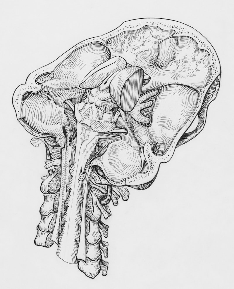
To visualize the velopharyngeal mechanism in motion, clinicians often rely on imaging techniques. Videofluoroscopy involves X-ray examination of the velum and pharyngeal walls during speech production, allowing the SLP and surgeon to see the extent and pattern of velopharyngeal closure or lack thereof. Alternatively, nasopharyngoscopy (or endoscopy) involves inserting a flexible camera through the nose to directly view the velopharyngeal port structure and function during speech tasks. These visualization methods are crucial for surgical planning, as they determine whether the VPI is due to poor palatal movement, lateral wall movement, or a combination, dictating the appropriate surgical intervention, such as pharyngeal flap or sphincter pharyngoplasty procedures.
Treatment Approaches: Medical, Surgical, and Behavioral
Treatment for rhinolalia is highly individualized and dictated by the underlying etiology identified during the diagnostic phase. For structural rhinolalia, particularly VPI, surgical intervention is often the primary and most effective approach. Procedures aim to physically close or narrow the velopharyngeal port, ensuring adequate separation of the oral and nasal cavities. Examples include the aforementioned pharyngeal flap (creating a bridge of tissue between the soft palate and the posterior pharyngeal wall) or sphincter pharyngoplasty (creating a ring of tissue to narrow the opening). In cases of hyponasality caused by disease, medical intervention such as steroid sprays or antibiotics, or surgical removal of obstructions like enlarged adenoids or polyps, is necessary before speech therapy can be effective.
For rhinolalia that persists post-surgically, is mild, or is functional/neurological in origin, speech-language therapy is the cornerstone of management. SLP goals focus on two main areas: improving the efficiency of velopharyngeal closure and modifying compensatory articulation patterns. Techniques aimed at improving closure might include exercises to increase the awareness and movement of the soft palate, often involving blowing or sucking exercises, though the efficacy of these non-speech tasks is debated. More effective are techniques that involve speech production itself, such as increasing oral pressure, utilizing slightly lowered head posture, and training the patient to produce sounds with greater oral effort.
Advanced behavioral strategies often incorporate instrumental feedback. For instance, biofeedback using a Nasometer allows the patient to visually monitor their nasalance score in real-time, helping them gain conscious control over their resonance balance. For patients with neurological deficits, therapy may focus on maximizing muscle tone and endurance. In situations where surgery is not possible or advisable, prosthetic devices, such as palatal lift appliances (to elevate a paralyzed soft palate) or obturators (to physically block a persistent gap), may be fitted by a specialized dental professional (prosthodontist) to mechanically improve resonance and speech clarity.
Prognosis and Long-Term Management
The prognosis for individuals with rhinolalia varies significantly depending on the underlying cause, the severity of the structural deficit, and the age of intervention. Rhinolalia resulting from temporary obstructions (e.g., adenoids or transient rhinitis) typically resolves completely following medical or surgical treatment. For rhinolalia stemming from VPI associated with cleft palate, the prognosis is generally good, provided that timely and appropriate surgery is followed by consistent, intensive speech therapy. Success in these cases often results in near-normal resonance and articulation, minimizing the long-term psychological impact.
Factors that negatively influence the long-term prognosis include severe neurological impairment (where muscle control may progressively decline), delayed intervention, and the establishment of severe, ingrained compensatory articulation patterns. If a child or adult has relied on glottal stops or pharyngeal fricatives for years to compensate for nasal air emission, these habits are exceptionally difficult to extinguish, even after the underlying resonance mechanism is corrected. Successful outcomes require strong commitment from the patient and family, consistent practice, and a carefully coordinated team approach involving all medical specialists.
Long-term management is crucial, particularly for individuals who have undergone complex surgical procedures or who have chronic neurological conditions. Follow-up evaluations by the SLP and ENT are necessary to monitor for potential regression or the development of new compensatory patterns. For children, monitoring continues through adolescence to ensure that resonance adapts appropriately as the facial skeleton and pharyngeal structures mature. In some instances, minor secondary surgical procedures may be required years after the initial intervention to fine-tune the velopharyngeal mechanism. Maintenance exercises and periodic check-ups are essential to ensure that the achieved improvements in vocal quality and intelligibility are sustained over the lifespan, allowing the individual to communicate effectively and confidently.