Radiographic Imaging: Visualizing the Hidden Mind
- Introduction to Roentgenogram
- The Underlying Mechanism of X-ray Imaging
- Historical Foundations and Pioneering Discoveries
- Evolution and Advancements in Radiography
- Practical Applications in Clinical Diagnosis
- Illustrative Case Study: Diagnosing a Fracture
- Broader Significance and Enduring Impact on Medicine
- Limitations and Complementary Imaging Modalities
- Connections to Broader Radiological Principles
Introduction to Roentgenogram
A roentgenogram, more commonly known as a radiograph or simply an X-ray, represents a foundational and indispensable medical imaging technique employed to visualize the internal structures of the body. This non-invasive diagnostic tool leverages the properties of X-rays, a form of electromagnetic radiation, to produce a two-dimensional projection image of bones, organs, and various soft tissues. The primary principle behind its operation involves the differential absorption of X-ray photons as they pass through tissues of varying densities, allowing for the creation of a shadow-like image on a detector. Since its inception, radiography has revolutionized medical diagnosis, providing clinicians with crucial insights into a wide array of pathological conditions, from skeletal fractures to certain types of tumors and pulmonary diseases, thereby significantly aiding in treatment planning and patient management.
The core utility of a roentgenogram lies in its ability to rapidly and cost-effectively provide vital diagnostic information. Unlike more complex imaging modalities, a standard X-ray examination is often quick to perform, making it invaluable in emergency settings where immediate assessment is critical. It offers a macroscopic view of internal anatomy, highlighting structural anomalies that might indicate disease or injury. While its capabilities for detailed soft-tissue differentiation are limited compared to advanced techniques like Magnetic Resonance Imaging (MRI) or Computed Tomography (CT), the roentgenogram remains the first-line imaging choice for numerous clinical presentations due to its accessibility, efficiency, and diagnostic efficacy for specific conditions, particularly those involving bone and air-filled spaces.
The Underlying Mechanism of X-ray Imaging
The fundamental mechanism underpinning the production of a roentgenogram is rooted in the interaction of X-ray photons with human tissues. An X-ray machine generates these high-energy photons by accelerating electrons towards a metallic target, typically tungsten, within an evacuated tube. When these electrons strike the target, their kinetic energy is converted into X-rays and heat. The emitted X-ray beam is then directed through the patient’s body, where it encounters various tissues, each possessing a unique atomic composition and density. This encounter is characterized by a process known as attenuation, which describes the reduction in the intensity of the X-ray beam as it passes through matter due to absorption and scattering.
The principle of differential absorption is paramount to image formation. Denser tissues, such as bones, contain higher concentrations of calcium and other elements with higher atomic numbers, making them more effective at absorbing X-ray photons. Consequently, fewer X-rays penetrate these structures and reach the detector. Conversely, less dense tissues, like muscle, fat, and air-filled organs (e.g., lungs), allow a greater proportion of X-rays to pass through. This variation in X-ray transmission creates a spatial pattern of radiation intensity that is then captured by an image receptor, which can be traditional photographic film or a digital detector. The areas where fewer X-rays pass through (e.g., bones) appear white or very light on the resulting image, while areas where many X-rays pass through (e.g., air) appear dark or black, with soft tissues appearing in various shades of gray in between, thus forming the characteristic shadow-like image of a roentgenogram.
Historical Foundations and Pioneering Discoveries
The journey of the roentgenogram began in 1895 with the groundbreaking discovery by German physicist Wilhelm Conrad Röntgen. Working in his laboratory in Würzburg, Röntgen was experimenting with cathode rays and vacuum tubes when he serendipitously observed a fluorescent glow emanating from a barium platinocyanide screen placed nearby, even when the tube was covered with opaque cardboard. He quickly deduced that a new, invisible form of radiation was being emitted, capable of penetrating solid objects and causing fluorescence. He dubbed these mysterious rays “X-rays” to denote their unknown nature. Within weeks of his discovery, Röntgen produced the first medical roentgenogram – an image of his wife Anna Bertha’s hand, clearly showing her bones and wedding ring. This image instantly captivated the scientific community and the public, heralding a new era in medicine.
The impact of Röntgen’s discovery was immediate and profound. Physicians rapidly recognized the immense diagnostic potential of X-rays, leading to their swift adoption in medical practice across the globe. By the early 20th century, X-ray machines were being installed in hospitals and clinics, fundamentally changing how doctors could diagnose internal conditions without invasive surgery. Early applications included identifying bone fractures, locating foreign objects within the body, and detecting lung diseases like tuberculosis. However, the initial enthusiasm was tempered by a lack of understanding regarding the biological effects of radiation. Early practitioners and patients often suffered severe burns and other radiation-induced injuries due to prolonged exposure, prompting the development of radiation protection protocols and improved equipment design over time to minimize risks and ensure safety for both patients and medical personnel.
Evolution and Advancements in Radiography
Following its initial discovery, radiography underwent continuous refinement and innovation. The early years focused on improving X-ray tube technology, enhancing image quality, and developing safer practices. The introduction of intensifying screens reduced patient dose by amplifying the effect of X-rays on photographic film, while the advent of contrast agents allowed for the visualization of hollow organs and blood vessels, expanding the diagnostic scope of conventional radiography significantly. These advancements solidified the roentgenogram’s role as a cornerstone of medical diagnosis throughout the first half of the 20th century, becoming an indispensable tool in orthopedics, emergency medicine, and general practice.
A significant evolutionary leap occurred in the 1970s with the development of computed tomography (CT) scans. As noted in the original historical context, CT represented a revolutionary departure from traditional 2D radiography by utilizing multiple X-ray projections and computer processing to create cross-sectional, three-dimensional images of the body. This provided superior anatomical detail and soft-tissue differentiation, which was a significant limitation of conventional roentgenograms. While CT scans offered greater accuracy and depth, they did not entirely replace roentgenograms. Instead, they complemented them, with each modality finding its niche based on diagnostic requirements, cost-effectiveness, and radiation dose considerations. The subsequent transition from film-based to digital radiography further modernized the field, offering advantages such as instant image acquisition, enhanced image manipulation capabilities, easier storage, and seamless sharing of images across healthcare networks, significantly improving workflow and diagnostic efficiency.
Practical Applications in Clinical Diagnosis
The roentgenogram continues to be a cornerstone in clinical diagnosis across various medical specialties due to its versatility and efficiency. Its most widespread application is in the assessment of the musculoskeletal system. For instance, in cases of suspected bone fractures, an X-ray can quickly confirm the presence, location, and severity of the break, guiding immediate treatment decisions. Beyond fractures, it is invaluable for identifying dislocations, assessing joint integrity, detecting bone tumors, and monitoring the progression of degenerative bone conditions like arthritis. In the realm of pulmonology, chest X-rays are routinely used to diagnose conditions such as pneumonia, tuberculosis, lung cancer, and pneumothorax (collapsed lung), by visualizing characteristic patterns of opacities or air accumulation within the lung fields.
Furthermore, roentgenograms play a crucial role in other areas of medicine. Dental radiography, for example, is indispensable for detecting cavities, assessing bone health, and planning orthodontic treatments. Abdominal X-rays can help identify bowel obstructions, kidney stones, or the presence of foreign bodies. Specialized forms of radiography, such as mammography, are vital for breast cancer screening and diagnosis. The ability of a roentgenogram to provide a rapid, macroscopic overview of internal structures makes it an ideal initial diagnostic tool in many clinical scenarios, often guiding the decision for more advanced imaging if greater detail or soft-tissue differentiation is required. Its relatively low cost and widespread availability also ensure its continued relevance as a first-line imaging modality in healthcare systems globally.
Illustrative Case Study: Diagnosing a Fracture
To illustrate the practical application of a roentgenogram, consider a common scenario: a patient presents to an emergency department after falling and experiencing severe pain and swelling in their wrist. The clinical suspicion is a potential bone fracture. The diagnostic process using a roentgenogram unfolds in a systematic manner to confirm or rule out this injury, providing critical information for subsequent management.
- Patient Positioning: The medical imaging technologist carefully positions the patient’s wrist on the X-ray table or against a detector plate. Typically, multiple views (e.g., anteroposterior, lateral, and oblique views) are taken to ensure comprehensive visualization of all bones and joints from different angles, as a fracture might be obscured in a single projection.
- X-ray Exposure: The technologist sets the appropriate X-ray parameters (voltage, current, exposure time) to optimize image quality while minimizing radiation dose. The X-ray beam is then briefly activated, passing through the patient’s wrist and onto the digital detector. The process is quick, often taking only seconds for each view.
- Image Processing and Review: Once the X-rays are acquired, the digital images are immediately processed and displayed on a monitor. A radiologist, a physician specializing in medical imaging interpretation, then meticulously examines the images. They look for discontinuities in the bone cortex, displacement of bone fragments, joint effusions, or other signs indicative of a fracture. In the case of a wrist fracture, the radiologist might identify a clear fracture line across the distal radius or ulna.
- Diagnosis and Treatment Planning: Based on the radiologist’s interpretation, a definitive diagnosis of a wrist fracture is made. This immediate visual confirmation allows the emergency physician or orthopedic specialist to formulate an appropriate treatment plan without delay. This might involve immobilization with a cast, reduction of displaced fragments, or, in more complex cases, surgical intervention. The roentgenogram thus provides the foundational evidence needed to guide critical medical decisions, ensuring timely and effective patient care.
Broader Significance and Enduring Impact on Medicine
The significance of the roentgenogram extends far beyond its direct diagnostic capabilities; it fundamentally transformed the practice of medicine and public health. Before its invention, physicians relied heavily on physical examination, patient history, and often invasive surgical exploration to diagnose internal conditions. Röntgen’s discovery provided the first non-invasive window into the human body, inaugurating the era of medical imaging and dramatically improving diagnostic accuracy, speed, and safety. This paradigm shift allowed for earlier detection of diseases, more precise surgical planning, and better monitoring of treatment efficacy, ultimately leading to improved patient outcomes and reduced morbidity and mortality from a wide range of conditions.
Despite the emergence of sophisticated imaging technologies such as CT, MRI, and ultrasound, the roentgenogram retains an enduring and critical role in modern healthcare. Its inherent advantages—low cost, wide availability, speed of acquisition, and ease of interpretation for specific conditions—make it an indispensable first-line diagnostic tool, particularly in emergency rooms, primary care settings, and resource-limited environments. It serves as a foundational imaging modality, often guiding the decision-making process for whether more advanced and often more expensive imaging is warranted. The roentgenogram’s legacy is evident in its continued widespread use, proving that a century-old technology can remain at the forefront of medical diagnostics due to its foundational utility and practical advantages.
Limitations and Complementary Imaging Modalities
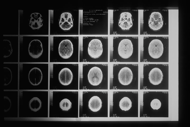
While the roentgenogram is invaluable for visualizing dense structures like bones and air-filled spaces, it possesses inherent limitations, particularly concerning the detailed assessment of soft tissues. As articulated in the original content, its effectiveness in depicting organs, muscles, ligaments, and the nervous system is significantly less than for hard tissues. This is because soft tissues have similar X-ray attenuation characteristics, resulting in poor contrast resolution and making it challenging to differentiate between various soft tissue structures or identify subtle pathologies within them. For instance, a conventional X-ray may not adequately show a torn ligament, a small tumor within an organ, or detailed abnormalities in the brain or spinal cord.
To overcome these limitations, modern medicine increasingly relies on a suite of complementary medical imaging techniques, each offering unique strengths. Computed Tomography (CT) scans, for example, provide detailed cross-sectional images with superior soft tissue contrast compared to conventional X-rays, making them excellent for evaluating internal organs, complex fractures, and vascular structures. Magnetic Resonance Imaging (MRI), which uses strong magnetic fields and radio waves instead of X-rays, excels at visualizing soft tissues like the brain, spinal cord, muscles, ligaments, and cartilage with exquisite detail, offering unparalleled contrast resolution. Ultrasound imaging, utilizing high-frequency sound waves, is particularly useful for dynamic imaging of organs, blood flow, and for evaluating soft tissue structures in real-time, especially in obstetrics and cardiology. These advanced modalities are not replacements but rather essential supplements to the roentgenogram, allowing clinicians to select the most appropriate imaging study based on the specific clinical question and the tissue of interest.
Connections to Broader Radiological Principles
The roentgenogram is not an isolated technique but is deeply embedded within the broader field of radiology and medical imaging. It represents the foundational principle of projection radiography, where a three-dimensional object is represented as a two-dimensional image. This fundamental concept underpins many other imaging modalities, even those that produce cross-sectional or volumetric data. Understanding the principles of X-ray generation, attenuation, and image formation in basic radiography provides a crucial framework for comprehending more complex X-ray-based techniques like fluoroscopy (real-time X-ray imaging) and computed tomography, which essentially involves multiple X-ray projections reconstructed into a 3D volume.
Furthermore, the roentgenogram connects to broader scientific principles, including the physics of the electromagnetic spectrum, radiation biology, and image processing. The X-rays used in radiography are a specific segment of the electromagnetic spectrum, characterized by their high energy and short wavelength, enabling them to penetrate matter. The study of how this radiation interacts with biological tissue, including its potential for harm and the necessary safety measures, is a core component of medical physics and radiation protection, directly stemming from the early experiences with X-rays. In essence, the roentgenogram serves as a gateway to understanding the entire discipline of medical imaging, illustrating fundamental concepts that are expanded upon and refined in more advanced diagnostic tools, making it an indispensable starting point for anyone studying the human body’s internal visualization.