Cognitive Scanning: How Your Brain Filters Hidden Realities
- Introduction to Scanning in Medical and Psychological Contexts
- Structural Brain Imaging Techniques
- Functional Neuroimaging: Mapping Brain Activity
- Electroencephalography (EEG) and Magnetoencephalography (MEG)
- Cognitive Scanning and Visual Search
- Applications in Clinical Psychology and Research
- Challenges and Ethical Considerations in Neuroimaging
- Future Directions in Scanning Technology
Introduction to Scanning in Medical and Psychological Contexts
The term scanning, within the medical and psychological lexicon, refers fundamentally to techniques employed for visualizing internal structures or functional processes, often without invasive procedures. Medically, this includes a diverse array of methodologies—radiological, magnetic, and radioisotopic—designed to create detailed images of the body or its constituent parts. A quintessential example is the utilization of a brain scan, such as an MRI, in analyzing a specific ailment, mapping structural integrity, or identifying the physiological correlates of psychological conditions. However, the application of scanning in psychology extends far beyond mere structural visualization; it serves as a critical bridge between observable behavior and the underlying neural architecture that gives rise to cognition, emotion, and action. The evolution of scanning technologies has transformed psychology from a purely behavioral science into one deeply rooted in neuroscience, allowing researchers to objectively correlate subjective mental states with measurable physical phenomena.
The psychological utility of scanning lies in its ability to offer objective data regarding the functionality and connectivity of the central nervous system. Prior to the advent of sophisticated neuroimaging, inferences about brain function were largely derived from studying lesion patients or post-mortem analysis, providing limited insight into real-time cognitive processing in healthy individuals. Modern scanning methods, conversely, allow for the dynamic observation of the brain while it is actively engaged in tasks such as memory retrieval, language comprehension, or emotional regulation. This capability has been instrumental in testing complex theoretical models of the mind, enabling researchers to localize cognitive functions to specific neural regions, thereby providing empirical validation for theories of modularity and distributed processing within the brain.
It is important to delineate the two primary conceptualizations of scanning within psychology. The first, and most technologically driven, involves neuroimaging technologies used to visualize the structure and function of the brain (e.g., fMRI, PET). The second refers to cognitive scanning, a core concept in visual attention and perception, defining the active, systematic search process executed by an individual across a visual field or through an internal memory representation. While structurally distinct, both uses share the common underlying goal of systematic exploration—either physically through technology or cognitively through attentional mechanisms—to gain information critical for diagnosis, research, or environmental understanding.
Structural Brain Imaging Techniques
Structural scanning techniques are primarily concerned with mapping the anatomy of the brain, differentiating between various tissue types, and identifying pathological changes such as tumors, hemorrhages, or atrophy. One of the earliest widely adopted methods was Computed Tomography (CT). CT scanning utilizes rotating X-ray beams and highly sensitive detectors to generate cross-sectional images, relying on differential absorption rates of X-rays by various tissues. Dense materials like bone appear bright, while soft tissues and cerebrospinal fluid show varying shades of gray. While CT remains invaluable in acute medical settings due to its speed and ability to detect fresh hemorrhage, its utility in detailed psychological research is limited by its comparatively lower soft tissue contrast and the inherent exposure of the patient to ionizing radiation.
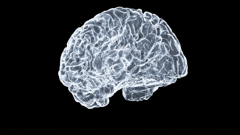
A significant leap forward in structural visualization was achieved with Magnetic Resonance Imaging (MRI). This powerful technique avoids ionizing radiation entirely, instead employing extremely strong magnetic fields and radiofrequency pulses to align and then perturb the hydrogen nuclei (protons) naturally present in the body’s water molecules. As these protons realign, they emit radio signals that are captured by the scanner and processed by sophisticated algorithms to create high-resolution images. MRI provides vastly superior contrast between different soft tissues, such as gray matter, white matter, and cerebrospinal fluid, making it the gold standard for detailed anatomical mapping of the brain in psychological and neurological research. This precision allows for highly accurate measurements of cortical thickness, white matter tract integrity, and subcortical structure volumes.
The insights gained from structural scanning are fundamental to understanding the neural basis of psychological disorders. For instance, researchers utilize specialized MRI sequences, such as Diffusion Tensor Imaging (DTI), to map the white matter tracts that connect different brain regions, providing critical data on structural connectivity. Abnormalities in these structural connections have been reliably implicated in developmental disorders like autism spectrum disorder and psychiatric conditions such as schizophrenia. By meticulously scanning and comparing the morphology of brain structures in clinical populations versus healthy controls, researchers can identify subtle yet significant anatomical markers that contribute to the manifestation and progression of complex psychological phenomena.
Functional Neuroimaging: Mapping Brain Activity
While structural scanning reveals the hardware of the brain, functional neuroimaging techniques are essential for observing the software—the dynamic processes of neural activity during cognitive engagement. These methods operate on the principle of neurovascular coupling, meaning that increased neuronal activity in a specific brain region is rapidly followed by a localized increase in cerebral blood flow and metabolism. The most widely adopted functional technique in psychology is Functional Magnetic Resonance Imaging (fMRI), which measures changes related to blood oxygenation, known as the Blood-Oxygen-Level Dependent (BOLD) signal. When neurons are active, they consume oxygen, but the subsequent rush of oxygenated blood overwhelms the local demand, leading to a temporary, measurable increase in the ratio of oxygenated to deoxygenated hemoglobin, which the fMRI scanner detects.
fMRI is highly valued for its excellent spatial resolution, allowing researchers to pinpoint the location of cognitive activity with high precision. It has been crucial in mapping the functional organization of the cortex, identifying regions responsible for specific functions like facial recognition (fusiform face area) or working memory (prefrontal cortex). Despite its strength in spatial localization, fMRI is inherently limited in its temporal resolution because the hemodynamic response it measures is sluggish, taking several seconds to peak after the initial neural event. This limitation necessitates the use of other scanning modalities when the research question requires tracking cognitive processes that occur on the millisecond scale.
Another pivotal functional scanning method is Positron Emission Tomography (PET). PET scanning involves injecting a small amount of a radioactive tracer (radiopharmaceutical) into the bloodstream, which is then taken up by metabolically active tissues. The most common tracer is fluorodeoxyglucose (FDG), which mimics glucose and measures regional cerebral glucose metabolism, a direct proxy for sustained neural activity. Unlike fMRI, PET excels at measuring specific molecular targets, such as the density and distribution of particular neurotransmitter receptors (e.g., dopamine or serotonin receptors), making it indispensable for pharmacological studies and the investigation of neurological disorders like Parkinson’s disease. Although PET involves exposure to low levels of radiation and generally offers poorer spatial and temporal resolution than modern fMRI, its unique capability to image specific biochemical pathways ensures its continued importance in psychopharmacological research.
Electroencephalography (EEG) and Magnetoencephalography (MEG)
To overcome the temporal limitations inherent in hemodynamic scanning techniques like fMRI and PET, researchers often turn to electrophysiological methods that measure the neural activity directly, rather than relying on the slower blood flow response. Electroencephalography (EEG) records the electrical potential fields generated by the synchronous firing of large populations of neurons. This is achieved by placing electrodes strategically across the scalp, which measure fluctuations in voltage over time. The primary strength of EEG is its extraordinary temporal resolution, capable of tracking neural events with millisecond precision, which is crucial for studying rapid processes such as sensory gating, auditory discrimination, and visual processing reaction times.
A key application of EEG is the study of Event-Related Potentials (ERPs). ERPs are averaged EEG responses time-locked to the presentation of a specific stimulus or the execution of a cognitive task. Distinct components of the ERP waveform, such as the P300 (related to stimulus evaluation) or the N400 (related to semantic processing), provide robust markers of specific cognitive stages. While EEG provides unparalleled temporal data, its spatial resolution is relatively poor; the electrical signals are smeared and distorted as they pass through the skull and scalp, making it challenging to precisely localize the source of the activity deep within the brain.
Magnetoencephalography (MEG) represents an advanced scanning technology that addresses the spatial ambiguity of EEG while retaining high temporal resolution. MEG measures the extremely faint magnetic fields produced by the same electrical currents responsible for the EEG signal. Because magnetic fields are not distorted by the skull or scalp, MEG offers significantly better spatial localization capabilities than EEG, especially for sources originating in the cortex. This makes MEG an invaluable tool for mapping cognitive processes in real-time, such as tracking the progression of auditory information through cortical language networks or identifying the precise onset and termination of neural oscillations associated with attention and memory maintenance. The synergy of combining high temporal resolution methods (EEG/MEG) with high spatial resolution methods (fMRI) allows for a more complete picture of brain function, capitalizing on the strengths of each respective scanning technique.
Cognitive Scanning and Visual Search
The secondary, yet equally vital, psychological definition of scanning pertains to the active, behavioral process of directing attention and gaze. Cognitive scanning is the systematic exploration of a visual scene or environment, crucial for information gathering, object recognition, and navigating complex spaces. This process is governed by a combination of bottom-up (stimulus-driven) factors and top-down (goal-driven) attentional control mechanisms. Research into visual search paradigms utilizes eye-tracking technology to meticulously record the sequence of fixations and saccades—the rapid movements of the eyes—that constitute the scanning pattern, revealing the strategies an individual employs to locate a target among distractors.
Psychologists differentiate between various types of visual search scanning. In a parallel search, the target seems to “pop out” immediately, regardless of the number of distractors, suggesting that the entire visual field is scanned simultaneously for a unique feature. Conversely, a serial search requires effortful, item-by-item inspection, characteristic of tasks where the target is defined by a conjunction of features (e.g., finding a red vertical line among red horizontal and green vertical lines). The efficiency of cognitive scanning is a critical measure of attentional capacity and processing speed, and deficits in systematic scanning are often symptomatic of clinical conditions or cognitive fatigue.
Deficits in cognitive scanning are prominent in neurological and psychological disorders. For example, patients suffering from Spatial Neglect Syndrome, typically following damage to the parietal lobe, exhibit a profound inability or failure to orient to or systematically scan the contralesional side of space, often behaving as if that half of the world does not exist. Similarly, individuals with specific reading difficulties or certain forms of attentional deficit disorder may show disorganized or inefficient scanning patterns during reading tasks or when searching for information on a page. Understanding the precise manner in which an individual scans their environment is paramount for developing effective rehabilitation strategies and compensatory training methods aimed at improving real-world functioning.
Applications in Clinical Psychology and Research
Scanning technologies have revolutionized clinical psychology by transforming diagnostic approaches and deepening the understanding of psychopathology. In clinical settings, structural scanning (MRI) is essential for ruling out organic causes of psychiatric symptoms, such as tumors or vascular incidents that might mimic psychological disorders. Functional scanning, however, offers greater insight into the mechanisms of illness by highlighting aberrant patterns of brain activity and connectivity. For instance, fMRI studies have identified altered functional connectivity within the Default Mode Network (DMN)—a set of brain regions active during internal thought—in patients with major depressive disorder and anxiety disorders, suggesting a neural basis for excessive rumination.
In research, scanning provides objective, quantifiable biomarkers that can monitor the effects of therapeutic interventions, both psychological and pharmacological. For example, a PET scan can be used to measure changes in neurotransmitter receptor occupancy before and after administering an antidepressant, providing crucial data on the drug’s mechanism of action and efficacy. Similarly, fMRI can track whether cognitive behavioral therapy (CBT) leads to measurable changes in the activation patterns of emotion regulation circuits, thus validating the neural effectiveness of the psychological treatment. This objective measurement moves the field closer to precision psychiatry, where treatments are tailored based on an individual’s unique neurobiological profile identified through scanning.
Beyond traditional clinical applications, scanning techniques are increasingly used in applied psychological domains, including consumer behavior and legal contexts. Neuroeconomic studies utilize fMRI to scan subjects’ brains while making decisions under risk, revealing the neural correlates of impulse buying or financial risk aversion. In forensic psychology, while highly controversial and subject to intense debate, techniques attempting to utilize fMRI for lie detection or assessing criminal responsibility aim to identify neural signatures associated with deception or impaired cognitive control, although these applications face significant ethical and reliability challenges that preclude their widespread judicial acceptance currently.
Challenges and Ethical Considerations in Neuroimaging
Despite the tremendous power of scanning technologies, their utilization in psychological research and clinical practice is fraught with methodological and ethical challenges. Methodologically, the interpretation of functional neuroimaging data relies heavily on complex statistical inference, often leading to the challenge of reverse inference—the flawed attempt to deduce the engagement of a specific cognitive process solely from the activation of a particular brain region. Furthermore, the inherent variability and noise in biological data necessitate large sample sizes and rigorous statistical corrections, contributing to the “replication crisis” experienced in some areas of neuroimaging research where initial findings fail to hold up under scrutiny.
Ethically, the use of advanced scanning poses significant dilemmas. Protecting participant autonomy requires robust processes for informed consent, especially when scanning individuals whose capacity for decision-making may be compromised (e.g., severe psychiatric patients or children). The potential for neuroprediction, where scanning data might reveal predispositions to future illness or behavior, raises concerns about privacy and discrimination. There is a fear that employers or insurance companies could potentially misuse imaging data to assess risk, leading to neurological exceptionalism and stigmatization based on an individual’s neural structure or functional patterns, irrespective of current health status or behavior.
Moreover, the high capital cost and maintenance expenses associated with advanced scanning equipment (especially high-field MRI and MEG systems) create issues of accessibility. These sophisticated tools are generally confined to large, well-funded research institutions and major medical centers, leading to significant disparities in research capabilities and clinical care availability across different regions and socioeconomic groups. Addressing these challenges requires establishing transparent data sharing protocols, developing standardized acquisition and analysis techniques, and engaging in proactive policy discussions concerning the societal implications of reading and interpreting the human brain.
Future Directions in Scanning Technology
The field of neuroimaging is continually advancing, promising even greater detail and accessibility in future scanning procedures. One major technological push involves the deployment of ultra-high field MRI scanners (7 Tesla and above). These machines generate images with significantly higher spatial resolution than standard clinical scanners (3T), allowing researchers to visualize smaller brain structures and subtle functional changes with unprecedented clarity, leading to a microscopic view of cortical organization and subcortical nuclei function.
A parallel development focuses on increasing portability and reducing the footprint of scanning devices. Efforts are underway to create more compact, potentially wearable EEG and MEG systems that utilize optically pumped magnetometers (OPM-MEG). These portable scanners would allow researchers and clinicians to study brain function in more naturalistic environments, moving the technology out of the restrictive shielding rooms and into real-world settings, thereby enhancing the ecological validity of psychological research findings. Furthermore, the development of specialized, low-cost scanning devices, such as functional Near-Infrared Spectroscopy (fNIRS), offers a non-invasive, more affordable method to measure regional blood flow changes in clinical settings where high-end equipment is unavailable.
Crucially, the sheer volume and complexity of data generated by modern scanning demand sophisticated analytical solutions. The future of scanning heavily relies on the integration of Artificial Intelligence (AI) and machine learning algorithms. AI is essential for identifying subtle patterns, classifying complex brain states, and developing robust computational biomarkers for early disease detection and personalized treatment planning. By combining multimodal scanning data—structural, functional, electrical, and metabolic—and processing it through advanced AI models, researchers anticipate the ability to predict individual responses to therapeutic interventions and to map the trajectory of mental health across the lifespan with far greater accuracy than is currently achievable.