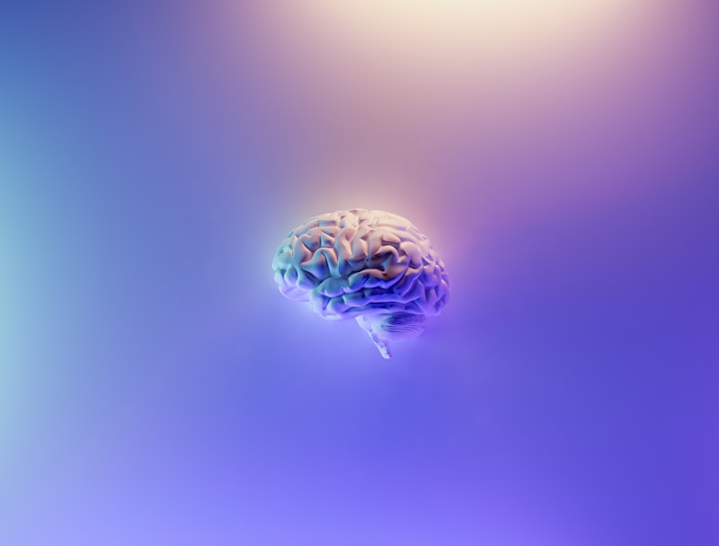
Sensory Neurons: Decoding How Your Brain Perceives Reality
The Core Definition of Sensory Neurons
A sensory neuron, often referred to as an afferent neuron, is a specialized nerve cell responsible for converting external or internal stimuli into electrical signals. This process, known as sensory transduction, forms the fundamental basis of how organisms perceive and respond to their environment. The primary function of these neurons is to transmit information from sensory receptors—which are sensitive to light, sound, touch, chemical changes, or temperature—toward the central nervous system (CNS).
The core principle governing the function of sensory neurons is the detection and encoding of environmental energy. Unlike motor neurons or interneurons, which primarily process or transmit signals within the CNS, sensory neurons initiate the neural pathway by acting as the interface between the body and the external or internal world. They essentially translate diverse physical and chemical energy forms into the common language of the nervous system: the electrochemical impulse. Without this initial conversion, the brain would be isolated, unable to receive the critical data necessary for survival, movement, and conscious perception.
These neurons are integral components of the peripheral nervous system (PNS) and are critical for all forms of sensation, ranging from simple reflexes to complex cognitive experiences. They operate by maintaining a resting membrane potential, and when a stimulus reaches a sufficient threshold, ion channels open, leading to depolarization and the generation of an action potential. This electrical signal then travels along the neuron’s axon toward the spinal cord or brain, carrying vital information about the nature, intensity, and location of the initial stimulus.
Classification and Morphology
Sensory neurons exhibit remarkable structural diversity, tailored to the specific type of stimulus they are designed to detect. Structurally, they are classified based on the arrangement of their processes—the dendrites and axons—relative to the cell body (soma). The most common type associated with general body sensation (touch, pain, temperature) is the pseudounipolar neuron. In this configuration, the dendrite and axon emerge from the soma as a single short process that immediately splits into two branches: one extending toward the periphery to receive sensory input, and the other projecting toward the central nervous system. The cell bodies of these pseudounipolar neurons are typically located outside the CNS in structures called ganglia, such as the dorsal root ganglia (DRG).
In contrast, specialized senses often utilize bipolar neurons, particularly those involved in olfaction, vision, and balance. Bipolar neurons possess two distinct processes: one axon and one dendrite, both extending directly from opposite sides of the soma. This structure facilitates highly localized and directional transmission of information, crucial for the precise processing required by organs like the retina. Furthermore, the specialized endings of sensory neurons—the sensory receptors—can be classified based on the stimulus modality they detect, including mechanoreceptors (touch, pressure), chemoreceptors (taste, smell), photoreceptors (light), and thermoreceptors (temperature).
The overall morphology dictates the speed and pathway of signal transmission. The long axons of many sensory neurons are myelinated, a feature provided by glial cells (Schwann cells in the PNS), which dramatically increases the conduction velocity of the electrical impulse. The degree of myelination and the diameter of the axon directly influence how quickly sensory data—such as immediate pain or proprioceptive feedback—can reach the processing centers in the brain, ensuring rapid and appropriate bodily responses to critical environmental changes.
The Historical Discovery of Neural Transmission
The understanding of sensory neurons evolved significantly during the late 19th and early 20th centuries, a period marked by revolutionary advances in neuroanatomy. Before this era, the nervous system was often conceived of as a continuous net or reticulum. The pivotal shift came with the pioneering work of Santiago Ramón y Cajal, who, utilizing the staining techniques perfected by Camillo Golgi, provided conclusive evidence for the discontinuous nature of the nervous system. This led to the formulation of the Neuron Doctrine, the foundational theory asserting that the nervous system is composed of discrete, individual cells (neurons).
Ramón y Cajal meticulously observed and illustrated the specific structures of different types of neurons, clearly identifying the afferent pathways associated with sensory input. He demonstrated that sensory information flowed in a specific direction—from the peripheral receiving structures (dendrites) through the axon toward the CNS—establishing the principle of dynamic polarization. This detailed anatomical mapping was crucial for distinguishing sensory neurons from other neural types and understanding their dedicated role as input conductors.
Further historical refinement involved understanding the electrical nature of the nerve impulse. Researchers like Luigi Galvani in the 18th century first observed that electrical stimulation could cause muscle contraction, hinting at the electrical basis of neural function. However, it was the 20th-century work of Hodgkin and Huxley on the squid giant axon that mathematically modeled the ionic basis of the action potential, providing the physiological explanation for how sensory transduction translates external energy into a viable signal that can be propagated along the sensory neuron’s length toward the brain.
The Mechanism of Sensory Transduction
Sensory transduction is the crucial biophysical process wherein the energy of a stimulus (e.g., pressure, light wavelength, chemical concentration) is converted into an electrical signal that the neuron can transmit. This process begins at the specialized receptor ending of the sensory neuron. When an adequate stimulus is applied, it causes a conformational change in membrane proteins, often leading to the opening or closing of ion channels specific to sodium, potassium, or calcium ions. This flux of ions alters the membrane potential locally, creating a graded potential.
The graded potential is proportional to the intensity of the stimulus; a stronger touch or brighter light generates a larger depolarization. Unlike the all-or-nothing nature of the action potential, the graded potential dissipates quickly over distance. However, if this graded potential reaches a specific region of the sensory neuron, typically the axon hillock or the initial segment of the axon, and exceeds the threshold of excitation, it triggers the generation of a full-fledged action potential. This threshold mechanism ensures that the nervous system only responds to meaningful or significant stimuli, filtering out background noise.
Once the action potential is initiated, it propagates rapidly along the axon without decrement, owing to the sequential opening of voltage-gated sodium channels. The frequency of these action potentials, rather than their amplitude, is the primary method by which the sensory neuron encodes stimulus intensity. A strong, sustained stimulus results in a high frequency of firing, while a weak stimulus results in infrequent firing. This frequency modulation allows the central nervous system to accurately interpret the magnitude and duration of the sensory event.
A Real-World Illustration: The Reflex Arc
A perfect practical example illustrating the function of sensory neurons is the patellar reflex, commonly known as the knee-jerk reflex. This is a monosynaptic reflex arc, meaning it involves only two neurons and one synapse, allowing for an incredibly fast, involuntary response that bypasses conscious brain processing. The sensory neuron is the initiating agent in this critical protective mechanism.
The process begins when a doctor taps the patellar tendon just below the knee cap. This sudden mechanical stretch serves as the stimulus.
-
Stimulus Detection: Specialized sensory receptors within the quadriceps muscle, known as muscle spindles, detect the stretch caused by the tap.
-
Sensory Transduction: The muscle spindles convert the mechanical stretch energy into a graded potential, which quickly reaches threshold and generates an action potential in the associated sensory neuron (a pseudounipolar neuron).
-
Afferent Transmission: The sensory neuron transmits the action potential along its axon directly into the spinal cord via the dorsal root.
-
Synaptic Transmission: Inside the spinal cord, the sensory neuron axon forms a direct synapse with a motor neuron that controls the same quadriceps muscle. (Note: Interneurons are involved in inhibiting antagonistic muscles, but the primary reflex path is direct.)
-
Motor Response: The motor neuron is immediately activated, sending an efferent signal back out to the quadriceps muscle, causing it to contract and the leg to kick forward.
This example clearly demonstrates the role of the sensory neuron as the essential input device. It is responsible for gathering the information about the external perturbation (the tap) and translating it into a neural signal that immediately initiates a physiological output (the kick). The speed and reliability of this sensory-motor circuit are entirely dependent on the efficiency of the sensory neuron’s ability to generate and transmit the initial action potential.
Significance and Therapeutic Impact
Sensory neurons hold immense significance in psychology and neuroscience, primarily because they are the gatekeepers of perception, pain, and homeostasis. In the field of psychology, understanding how sensory information is encoded, filtered, and processed is foundational to the study of perception and psychophysics—the relationship between physical stimuli and sensory experience. Dysfunctions in sensory processing, such as hypersensitivity or hyposensitivity, are central to understanding conditions like autism spectrum disorder or sensory processing disorder.
Clinically, sensory neuron research is crucial for understanding and treating neurological disorders, particularly those involving chronic pain. Conditions like peripheral neuropathy, often caused by diabetes or chemotherapy, involve damage to the sensory neurons located in the peripheral nervous system. This damage can lead to phantom sensations, numbness, or debilitating chronic pain due to the inappropriate or exaggerated firing of damaged sensory axons.
Therapeutic interventions based on sensory neuroscience aim to modulate the activity of these neurons. For instance, novel pain management strategies focus on inhibiting the release of pain-signaling neurotransmitters at the synapse between the sensory neuron and the spinal cord interneurons, or on developing drugs that stabilize the ion channels responsible for the generation of the action potential in nociceptors (pain sensory neurons). Furthermore, research into restoring sensory function after spinal cord injury often involves understanding how to bridge the gap in sensory pathways and encourage the regeneration of long afferent neuron axons.
Connections to Related Neural Systems
Sensory neurons do not operate in isolation; they are part of a tripartite system required for complete nervous system function. This subfield is broadly categorized under Physiological Psychology or Neuroscience. The two primary counterparts to the sensory neuron are the motor neuron and the interneuron. The relationship between these three types defines the structure of the nervous system.
-
Motor Neurons (Efferent): While sensory neurons carry signals toward the CNS (afferent), motor neurons carry signals away from the CNS to effector organs like muscles and glands (efferent). They execute the command formulated in response to the sensory input.
-
Interneurons: Interneurons constitute the vast majority of neurons in the brain and spinal cord. They act as intermediaries, receiving input from sensory neurons and integrating, processing, and relaying that information to motor neurons or other interneurons. They are responsible for the complex decision-making and modulation that occurs between stimulus input and behavioral output.
The functional connection between sensory input and behavioral output is managed by circuits, such as the reflex arc previously mentioned, or more complex polysynaptic pathways governing voluntary movement and conscious thought. Furthermore, sensory signaling is deeply related to the concept of the Neuron Doctrine, as it highlights how discrete cells interact sequentially to process information. Sensory neurons provide the initial data stream, which is then refined and acted upon by the central processing units (interneurons) before a command is issued by the motor units. This coordinated relay system ensures that the organism can accurately perceive the world and generate appropriate, adaptive responses.