Sleep Apnea: Reclaim Your Cognitive Clarity
Scientifically Reviewed · Dr. Marwa Abd-Alazim · September 1, 2026
Review Criteria & Clinical Standards
This content undergoes rigorous scientific peer-review and medical editorial standards at Arab Psychology Network to ensure clinical accuracy, validity, and compliance with evidence-based guidelines from leading psychological and healthcare authorities (APA / WHO).
Introduction and Definition of Sleep Apnea
Sleep apnea, often referred to as sleep apnoea, is a pervasive and potentially serious sleep disorder characterized by repeated cessation or significant reduction in airflow during sleep. This interruption, known as an apneic event, occurs when an individual stops breathing while they are asleep, typically lasting ten seconds or longer, and can happen scores or even hundreds of times throughout a single night. These episodes severely fragment the normal sleep architecture, preventing the individual from achieving the restorative deep sleep necessary for optimal physical and cognitive function. The defining feature of this disorder is the pathological instability of respiration during sleep, which leads to chronic intermittent hypoxia and hypercapnia, consequences that have profound implications for cardiovascular and metabolic health beyond simple sleep deprivation.
The most common manifestation, Obstructive Sleep Apnea (OSA), specifically involves the mechanical blockage of the upper airway, resulting from the complete or partial collapse of soft tissues, including the tongue, uvula, and pharyngeal walls, or obstruction by mucous secretions. During normal sleep, muscle tone naturally decreases, but in susceptible individuals, this relaxation allows the structures surrounding the pharynx to occlude the passage, halting air movement despite continued efforts by the diaphragm and chest muscles to inhale. The cessation of breathing is typically a startling event, often terminated violently with a loud, explosive snore, a gasp, or a sudden, involuntary body jerk, which momentarily arouses the individual just enough to restore muscle tone and reopen the airway.
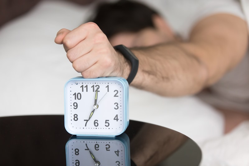
While the immediate resumption of breathing may seem successful, the constant cycle of oxygen desaturation, carbon dioxide retention, and brief arousals places tremendous stress on the body’s homeostatic mechanisms. These physiological insults prevent consolidated sleep, directly resulting in the hallmark clinical symptom: excessive **daytime sleepiness**. The quality of sleep is profoundly poor, characterized by highly fragmented non-REM and REM cycles, meaning the individual never truly enters sustained periods of deep, restorative rest. This chronic fatigue significantly impairs daily functioning, affecting concentration, mood regulation, reaction time, and overall quality of life, making the identification and treatment of sleep apnea crucial for public health and individual well-being.
Classification and Types of Sleep Apnea
Sleep apnea is generally categorized into three distinct types based on the underlying mechanism of respiratory failure: Obstructive Sleep Apnea (OSA), Central Sleep Apnea (CSA), and Mixed Sleep Apnea (MSA). Understanding these classifications is critical for accurate diagnosis and the selection of appropriate therapeutic interventions. **Obstructive Sleep Apnea** is overwhelmingly the most prevalent form, accounting for approximately 84 percent of all diagnosed cases. It is characterized by persistent respiratory effort against a physically blocked or narrowed upper airway, meaning the brain is actively attempting to breathe, but the mechanical path for air is sealed off by tissue or other substances. The classic clinical presentation involves loud, habitual snoring followed by silence, only to be broken by a loud gasp or snore as the airway briefly reopens.
In contrast to the mechanical failure seen in OSA, **Central Sleep Apnea** (CSA) involves a failure of the respiratory control center in the brainstem. In CSA, the brain temporarily ceases to send signals to the muscles of respiration—the diaphragm and chest wall—meaning there is neither airflow nor respiratory effort during the apneic event. This form is often associated with specific medical conditions, such as severe heart failure, stroke, or high-altitude exposure, or it may be induced by certain medications, particularly opioid pain relievers. The clinical presentation of CSA differs from OSA in that snoring is usually less prominent or absent, and the primary complaint may be insomnia or the observation of long respiratory pauses without the struggle characteristic of obstruction.
The third category, **Mixed Sleep Apnea** (MSA), incorporates features of both Obstructive and Central types within the same apneic episode or across the duration of the sleep study. Typically, a mixed event begins as a Central event, characterized by a lack of respiratory effort, and then transitions into an Obstructive event, where airway collapse occurs and the patient begins demonstrating respiratory effort against the blockage. While less common than pure OSA, MSA requires careful diagnosis as effective treatment often requires addressing both the mechanical obstruction and the underlying instability in central respiratory drive, sometimes necessitating specialized adaptive servo-ventilation (ASV) devices rather than standard continuous positive airway pressure (CPAP).
Etiology and Risk Factors
The development of Sleep Apnea is multifactorial, arising from a complex interplay of anatomical predispositions, physiological vulnerabilities, and lifestyle factors. The single most significant and modifiable risk factor for OSA is **obesity**, particularly the deposition of adipose tissue around the neck and pharyngeal structures. Increased neck circumference leads to narrowing of the collapsible segment of the pharynx, reducing the space available for airflow and increasing the likelihood of tissue collapse when muscle tone naturally diminishes during sleep. This anatomical narrowing is compounded by factors such as age, as the elasticity and muscular tone of the pharyngeal tissues decrease over time, rendering the airway more susceptible to collapse in middle-aged and older adults.
Anatomical features unrelated to body weight also contribute significantly to risk. Individuals with retrognathia (a recessed lower jaw), a large tongue (macroglossia), enlarged tonsils or adenoids (particularly common in children), or a narrow, high-arched palate are structurally predisposed to airway obstruction. Furthermore, genetic factors play an undeniable role; a family history of sleep apnea suggests inherited craniofacial characteristics or pharyngeal neuromuscular control issues that heighten vulnerability. Endocrine disorders, most notably hypothyroidism and acromegaly, can lead to tissue swelling in the upper airway, exacerbating existing risk factors and contributing to the severity of the apneic events.
Lifestyle and substance use also dramatically influence the frequency and severity of apneic episodes. The consumption of **alcohol** or the use of sedating medications, such as benzodiazepines or certain muscle relaxants, significantly diminishes the responsiveness and tone of the upper airway muscles. This chemical relaxation increases the degree of pharyngeal collapse, leading to longer and more severe periods of apnea and hypopnea. Smoking is also a recognized risk factor, as it causes inflammation and swelling of the upper airway lining, further contributing to narrowing and obstruction. Addressing these modifiable risk factors is often the foundational step in the comprehensive management plan for patients presenting with sleep apnea.
Pathophysiology of Airway Collapse
The pathophysiology of Obstructive Sleep Apnea centers on the delicate balance between the forces that maintain airway patency and the negative intraluminal pressure generated during inspiration. When an individual inhales, the rapid movement of air through the throat naturally creates a negative pressure, essentially a vacuum effect, which acts to pull the pharyngeal walls inward. In healthy individuals, the airway dilator muscles—primarily the genioglossus, which protrudes the tongue, and the tensor palatini—are sufficiently active during sleep to counteract this negative pressure, ensuring the airway remains open. However, in individuals with OSA, the critical closing pressure of the airway is too high, meaning even modest negative pressure causes collapse.
During the transition to sleep, particularly during REM sleep when muscle atonia is maximal, the central nervous system significantly reduces the activity of these vital pharyngeal dilator muscles. If the patient has pre-existing anatomical narrowing—due to excess tissue from obesity, inflammation, or structural anomalies—this diminished muscular tone is insufficient to stabilize the airway against the negative inspiratory pressure. The resulting collapse leads to an **apneic event**, which is characterized by the complete cessation of airflow, or a hypopneic event, which is a partial reduction in airflow accompanied by oxygen desaturation or arousal. This blockage by tissue or mucous prevents the necessary gas exchange.
The immediate consequence of the apneic event is a drop in blood oxygen saturation (hypoxia) and a rise in blood carbon dioxide levels (hypercapnia). These chemical changes are sensed by peripheral and central chemoreceptors, which rapidly trigger a sympathetic nervous system response. This alarm signal causes an abrupt, though often unconscious, microarousal from sleep, frequently resulting in the characteristic body jerk or loud snore. This arousal restores muscle tone to the pharyngeal dilators, momentarily reopening the airway and allowing breathing to resume. However, as the individual falls back asleep, muscle tone again decreases, and the cycle of collapse, hypoxia, and arousal repeats itself, potentially hundreds of times per night, leading to chronic sleep fragmentation and the cascade of daytime symptoms.
Clinical Manifestations and Daytime Consequences
The clinical manifestations of sleep apnea are broadly categorized into nocturnal and diurnal symptoms, reflecting the disruption of sleep and the resulting physiological strain. Nocturnal symptoms are often reported by the bed partner and include loud, habitual, and bothersome snoring, frequently interrupted by periods of silence (apneas) followed by gasping, snorting, or choking sounds as the individual struggles to breathe. Other common nocturnal complaints include restless sleep, frequent awakenings, nocturnal sweating, and the necessity of waking up to urinate (nocturia), all consequences of the body’s physiological distress during the apneic events. The cessation of breathing is abruptly stopped by a reflexive body jerk or a loud snore as the brain forces an arousal to restore airway patency.
The most debilitating and commonly reported diurnal consequence is the excessive **daytime sleepiness**, a direct result of the profoundly poor quality sleep and severe fragmentation of the sleep architecture. This sleepiness is often pathological, interfering with work, driving, and social activities. Patients may report falling asleep unintentionally during passive activities, such as watching television or reading, or even during highly engaging activities, such as driving or conversations. This hypersomnolence is measured using scales like the Epworth Sleepiness Scale and serves as a primary indicator of disease severity and treatment efficacy.
Beyond simple fatigue, the chronic sleep deprivation and repetitive cycles of oxygen desaturation lead to significant cognitive and psychological impairment. Patients frequently experience difficulty concentrating, memory problems, reduced vigilance, and impaired executive function. Mood disturbances are also common, including irritability, anxiety, and clinical depression, all of which substantially diminish overall quality of life. Furthermore, the nocturnal sympathetic surges contribute to long-term systemic consequences, dramatically increasing the risk of hypertension, atrial fibrillation, coronary artery disease, insulin resistance, and stroke, cementing sleep apnea as a major public health concern requiring thorough medical intervention.
Diagnosis and Assessment
The definitive diagnosis of sleep apnea relies on a comprehensive clinical evaluation combined with objective physiological monitoring of sleep, breathing, and related parameters. The initial assessment involves a detailed history, focusing on subjective symptoms like snoring, observed apneas, and the severity of daytime sleepiness. If sleep apnea is clinically suspected, the gold standard diagnostic procedure remains in-laboratory **Polysomnography** (PSG), a detailed overnight study conducted in a specialized sleep center.
During PSG, various physiological signals are recorded simultaneously throughout the night. Key measurements include electroencephalography (EEG) to determine sleep stages and arousals, electrooculography (EOG) to monitor eye movements, electromyography (EMG) to track muscle activity, and electrocardiography (ECG) for cardiac rhythm. Crucially, respiratory parameters are monitored using nasal pressure transducers and thermistors to measure airflow, belts placed around the chest and abdomen to record respiratory effort (distinguishing between OSA and CSA), and pulse oximetry to track oxygen saturation levels. The results of the PSG allow clinicians to calculate the Apnea-Hypopnea Index (AHI), which is the average number of apneas and hypopneas occurring per hour of sleep, classifying severity as mild (AHI 5–15), moderate (AHI 15–30), or severe (AHI > 30).
In appropriate patient populations, particularly those with a high pre-test probability of moderate to severe OSA and no significant comorbidities, **Home Sleep Apnea Testing** (HSAT) may be utilized. HSAT devices are simpler, portable monitors that measure core parameters such as airflow, respiratory effort, and oxygen saturation. While HSAT is convenient and cost-effective, it does not typically measure sleep staging (EEG), meaning it may underestimate AHI or miss less severe cases. The interpretation of both PSG and HSAT must be performed by qualified sleep specialists who integrate the objective data with the clinical picture to formulate an accurate diagnosis and treatment plan, ensuring that the critical underlying cause of the patient’s sleep fragmentation and subsequent daytime symptoms is identified.
Treatment Modalities and Management
The management of sleep apnea is highly individualized, depending on the severity of the disorder, the patient’s underlying etiology, and adherence capabilities. For most patients with moderate to severe Obstructive Sleep Apnea, the cornerstone of therapy is **Continuous Positive Airway Pressure** (CPAP). This device delivers pressurized air through a mask worn over the nose or mouth, creating a pneumatic splint that keeps the upper airway patent throughout the night, overcoming the tendency for tissue collapse by maintaining a pressure higher than the critical closing pressure of the pharynx. Consistent and correct usage of CPAP almost universally eliminates apneas and hypopneas, normalizing blood oxygen levels and restoring the quality of sleep, thereby resolving the debilitating excessive daytime sleepiness.
For patients who cannot tolerate CPAP, or for those with mild to moderate OSA, alternative therapies are considered. **Oral Appliance Therapy** (OAT) involves the use of custom-fitted mandibular advancement devices (MADs), which are designed by dentists to reposition the lower jaw and tongue forward. This mechanical repositioning helps to increase the cross-sectional area of the pharyngeal airway, significantly reducing the frequency of obstructive events. OAT is generally well-accepted by patients but is less effective than CPAP for severe cases. Additionally, specific lifestyle modifications, such as achieving weight loss, avoiding alcohol and sedatives before bedtime, and positional therapy (avoiding sleeping on the back), are essential adjunct treatments that can reduce disease severity in many patients.
Surgical interventions are typically reserved for patients who have failed conservative therapies or those with specific anatomical abnormalities that are amenable to correction. Surgical options range from targeted procedures, such as uvulopalatopharyngoplasty (UPPP) or radiofrequency ablation of the soft palate, to more complex maxillomandibular advancement (MMA) surgery, which physically moves the entire jaw structure forward to create a larger airway space. For patients with Central Sleep Apnea, treatment often focuses on managing the underlying cardiac or neurological condition, and specialized devices like Adaptive Servo-Ventilation (ASV) may be required to stabilize the ventilatory pattern during sleep by providing customized pressure support tailored to the patient’s instantaneous breathing needs.