Motor Control: Navigating the Psychology of Spasticity
- Introduction and Definition of Spasticity
- Pathophysiology: The Role of Upper Motor Neuron Damage
- Clinical Manifestations and Assessment
- Etiology: Primary Causes of Upper Motor Neuron Damage
- Differentiation from Rigidity and Other Hypertonias
- Comprehensive Management and Treatment Modalities
- Impact on Quality of Life and Prognosis
Introduction and Definition of Spasticity
Spasticity represents a significant and often debilitating motor disorder that arises from damage to the central nervous system, specifically involving the descending pathways known as the upper motor neurons (UMNs). This condition is clinically defined by an increased state of resting muscle tension, or hypertonia, which manifests as a pronounced and involuntary resistance when the affected muscle group is subjected to passive stretching. Crucially, this resistance is a velocity-dependent phenomenon, meaning the force required to stretch the muscle increases proportionally to the speed at which the movement is attempted. This dynamic characteristic is central to its definition and differentiates it from other forms of muscle stiffness. Spasticity is recognized as a core component of the upper motor neuron syndrome, resulting directly from the loss of regulatory, inhibitory signals normally transmitted from the brain and brainstem down to the spinal cord circuits.
The widely accepted clinical definition, articulated by Lance in 1980, describes spasticity as a motor disorder characterized by a velocity-dependent increase in tonic stretch reflexes, often accompanied by exaggerated tendon jerks (hyperreflexia), resulting from the hyperexcitability of the stretch reflex. This hyperexcitability is caused by an underlying imbalance in the spinal motor circuits, where excitatory input dominates due to the diminished influence of descending inhibitory tracts. In practical terms, this means that even minor sensory stimuli or attempts at movement can trigger powerful, uncontrolled muscle contractions. The condition significantly compromises voluntary motor control, leading to difficulties in initiating movement, performing coordinated tasks, and maintaining appropriate posture, thereby severely restricting the individual’s functional independence and mobility.
While the term spasticity is often used interchangeably with general stiffness, precision requires acknowledging that it is a reflex-driven disorder rather than a simple mechanical issue. The exaggerated reflexes are a direct consequence of the loss of UMN modulation, which normally dampens the sensitivity of the muscle spindle and the subsequent firing of alpha motor neurons. The clinical manifestation often includes characteristic patterns of muscle involvement, such as a predisposition for flexor muscles in the upper limbs and extensor muscles in the lower limbs to become hypertonic, contributing to specific, often restrictive, postures. A comprehensive understanding of spasticity must therefore integrate both the neural mechanisms of disinhibition and the resulting biomechanical consequences on the musculoskeletal system.
Pathophysiology: The Role of Upper Motor Neuron Damage
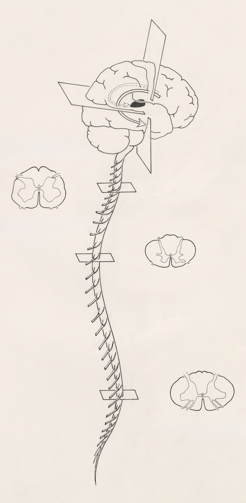
The root cause of spasticity lies in the damage or lesion to the upper motor neurons (UMNs), which encompass the motor pathways descending from the cerebral cortex and brainstem, primarily through the corticospinal tract, to the lower motor neurons (LMNs) in the spinal cord. These UMN pathways are essential for executing voluntary movement and, critically, for providing continuous inhibitory control over the spinal reflexes. When a lesion occurs—whether due to stroke, trauma, or disease—this descending inhibition is lost. The subsequent lack of modulation leaves the spinal motor neurons and the associated interneurons in a state of chronic hyperexcitability. This neurological disinhibition leads to the characteristic signs of the UMN syndrome, including muscle weakness (paresis), loss of fine motor control, and the development of pathological hypertonia known as spasticity.
The mechanism of hyperexcitability involves several interconnected changes within the spinal cord circuitry. Following UMN damage, there is a reorganization and functional alteration of synaptic connections. Specifically, the alpha motor neuron pool, which innervates the skeletal muscle fibers, becomes overly sensitive to excitatory input from the muscle spindles, which are the sensory receptors responsible for detecting muscle stretch. The removal of descending control shifts the balance heavily toward excitatory neurotransmitter release, leading to sustained depolarization of the motor neurons. This heightened state of readiness means that even a rapid, passive stretch—the specific movement that defines the velocity-dependence—triggers an abnormally strong and prolonged firing of the motor units, resulting in the sudden, often forceful resistance observed clinically.
Furthermore, chronic spasticity induces secondary, structural changes in the muscle tissue itself. Over time, sustained, involuntary contraction leads to alterations in muscle fiber properties, including increased stiffness, loss of sarcomeres, and replacement of muscle tissue with fibrous, non-contractile connective tissue, a process known as fibrosis. These biomechanical changes exacerbate the functional limitations imposed by the neural damage, often resulting in fixed joint deformities called contractures, which become resistant to pharmacological treatment. The complex interplay between neural disinhibition (primary cause) and muscle tissue adaptation (secondary effect) underscores the progressive and challenging nature of managing chronic spasticity, demanding interventions that address both the neurological source and the musculoskeletal consequences.
Clinical Manifestations and Assessment
The clinical presentation of spasticity is marked by several key signs that allow for its accurate diagnosis and grading. The most essential sign is the velocity-dependent increase in muscle tone. During a physical examination, the clinician passively moves the patient’s limb; if resistance is felt only when the movement is performed quickly, spasticity is confirmed. A frequent accompanying phenomenon is the “clasp-knife” response, particularly evident in severe cases: an initial high resistance during rapid stretch suddenly gives way, similar to the action of opening a folding knife. This collapse is thought to be due to autogenic inhibition mediated by Golgi tendon organs once the tension reaches a critical threshold.
Associated findings invariably include hyperreflexia, characterized by exaggerated deep tendon reflexes (DTRs). The neurological examination typically reveals an expanded reflexogenic zone, where tapping an area far from the tendon may still elicit a reflex response. Furthermore, clonus—rhythmic, involuntary, sustained muscle contractions, usually elicited by a sudden stretch and maintained pressure on the tendon—is highly characteristic of severe spasticity, most commonly observed at the ankle. These exaggerated reflexes are direct evidence of the hyperexcitable spinal motor pools that define the condition. Spasticity can also manifest as involuntary, painful muscle spasms, which are often triggered by external stimuli such as temperature changes, positional shifts, or visceral distension (e.g., a full bladder).
To objectively quantify the severity of spasticity and monitor treatment effectiveness, clinicians rely on standardized scales. The most prevalent tool is the Modified Ashworth Scale (MAS), which grades muscle tone from 0 (no increase in tone) to 4 (the limb is rigid in flexion or extension). While widely used for its simplicity, the MAS has limitations, particularly its inability to distinguish true spasticity from non-reflexive stiffness and its lack of explicit measurement of velocity dependence. Therefore, specialized tools such as the Modified Tardieu Scale (MTS) are sometimes employed; the MTS measures the resistance at two distinct speeds (V1, slow stretch; V3, fast stretch) and identifies the specific angle at which the muscle “catches” the resistance, providing a more detailed picture of the dynamic component and assisting in differentiating fixed contractures from dynamic hypertonia.
Etiology: Primary Causes of Upper Motor Neuron Damage
Spasticity is not a primary disease but rather a common sequela of various central nervous system injuries or disorders that compromise the integrity of the descending motor pathways. The most frequent cause globally is cerebrovascular accident (stroke), particularly ischemic or hemorrhagic events that affect the primary motor cortex, internal capsule, or brainstem. Post-stroke spasticity typically develops weeks or months following the acute insult and significantly impedes functional recovery and rehabilitation efforts. The pattern of spasticity often reflects the typical motor synergies associated with hemispheric lesions, such as flexor dominance in the affected upper limb and extensor dominance in the lower limb.
Another major etiological category is spinal cord injury (SCI). Following a complete or incomplete transection of the spinal cord, spasticity commonly develops in segments below the level of the lesion. In this context, the isolated spinal cord circuitry, deprived of supraspinal inhibitory control, becomes locally hyperexcitable. Spasticity in SCI patients can present as generalized spasms, which are often triggered by external sensory stimuli, necessitating careful management of environmental factors and personal care routines. While sometimes useful for providing transient trunk stability for transfers, SCI spasticity is typically debilitating, causing pain, sleep disruption, and difficulty with positioning and hygiene.
Furthermore, chronic and progressive neurological diseases frequently lead to spasticity. Multiple Sclerosis (MS), an autoimmune demyelinating disease, commonly involves the corticospinal tracts, resulting in widespread and often fluctuating spasticity that affects up to 80% of patients. In pediatric populations, Cerebral Palsy (CP), resulting from a non-progressive insult to the developing brain, is the leading cause of chronic spasticity, classified as spastic CP. Other relevant causes include traumatic brain injury (TBI), cerebral hypoxia following cardiac arrest, and space-occupying lesions such as tumors that compress or destroy UMN fibers in the brain or spinal cord, underscoring the diverse range of neurological insults that can lead to this motor syndrome.
Differentiation from Rigidity and Other Hypertonias
Accurate clinical differentiation is crucial because spasticity must be distinguished from other forms of increased muscle tone, particularly rigidity, as the treatment approaches for these conditions are fundamentally different. Rigidity is characteristically associated with extrapyramidal disorders, primarily those affecting the basal ganglia, such as Parkinson’s disease. Unlike spasticity, rigidity is non-velocity dependent; the resistance to passive movement remains constant regardless of the speed of the movement. It often involves the co-contraction of both agonist and antagonist muscles simultaneously, leading to a continuous resistance throughout the range of motion, described as “lead-pipe” rigidity, or if combined with tremor, “cogwheel” rigidity.
Spasticity, conversely, is defined by its velocity dependence and is strictly a manifestation of damage to the pyramidal (corticospinal) tracts, leading to reflex hyperexcitability. This distinction is paramount: spasticity is a dynamic, reflex-mediated hypertonia, while rigidity is a sustained, continuous hypertonia. For instance, testing for spasticity involves rapid limb movement to elicit the stretch reflex, whereas testing for rigidity involves slow, smooth movement. Furthermore, spasticity often exhibits the clasp-knife phenomenon and associated signs like hyperreflexia and clonus, which are typically absent in pure rigidity.
Other forms of hypertonia require exclusion as well. Dystonia involves sustained or intermittent muscle contractions causing abnormal, often twisting, repetitive movements or postures, sometimes task-specific, and is typically a result of basal ganglia or associated network dysfunction. Another form is paratonia (gegenhalten), often seen in diffuse frontal lobe or cortical damage, where the resistance to passive movement is highly dependent on the amount of force applied by the examiner, representing an involuntary inability to relax rather than a reflex disorder. The precise identification of the type of hypertonia dictates whether the patient requires agents targeting GABA receptors (for spasticity) or dopaminergic agents (for Parkinsonian rigidity), emphasizing the clinical necessity of a meticulous neurological examination.
Comprehensive Management and Treatment Modalities
The management of spasticity is complex and requires an integrated, multidisciplinary approach involving physical therapy, pharmacological agents, and sometimes surgical interventions, all aimed at reducing pain, preventing long-term complications, and maximizing functional outcomes. Physical and occupational therapy form the cornerstone of non-pharmacological management. This involves rigorous, prolonged stretching protocols to minimize muscle shortening and prevent contractures, the use of passive range of motion exercises, and the application of orthotic devices (braces or splints) to maintain limbs in functional, neutral positions, especially during rest. Therapy also focuses on strengthening antagonist muscles to improve functional balance and utilizing techniques like constraint-induced movement therapy or functional electrical stimulation to retrain motor control.
Pharmacological treatments are employed when spasticity significantly limits function or causes pain. Oral medications, which act systemically, are often the first line of defense. These include Baclofen, a GABA-B receptor agonist that enhances presynaptic inhibition in the spinal cord; Tizanidine, an alpha-2 adrenergic agonist that acts on spinal interneurons to inhibit reflex activity; and less commonly, benzodiazepines, which enhance GABA-A signaling. A major challenge with oral agents is dose-limiting side effects, such as generalized weakness, sedation, and cognitive fog, which can impede rehabilitation progress. For focal or regional spasticity, Botulinum toxin (BoNT) injections are highly effective, providing temporary relief by blocking acetylcholine release at the neuromuscular junction of the specific hyperactive muscle groups, with effects lasting typically three to four months.
For individuals suffering from severe, generalized spasticity—particularly those with spinal cord injury or advanced MS—who do not respond adequately to oral medications, advanced neurosurgical interventions may be necessary. The gold standard for widespread refractory spasticity is the implantation of a pump for continuous delivery of Intrathecal Baclofen (ITB) directly into the cerebrospinal fluid. This method achieves high concentrations of the drug at the spinal cord level while minimizing systemic side effects, often resulting in dramatic improvements in tone and comfort. In pediatric cases, particularly severe Cerebral Palsy, Selective Dorsal Rhizotomy (SDR), a procedure that surgically cuts specific sensory nerve roots, may be performed to permanently reduce the afferent input driving the reflex hyperexcitability, though this is reserved for carefully selected candidates.
Impact on Quality of Life and Prognosis
The burden of spasticity extends far beyond motor impairment, significantly degrading the patient’s quality of life (QoL). Chronic, sustained muscle contractions often lead to severe and intractable pain, which, combined with the discomfort and movement restrictions, frequently causes severe sleep disturbances and chronic fatigue. The presence of high muscle tone makes basic activities of daily living (ADLs)—such as hygiene, dressing, and transfers—extremely difficult or impossible to perform independently, leading to profound reliance on caregivers. This dependency, combined with the reduced ability to participate in social and vocational activities, often precipitates secondary psychosocial issues, including clinical depression, anxiety, and social isolation.
The prognosis associated with spasticity is highly dependent on the underlying etiology and the effectiveness of early management. For static lesions like stroke or TBI, the degree of spasticity often stabilizes after the first year, and the long-term prognosis hinges on the success of intensive, ongoing rehabilitation aimed at maintaining flexibility and function. Conversely, in progressive conditions like Multiple Sclerosis, spasticity is often a gradually worsening symptom, necessitating continuous adjustment of treatment protocols. The key prognostic factor across all etiologies is the prevention of fixed contractures; once a joint is structurally fixed by shortened tendons and muscles, the functional deficit becomes irreversible and limits the efficacy of all pharmaceutical and interventional treatments aimed at dynamic tone reduction.
Future directions in research focus on leveraging neuroplasticity and developing targeted pharmacological agents. Advances in technology, such as sophisticated robotic devices and brain-computer interfaces, hold promise for modulating the abnormal spinal circuitry and restoring more normalized motor patterns. Furthermore, research into optimizing the timing and combination of BoNT injections with intensive physical therapy is continuously refined. Ultimately, successful management requires viewing spasticity not merely as excessive tone, but as a complex disruption of the motor control system that demands continuous therapeutic vigilance to maximize functional independence, reduce pain, and enhance the overall psychological well-being of the affected individual.