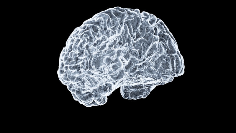
Cerebral Anatomy: Understanding Brain Fluid Obstruction
Introduction to Stenosis of Aqueduct of Sylvius
The condition known as Stenosis of Aqueduct of Sylvius (SAS) represents a critical neurological disorder characterized by the narrowing or complete occlusion of the cerebral aqueduct, a vital channel responsible for connecting the third and fourth ventricles within the brain. This anatomical constriction critically impedes the normal flow and circulation of Cerebrospinal Fluid (CSF), leading inexorably to the accumulation of fluid proximal to the blockage and resulting in obstructive hydrocephalus. Hydrocephalus, meaning “water on the brain,” is the primary pathological consequence of SAS, causing increased intracranial pressure and potentially severe neurological damage if left untreated. While SAS is recognized predominantly as a congenital or familial disorder, often presenting dramatically at birth, clinical evidence confirms that this condition is heterogeneous in its presentation, with significant cases developing later in life, sometimes emerging symptomatically long after the patient has reached adulthood. Understanding the varying etiology and clinical timeline is paramount for effective diagnosis and intervention, distinguishing between forms that arise from genetic predisposition and those that are acquired later due to secondary factors such as infection or hemorrhage.
SAS is not a uniformly acquired condition; a significant proportion of cases are attributed to a familial or hereditary disorder. Specifically, certain forms of congenital aqueductal stenosis are transmitted through an X-linked recessive gene, highlighting a clear genetic component that influences neurological development in utero. This genetic linkage implies a higher prevalence and specific inheritance pattern affecting male offspring primarily, though female carriers may also exist. The severity of the clinical manifestation is highly dependent on the degree of stenosis and the timing of the onset of CSF obstruction. In instances where the obstruction is severe and congenital, the resulting pressure changes rapidly overwhelm the developing fetal and neonatal skull, necessitating immediate medical intervention. Conversely, cases that manifest in adulthood typically present with a more insidious onset of symptoms, often mimicking other neurological conditions, making accurate and timely diagnosis a significant clinical challenge.
Etiology and Genetic Basis
The underlying cause of Stenosis of Aqueduct of Sylvius can be broadly categorized into congenital (developmental) and acquired forms. The congenital form, particularly that which is hereditary, is strongly associated with an X-linked recessive inheritance pattern. The gene most commonly implicated in this specific type of stenosis is the L1 Cell Adhesion Molecule gene (L1CAM), which is located on the X chromosome. Mutations in the L1CAM gene lead to faulty development of structures within the central nervous system, including defective formation or subsequent gliosis (scarring) within the narrow lumen of the aqueduct. Because this is an X-linked recessive disorder, it primarily affects males who inherit the mutated gene, while females are often asymptomatic carriers. This genetic predisposition underscores the importance of detailed family history screening when diagnosing infants presenting with congenital hydrocephalus.
While the genetic component is critical for understanding the familial nature of some cases, many instances of SAS are acquired, particularly those that emerge later in life or are sporadic rather than hereditary. Acquired stenosis results from pathological processes that cause inflammation, scarring, or direct compression of the aqueduct. Common secondary causes include intrauterine or postnatal infections, such as those caused by Toxoplasmosis or cytomegalovirus, which can lead to ependymal inflammation and subsequent obliteration of the aqueduct lumen through reactive gliosis. Furthermore, hemorrhagic events, often stemming from prematurity or trauma, can deposit debris that physically blocks the aqueduct or initiates a scarring process. Lastly, the presence of localized masses, such as tumors (e.g., pineal region tumors) or vascular malformations, can exert extrinsic pressure, functionally narrowing the passage and mimicking true intrinsic stenosis, thereby causing obstructive hydrocephalus.
The interplay between genetic susceptibility and environmental factors determines the precise pathology observed. For example, some individuals may possess a genetically narrower aqueduct, making them inherently more susceptible to developing symptomatic stenosis following a mild inflammatory insult that might be tolerated by an individual with a normally sized aqueduct. The complexity of the etiology mandates a thorough diagnostic workup, including advanced neuroimaging and, in familial cases, genetic testing, to differentiate primary developmental defects from secondary acquired obstructions. This differentiation is not merely academic; it significantly influences the predicted prognosis and the choice of therapeutic intervention.
Pathophysiology of Obstructive Hydrocephalus
The Aqueduct of Sylvius, also known as the cerebral aqueduct, is a slender channel running through the midbrain, serving as the sole conduit for CSF to pass from the third ventricle above to the fourth ventricle below. It is the narrowest point in the entire ventricular system, making it highly vulnerable to obstruction. When stenosis occurs—whether congenital due to developmental malformation or acquired due to gliosis or compression—the passage of CSF is significantly restricted or completely halted. Since CSF is continuously produced primarily by the choroid plexuses in the lateral and third ventricles, the blockage results in a relentless build-up of fluid in the spaces proximal to the stenosis. This accumulation leads to the dilation of the lateral ventricles and the third ventricle, a condition scientifically termed non-communicating or obstructive hydrocephalus.
The sustained accumulation of CSF exerts profound pressure on the surrounding brain parenchyma, leading to mechanical compression and potentially ischemia of the highly sensitive periventricular white matter tracts. In infants, whose cranial sutures are not yet fused, this increased pressure manifests externally as a progressive enlargement of the head at birth, or progressive macrocephaly, as the skull expands to accommodate the rising fluid volume. In older children and adults, however, the rigid, fused skull prevents external expansion, causing the pressure to be directed inward, resulting in severe symptoms related to elevated intracranial pressure (ICP). Chronic or acute elevation of ICP can disrupt normal neuronal function, damage nerve fibers, and impede cerebral blood flow, leading to irreversible neurological deficits.
The severity of the resulting hydrocephalus is directly proportional to the degree and rapidity of the obstruction. A complete and sudden obstruction causes an acute crisis characterized by rapidly escalating ICP, often requiring emergency intervention. Conversely, partial or slowly developing stenosis, which is often seen in adult-onset cases, allows for some compensatory mechanisms, such as increased CSF absorption or slower production rates, though symptoms of chronic hydrocephalus—such as gait disturbances and cognitive decline—will eventually emerge due to persistent, subtle pressure elevation. The mechanical forces exerted by the widening ventricles can stretch and damage the corpus callosum and the overlying cerebral cortex, fundamentally altering brain structure and function, which necessitates swift and effective CSF diversion.
Clinical Presentation: Congenital Onset
When Stenosis of Aqueduct of Sylvius is congenital, the clinical presentation is often dramatic and recognizable shortly after birth or within the first few months of life. The most defining and urgent physical sign is the progressive enlargement of the head, or macrocephaly, reflecting the rapidly increasing volume of trapped CSF within the compliant neonatal skull. Pediatricians and neonatologists meticulously track head circumference measurements, and a trajectory that crosses percentile lines upward is highly suggestive of pathological hydrocephalus. Along with macrocephaly, physical examination often reveals other classic signs of elevated intracranial pressure in infants.
These signs include a bulging and tense fontanelle, the separation of cranial sutures, and prominent scalp veins, all visual indicators of the underlying pressure. Neurologically, infants may exhibit the “setting-sun” sign, where the eyes are depressed downward due to pressure on the midbrain structures. Feeding difficulties, excessive irritability, poor head control, and developmental delay are also common findings, indicating compromise of normal brain function. If the stenosis is severe and untreated, the relentless pressure can lead to severe cognitive impairment, visual disturbances, and motor deficits, emphasizing the critical need for prompt neonatal diagnosis and surgical intervention to relieve the pressure and mitigate long-term damage. The identification of these symptoms forms the cornerstone of early diagnosis for the hereditary, X-linked forms of the disorder.
Clinical Presentation: Adult-Onset Cases
A crucial aspect of Stenosis of Aqueduct of Sylvius, often overlooked in generalized discussions, is the fact that the disorder can and does develop symptomatically after adulthood. These adult-onset cases may represent congenital stenosis that was compensated for years, or they might be truly acquired due to secondary events like minor hemorrhage, inflammatory processes, or previously asymptomatic small tumors that gradually grow to obstruct the aqueduct. Unlike the dramatic presentation in infancy, adult symptoms are often nonspecific and chronic, leading to potential misdiagnosis as psychiatric issues, migraine disorders, or normal pressure hydrocephalus (NPH).
The primary complaints in adult patients revolve around the symptoms of moderately increased intracranial pressure within the confined space of a rigid skull. These include chronic, often positional, headaches that are typically worse upon waking; episodes of nausea and vomiting; and specific visual changes, such as blurring or diplopia, often correlated with papilledema visible on fundoscopic examination. Furthermore, adult-onset hydrocephalus caused by SAS frequently presents with a triad of neurological symptoms known colloquially as the Hakim triad, though most characteristic of NPH, these symptoms are relevant here: gait disturbance (a wide-based, shuffling walk), urinary incontinence, and progressive cognitive decline, affecting memory, processing speed, and executive function.
The insidious nature of the adult presentation means that the exact timing of the pathological obstruction is often difficult to pinpoint. The symptoms may wax and wane, leading to delays in seeking specialized care. However, because the aqueduct is the bottleneck, the resulting hydrocephalus is purely obstructive, creating an urgent need for definitive treatment to prevent permanent neurological deterioration. Recognition that Stenosis of Aqueduct of Sylvius can occur after adulthood is vital for clinicians evaluating patients presenting with chronic, unexplained neurological deficits and headaches, especially when routine imaging reveals ventricular enlargement.
Diagnostic Procedures
Accurate diagnosis of Stenosis of Aqueduct of Sylvius relies heavily upon advanced neuroimaging techniques, which allow clinicians to visualize the ventricular system, the aqueduct itself, and the surrounding brain structures. The gold standard imaging modality is Magnetic Resonance Imaging (MRI), which provides superior soft-tissue detail and multiplanar capability essential for confirming the diagnosis. MRI scans clearly demonstrate the disproportionate dilation of the lateral and third ventricles, while the fourth ventricle remains normal or small, confirming the non-communicating nature of the hydrocephalus. Crucially, specific MRI sequences, such as Cine-Phase Contrast MRI, can be utilized to visualize CSF flow dynamics, confirming the absence or severe reduction of fluid movement through the aqueduct, thereby establishing the functional obstruction.
While MRI is preferred for definitive diagnosis, Computed Tomography (CT) scans remain valuable, particularly in acute or emergency settings, or when MRI is contraindicated. CT scans provide rapid assessment of ventricular size and are excellent for identifying acute hemorrhage or the presence of significant calcifications, which may be secondary indicators of infectious etiology or long-standing pressure changes. However, CT scans are often insufficient to visualize the aqueductal lumen itself or differentiate between intrinsic stenosis (e.g., due to gliosis) and extrinsic compression (e.g., due to a small mass).
In cases where a familial or hereditary basis is suspected, especially those presenting congenitally, genetic testing focusing on the L1CAM gene is highly recommended. Genetic counseling is essential for families identified with the X-linked recessive form of the disorder to assess recurrence risk for future pregnancies. Finally, in some instances, endoscopic evaluation during a surgical procedure may be necessary to directly visualize the aqueduct, confirm the stenosis, and potentially proceed immediately with treatment, such as a third ventriculostomy. The combination of clinical presentation, high-resolution MRI, and genetic confirmation ensures the most precise identification of the underlying pathology.
Management and Treatment Strategies
The management of Stenosis of Aqueduct of Sylvius is fundamentally surgical, focused entirely on relieving the obstructive pressure caused by the trapped Cerebrospinal Fluid (CSF) and establishing a new pathway for CSF flow. Pharmacological treatments are largely ineffective in treating mechanical obstructions. The two primary surgical approaches employed are CSF shunting and endoscopic third ventriculostomy (ETV).
The traditional and highly effective method for treating obstructive hydrocephalus is the placement of a ventriculoperitoneal (VP) shunt. This procedure involves inserting a catheter into one of the dilated lateral ventricles and tunneling a pressure-regulated valve and tubing subcutaneously down to the peritoneal cavity in the abdomen, where the excess CSF is safely absorbed. Shunting provides immediate and reliable pressure relief, drastically reducing the symptoms associated with elevated intracranial pressure. While highly successful, shunts are mechanical devices prone to complications, including obstruction, infection, and mechanical failure, requiring ongoing monitoring and potential revision surgeries throughout the patient’s lifetime.
A increasingly favored alternative, particularly in cases of primary aqueductal stenosis, is the Endoscopic Third Ventriculostomy (ETV). This minimally invasive neurosurgical procedure involves using an endoscope to enter the third ventricle and create a small, controlled opening in the floor of the ventricle. This opening allows the trapped CSF to bypass the blocked Aqueduct of Sylvius entirely, diverting the fluid directly into the subarachnoid space (the cisterns) where it can be naturally reabsorbed. ETV offers the significant advantage of treating the hydrocephalus without implanting a permanent foreign body, thus eliminating the risks of shunt dependence and chronic shunt complications. However, ETV efficacy is dependent on the patient’s anatomy and age, and it is generally more successful in older children and adults than in neonates.
Prognosis and Long-Term Outlook
The long-term prognosis for individuals diagnosed with Stenosis of Aqueduct of Sylvius is highly variable and depends on several critical factors, primarily the age of onset, the timing of surgical intervention, and the successful management of the hydrocephalus. For congenital cases, particularly those caused by the X-linked recessive gene, prompt diagnosis and surgical relief of the hydrocephalus in infancy are crucial for maximizing neurodevelopmental outcomes. Children treated early often achieve near-normal intelligence and motor function, though lifelong follow-up is mandatory due to the potential for shunt malfunction or gradual cognitive deficits.
Conversely, delayed diagnosis or treatment, especially in infants where brain plasticity is rapidly changing, can lead to severe and irreversible neurological damage, including profound intellectual disability, visual impairment, and spasticity. For those individuals whose disorder develops or becomes symptomatic in adulthood, the prognosis is often more favorable regarding intellectual outcome, as the brain has already matured. However, chronic symptoms such as gait ataxia and memory impairment may require longer rehabilitation even after successful CSF diversion.
A significant challenge impacting the long-term outlook for all patients is the management of the chosen treatment modality. Patients who undergo VP shunting require continuous neurosurgical monitoring due to the high lifetime risk of shunt revision. Factors such as infection, migration, or blockage necessitate repeated hospitalizations and surgeries, which introduce additional risks. Patients successfully treated with ETV may still experience failure of the ventriculostomy over time, necessitating a conversion to shunting. Ultimately, while modern surgical techniques offer excellent control over the elevated intracranial pressure, Stenosis of Aqueduct of Sylvius remains a chronic condition requiring specialized, multidisciplinary care throughout the patient’s life to ensure the highest possible quality of life and functional independence.