Sympathetic Chain: The Biological Roots of Your Stress Response
- Introduction and Definition of the Sympathetic Chain
- Anatomical Location and Structure
- Composition: The Sympathetic Ganglia
- Connectivity and Pre- and Postganglionic Neurons
- Physiological Role in the Autonomic Nervous System
- Pathways of Sympathetic Outflow
- Clinical Significance and Related Conditions
- Developmental Origin and Maturation
- Comparison with the Parasympathetic System (Integration)
Introduction and Definition of the Sympathetic Chain
The sympathetic chain, also known as the paravertebral ganglia or the sympathetic trunk, represents a fundamental and highly organized component of the autonomic nervous system (ANS). This intricate structure serves as the primary conduit for sympathetic nervous system signals originating in the central nervous system (CNS) to reach their various target organs throughout the body. Structurally, the chain is a bilateral, beadlike arrangement of neuronal clusters, or ganglia, running longitudinally alongside the vertebral column. Its critical function is to facilitate the rapid and coordinated responses associated with the ‘fight or flight’ mechanism, allowing the organism to quickly adapt to stressful or demanding environmental conditions. Understanding the sympathetic chain is essential for grasping the complexities of visceral control, homeostasis maintenance, and the physiological responses that govern energy expenditure and defensive postures. This anatomical marvel illustrates the efficiency of the nervous system in rapidly distributing diffuse signals necessary for systemic mobilization, contrasting sharply with the more localized effects typically observed in the somatic nervous system.
The defining characteristic of the sympathetic chain is its highly segmented and interconnected nature. It consists of approximately 21 to 23 pairs of ganglia arranged segmentally, typically mirroring the vertebral segments from the cervical region down to the coccyx. This arrangement ensures that the preganglionic fibers exiting the thoracolumbar spinal cord segments (T1 to L2 or L3) have immediate access to a wide array of postganglionic neurons, allowing for signal divergence and mass activation, a hallmark of sympathetic responses. Unlike the ganglia of the parasympathetic system, which are often located near or within the target organs, the sympathetic chain maintains this close proximity to the CNS, enabling quick synaptic relay and subsequent widespread distribution of efferent signals. This strategic positioning optimizes the speed and reach of sympathetic outflow, crucial for instantaneous physiological adjustments such as increased heart rate, bronchodilation, and peripheral vasoconstriction during moments of perceived danger or extreme exertion.
Historically, the study of the sympathetic chain unlocked profound insights into the involuntary control mechanisms governing visceral function. Early neuroanatomists recognized this structure as the central processing relay for the visceral motor system, distinguishing it from the pathways controlling skeletal muscle. The term ‘beadlike chain’ accurately describes its macroscopic appearance, where the individual ganglia resemble beads strung along a thread formed by the interconnecting nerve fibers. Functionally, it acts as a crucial synapse point where preganglionic sympathetic neurons, utilizing acetylcholine as their neurotransmitter, excite postganglionic neurons that then project toward target tissues, typically utilizing norepinephrine (noradrenaline). This two-neuron system, initiated within the intermediolateral cell column of the spinal cord and relayed through the sympathetic chain, establishes the foundation for autonomic regulation, influencing everything from pupil size and glandular secretions to gastrointestinal motility and cardiovascular dynamics, all operating below the level of conscious awareness.
Anatomical Location and Structure
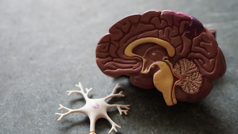
The anatomical placement of the sympathetic chain is remarkably precise and critical to its function. It is situated bilaterally, running parallel and anterolateral to the vertebral column, extending from the base of the skull (C1 level) down to the coccyx, where the two chains often merge into a single, small terminal ganglion known as the ganglion impar. This paravertebral location ensures that the short preganglionic fibers exiting the spinal cord via the ventral roots can quickly enter the chain. The chain is housed within the retroperitoneum in the abdominal region and deep within the neck and thorax, offering a degree of protection while maintaining essential proximity to the spinal nerves with which it constantly communicates. Crucially, the chain is not uniform; it is conventionally divided into cervical, thoracic, lumbar, and sacral segments, each housing specific ganglia responsible for innervating different body regions, highlighting the segmented organizational principle of the peripheral nervous system.
In the cervical region, the chain is typically condensed into three major ganglia: the superior cervical ganglion, the middle cervical ganglion, and the inferior cervical ganglion (often fused with the first thoracic ganglion to form the stellate ganglion). The superior cervical ganglion is the largest and is pivotal for sympathetic innervation to the head and neck, including the eye, salivary glands, and cerebral vasculature. Moving caudally, the thoracic region hosts 11 or 12 ganglia, which are the most numerous and receive the bulk of the sympathetic outflow from the spinal cord (T1-T12). Many of the preganglionic fibers entering the thoracic ganglia do not synapse immediately but instead continue through the chain to form the splanchnic nerves, which project to prevertebral ganglia closer to the abdominal viscera. This arrangement demonstrates the complex routing capabilities inherent in the chain, allowing signals to bypass immediate synapse points and reach distant targets.
The structural organization involves strong fibrous connections both between adjacent ganglia and between the chain and the spinal nerves. These connections are mediated by the rami communicantes. Specifically, the white rami communicantes, composed of myelinated preganglionic axons, connect the spinal nerve (T1-L2/L3) to the corresponding paravertebral ganglion, serving as the entry point for sympathetic signals. Conversely, the gray rami communicantes, containing unmyelinated postganglionic axons, exit the ganglia at all spinal levels (C1 to Co1) to join the spinal nerves. These postganglionic fibers then travel within the somatic spinal nerves to innervate peripheral structures such as sweat glands, arrector pili muscles, and the vasculature of the limbs and body wall. The consistent presence of gray rami communicantes at every spinal segment underscores the necessity of sympathetic input for regulating peripheral circulation and thermoregulation across the entire body surface.
Composition: The Sympathetic Ganglia
The fundamental units of the sympathetic chain are the paravertebral ganglia. Each ganglion is essentially a relay station composed primarily of the cell bodies of postganglionic sympathetic neurons, along with associated supporting cells like glial cells and satellite cells. These ganglia are structurally encapsulated by connective tissue, providing mechanical protection and structural integrity. Within the ganglion, the synaptic transmission occurs: the axon terminal of the preganglionic neuron releases acetylcholine (ACh), which binds to nicotinic receptors on the dendrites and cell body of the postganglionic neuron, initiating an action potential in the latter. This synaptic efficiency is paramount for the rapid propagation of sympathetic impulses required for immediate physiological adjustments.
The morphology and size of these ganglia vary significantly depending on their location and the density of the neuronal population they house. For example, the superior cervical ganglion is significantly larger than the ganglia found in the lower lumbar or sacral segments, reflecting the extensive area of the head and neck that it innervates. The organization within the ganglion is not merely a random cluster; the neurons are often topographically arranged, meaning that specific groups of postganglionic neurons within a single ganglion project to distinct target organs. This highly organized internal structure allows for a degree of specificity within the otherwise diffuse sympathetic outflow, although divergence (where one preganglionic neuron synapses with multiple postganglionic neurons) remains a prominent feature, enabling widespread activation.
Furthermore, the ganglia of the sympathetic chain contain specialized cells that contribute to modulation and regulation of synaptic transmission. Small intensely fluorescent (SIF) cells, which are neuroendocrine cells often containing dopamine or other catecholamines, are present and can locally modulate the activity of the principal postganglionic neurons. While the primary transmission is cholinergic (ACh acting on nicotinic receptors), the environment within the ganglion is complex, involving various peptides and modulators that fine-tune the output. The collective function of these structural components ensures that the signal originating in the CNS is efficiently and appropriately relayed and amplified before being distributed to effector tissues, thereby maintaining the homeostatic balance necessary for survival and responsiveness.
Connectivity and Pre- and Postganglionic Neurons
The operation of the sympathetic chain relies entirely on the precise anatomical relationship between preganglionic and postganglionic neurons. The cell bodies of the preganglionic neurons are located exclusively within the spinal cord, specifically within the intermediolateral cell columns (IML) of the gray matter, spanning segments T1 through L2 or L3—this is why the sympathetic system is often referred to as the thoracolumbar division. These axons exit the spinal cord via the ventral roots, join the spinal nerves briefly, and then peel off to enter the sympathetic chain via the white rami communicantes. Upon entering the chain, a single preganglionic fiber has three primary choices regarding its pathway, demonstrating the immense potential for signal dispersal and complex routing inherent to the sympathetic architecture.
The three possible fates of a preganglionic axon upon reaching the sympathetic chain are critical for defining the scope of sympathetic action. First, the fiber may synapse immediately with a postganglionic neuron at the same level of entry. Second, the fiber may ascend or descend within the chain to synapse with a postganglionic neuron in a ganglion located at a different vertebral level. This ascending and descending capability is essential for supplying sympathetic innervation to regions outside the thoracolumbar outflow area, such as the head (requiring ascent to the cervical ganglia) and the pelvic organs (requiring descent to the sacral ganglia). Third, the fiber may pass straight through the sympathetic chain without synapsing, continuing as a splanchnic nerve (e.g., greater, lesser, or least splanchnic nerves) to terminate in a prevertebral ganglion (e.g., celiac, superior mesenteric) located closer to the abdominal viscera.
Regardless of the pathway taken, the postganglionic neurons, whose cell bodies reside in either the paravertebral or prevertebral ganglia, receive the synaptic input and project their axons to the final target organs. These postganglionic axons are generally long and unmyelinated. When projecting from the paravertebral chain to peripheral structures (skin, blood vessels), they use the gray rami communicantes to rejoin the spinal nerves. At the effector organs (e.g., heart, smooth muscle, glands), these neurons typically release norepinephrine, which acts on adrenergic receptors, mediating the characteristic sympathetic effects. The high ratio of preganglionic to postganglionic neurons (often 1:10 or higher) ensures that a single signal from the CNS can activate a large number of effector cells simultaneously, reinforcing the concept of diffuse sympathetic response.
Physiological Role in the Autonomic Nervous System
The sympathetic chain is the central operational hub for the sympathetic nervous system, which is predominantly responsible for mediating the body’s responses to stress, emergency, and physical activity—a state commonly summarized as the “fight or flight” response. This system mobilizes energy reserves and directs resources away from maintenance functions (like digestion and rest) toward immediate survival needs (like muscle performance and heightened sensory awareness). The rapid relay provided by the sympathetic chain ensures that these widespread physiological changes occur almost instantaneously upon detection of a threat or demand, illustrating a critical evolutionary adaptation for survival in dynamic environments.
Key physiological actions regulated through the sympathetic chain include profound changes in the cardiovascular and respiratory systems. Sympathetic output causes an increase in heart rate (chronotropy) and force of contraction (inotropy), preparing the circulation for increased demand. Simultaneously, it mediates widespread vasoconstriction in the visceral organs and the skin, diverting blood flow toward the skeletal muscles and the brain. In the lungs, sympathetic fibers cause bronchodilation, increasing the diameter of the airways and maximizing oxygen intake. These responses are mediated by postganglionic fibers released from the thoracic ganglia or related cervical ganglia that project directly to the heart, blood vessels, and bronchi, ensuring systemic readiness for intense physical exertion or confrontation.
Beyond immediate physical mobilization, the sympathetic chain also governs less obvious but equally vital functions related to homeostasis. It controls the tonic activity of smooth muscle in the walls of various blood vessels, regulating blood pressure dynamically. Furthermore, it plays a crucial role in thermoregulation by activating sweat glands (unique in that the postganglionic sympathetic fibers here release ACh, not norepinephrine) and initiating piloerection (raising of hairs). It also inhibits non-essential functions, such as suppressing motility and secretion in the gastrointestinal tract and reducing urine production. Thus, the sympathetic chain operates not merely in emergencies but constantly modifies baseline organ activity to maintain internal stability in response to changing internal and external demands, fulfilling its role as a key modulator of involuntary bodily functions.
Pathways of Sympathetic Outflow
The organization of sympathetic outflow through the paravertebral chain is complex and involves four primary pathways, depending on the target location—body wall/limbs, head/neck, thorax, or abdomen/pelvis. Understanding these distinct routes is essential for appreciating the specificity that can sometimes override the general mass activation characteristic of the system. The path taken is determined at the point of entry into the chain, usually via the white rami communicantes (T1-L2/L3), and dictates whether the signal synapses locally, ascends/descends, or passes through to a prevertebral ganglion.
For targets in the body wall, limbs, and superficial structures (e.g., sweat glands, skin blood vessels), the preganglionic fiber enters the chain and synapses at the same level or travels vertically to synapse in a different ganglion. The postganglionic fibers then exit the chain via the gray rami communicantes to join the spinal nerve at that level, traveling peripherally to their targets. For innervation of the head, the preganglionic fibers originating in the upper thoracic segments (T1-T4) must ascend the chain without synapsing until they reach the superior cervical ganglion, where they synapse. Postganglionic fibers then follow large arteries (like the carotid artery) to reach structures of the orbit, nasal cavity, and salivary glands, illustrating a dependence on arterial pathways for distribution to the skull.
Innervation of the thoracic viscera (heart, lungs, esophagus) involves preganglionic fibers that enter the chain and synapse within the thoracic or cervical ganglia. The postganglionic fibers then leave the chain directly as visceral branches, forming plexuses (e.g., the cardiac and pulmonary plexuses) near their target organs. Finally, the abdominal and pelvic viscera receive input primarily through the splanchnic nerves. These are bundles of preganglionic fibers that traverse the sympathetic chain without synapsing and exit the chain to form the greater, lesser, and least splanchnic nerves. These nerves project to the prevertebral ganglia (such as the celiac and superior mesenteric ganglia), where the synapse occurs. The postganglionic fibers then travel the short distance from the prevertebral ganglion to the abdominal organs, providing the necessary sympathetic control for digestion, glandular secretion, and smooth muscle tone in the lower body.
Clinical Significance and Related Conditions
Disruptions to the function or integrity of the sympathetic chain can lead to significant clinical syndromes, underscoring its essential role in maintaining systemic homeostasis. Because the chain controls crucial functions like blood pressure regulation, pupil size, and sweating, damage to specific segments results in predictable clinical manifestations. Perhaps the most famous syndrome associated with sympathetic chain damage is Horner’s syndrome, which results from a lesion affecting the sympathetic pathway to the head, typically involving the superior cervical ganglion or the preganglionic fibers ascending toward it (originating T1-T3). Symptoms include miosis (constricted pupil), ptosis (drooping eyelid), and anhidrosis (absence of sweating) on the affected side of the face, clearly demonstrating the specific functions governed by the upper segments of the chain.
Furthermore, surgical procedures involving the sympathetic chain, known as sympathectomy, have historically been performed to treat conditions related to excessive sympathetic activity. For instance, chemical or surgical ablation of segments of the chain has been used to manage severe hypertension, although this practice has largely been superseded by pharmacological treatments. More recently, thoracic sympathectomy is occasionally used to treat severe palmar hyperhidrosis (excessive sweating of the hands) or certain types of debilitating vascular disorders, such as Raynaud’s phenomenon, where excessive vasoconstriction impairs peripheral blood flow. These interventions highlight the chain’s potent control over peripheral vasculature and glandular function, demonstrating that targeted disruption can alleviate symptoms caused by autonomic overactivity.
Conditions involving chronic pain, such as complex regional pain syndrome (CRPS), often involve dysfunction of the sympathetic nervous system, sometimes necessitating diagnostic procedures to determine if the pain is sympathetically maintained. The sympathetic chain’s intimate anatomical relationship with the spine also makes it vulnerable to injury from trauma, spinal pathology, or tumors. Damage to the lower thoracic or lumbar segments can affect visceral pain pathways or cause disruptions in pelvic organ function. Therefore, neurological and surgical evaluations often require a detailed understanding of the precise mapping of the sympathetic chain to localize pathologies and implement effective treatments that either block or modulate the signals being relayed through this vital neuronal highway.
Developmental Origin and Maturation
The development of the sympathetic chain is a remarkable process that begins early in embryonic life, derived primarily from the neural crest cells. These pluripotent cells migrate extensively throughout the embryo, and a specific population of them settles along the path lateral to the developing neural tube (spinal cord). These cells aggregate to form the initial rudimentary ganglia of the sympathetic trunk. This migratory origin explains the widespread distribution of sympathetic neurons throughout the body, as neural crest cells are responsible for generating many components of the peripheral nervous system, including sensory neurons and enteric nervous system neurons.
As the ganglia form, they must establish precise connections with the central nervous system. Axons from the preganglionic neurons, which differentiate within the intermediolateral cell column of the spinal cord (T1-L2), must grow outward to locate and synapse with the postganglionic neurons in the nascent paravertebral ganglia. This process of axonal guidance and target recognition is mediated by complex molecular cues and growth factors. Simultaneously, the interconnecting fibers that link adjacent ganglia, allowing for vertical travel within the chain, are established, solidifying the continuous, beadlike structure characteristic of the adult chain. Errors in this migration or connectivity process can lead to congenital autonomic dysfunction.
Maturation involves not only structural establishment but also the acquisition of functional characteristics, particularly the switch in neurotransmitter synthesis and receptor expression. While early postganglionic neurons might transiently express cholinergic characteristics, they eventually mature into adrenergic neurons, synthesizing and releasing norepinephrine, except for the postganglionic neurons innervating sweat glands. The continuous refinement and plasticity of the sympathetic chain throughout childhood and adolescence ensure that the autonomic responses are finely tuned to the increasing complexity and demands of the developing organism, securing the reliable function of the fight or flight mechanism throughout life. The robust, highly interconnected nature of the chain is a testament to this precise developmental sequence.
Comparison with the Parasympathetic System (Integration)
To fully appreciate the role of the sympathetic chain, it is crucial to contrast its structure and function with its counterpart, the parasympathetic nervous system. While both divisions constitute the autonomic nervous system, their anatomical organization, neurotransmitters, and physiological goals are fundamentally different. The sympathetic chain, located proximally to the CNS (paravertebral location), facilitates divergence and widespread, mass activation, utilizing short preganglionic fibers and long postganglionic fibers. This design supports its role in rapid mobilization and energy expenditure (catabolism), hence its thoracolumbar origin.
Conversely, the parasympathetic division originates in the craniosacral regions (cranial nerves and S2-S4 spinal segments) and lacks a unified paravertebral chain structure. Its ganglia are typically located much closer to or embedded within the target organs (intramural ganglia). This distal placement means the parasympathetic system features long preganglionic fibers and very short postganglionic fibers. Functionally, this structure promotes highly localized, discrete responses—ideal for its role in ‘rest and digest’ activities, conservation of energy, and maintenance (anabolism). Furthermore, both pre- and postganglionic parasympathetic neurons primarily utilize acetylcholine (ACh) as their neurotransmitter, contrasting with the sympathetic system’s reliance on norepinephrine at the target synapse.
Despite these differences, the two systems often exert antagonistic control over the same visceral organs, creating a dynamic balance known as autonomic tone. For example, the sympathetic chain increases heart rate, while the parasympathetic input decreases it. However, in some instances, their actions are cooperative, such as in sexual response. The sympathetic chain, through its structural efficiency, ensures that the body can quickly override parasympathetic maintenance when faced with stress, providing a powerful regulatory mechanism that integrates both central command and peripheral feedback to sustain optimal physiological function. The sympathetic chain is thus the physical embodiment of the body’s readiness for action.