Sympathetic Ganglion: How Your Body Drives Stress Responses
- Anatomical Definition and Fundamental Role of the Sympathetic Ganglion
- Microscopic Structure and Neurochemical Transmission
- Classification: Paravertebral and Prevertebral Ganglia
- Regional Organization of the Sympathetic Chain
- Physiological Impact on Major Organ Systems
- Developmental Origins and Histogenesis
- Clinical Relevance and Therapeutic Intervention
Anatomical Definition and Fundamental Role of the Sympathetic Ganglion
The sympathetic ganglion represents a cornerstone structure within the architecture of the Autonomic Nervous System (ANS), specifically serving as a crucial relay point for efferent signals originating from the central nervous system destined for peripheral target organs. By definition, a sympathetic ganglion is an aggregation of neuronal cell bodies situated outside the central nervous system, and its specific identity is derived from its inseparable membership in the sympathetic chain, also known as the sympathetic trunk or paravertebral chain. This chain is a bilateral, segmented structure running parallel and adjacent to the vertebral column, extending cranially from the base of the skull down to the coccyx. This strategic placement allows the sympathetic division to exert widespread, rapid control over visceral functions, facilitating the body’s adaptive responses necessary for homeostasis and, most famously, the acute stress response known as “fight or flight.”
Functionally, the sympathetic ganglion acts as the obligatory site of synapse between the preganglionic neurons, which originate in the intermediolateral cell column of the spinal cord (specifically segments T1 through L2 or L3), and the postganglionic neurons, which extend their axons to innervate smooth muscle, cardiac muscle, and glandular tissue throughout the body. The fundamental process occurring within the ganglion involves the transformation of a preganglionic signal, characterized by the release of acetylcholine (ACh), into a postganglionic signal, which typically utilizes norepinephrine (NE) as its primary neurotransmitter. This transition is regulated by nicotinic receptors located on the cell bodies of the postganglionic neurons, ensuring a swift and reliable transmission essential for the sympathetic system’s role in rapid physiological adjustments, such as increasing heart rate or diverting blood flow away from non-essential organs during periods of stress or perceived threat.
The structural organization of these ganglia, particularly their arrangement within the robust sympathetic trunk, allows for an extraordinary degree of signal divergence and simultaneous activation of multiple target systems. A single preganglionic neuron entering the chain often synapses with numerous postganglionic neurons within the same ganglion, or alternatively, the preganglionic fiber may ascend or descend the chain to synapse in ganglia at different vertebral levels. This extensive branching and distribution network is the physiological basis for the characteristic widespread and non-discrete activation associated with the sympathetic response, contrasting sharply with the more localized effects typically observed in the parasympathetic division. Understanding the precise anatomical location and neurochemical function of the sympathetic ganglion is paramount to grasping the mechanisms governing systemic metabolic regulation, cardiovascular control, and thermoregulation.
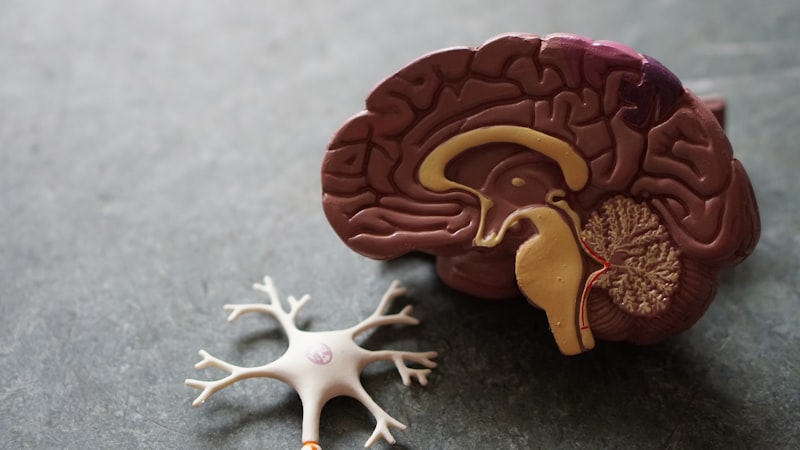
Microscopic Structure and Neurochemical Transmission
At the cellular level, the sympathetic ganglion is a complex microenvironment dominated by the cell bodies of multipolar postganglionic neurons, which are typically large and possess multiple dendritic processes. These neuronal somata are closely invested by specialized non-neuronal cells known as satellite glial cells (SGCs), which provide metabolic support, maintain the extracellular environment, and regulate neurotransmission efficacy by controlling ion balance and uptake. The ganglia also contain an extensive capillary network to support the high metabolic demand of continuous signal processing. The density of these structures underscores the ganglion’s role not merely as a passive relay station but as a critical integration center where signals are modulated before being relayed to the effector organs, potentially influenced by various circulating hormones and locally released neuromodulators.
The process of synaptic transmission within the sympathetic ganglion is cholinergic, meaning it is mediated by the neurotransmitter acetylcholine (ACh). When an action potential arrives via the myelinated preganglionic B-fiber, ACh is released into the synaptic cleft. This ACh rapidly binds to nicotinic acetylcholine receptors (nAChRs) located on the membranes of the postganglionic neurons. These receptors are ligand-gated ion channels; their activation causes an influx of sodium ions, leading to a rapid depolarization—an excitatory postsynaptic potential (EPSP)—which, if sufficient to reach threshold, generates an action potential in the postganglionic C-fiber. This fast, ionotropic transmission ensures minimal latency in the sympathetic reflex arc, which is crucial for instantaneous responses to environmental changes or physiological demands, such as sudden changes in posture or temperature.
Furthermore, while the primary fast excitatory transmission is mediated by nicotinic receptors, the synaptic machinery of the sympathetic ganglion is often modulated by slower components involving muscarinic receptors (metabotropic) and various neuropeptides co-released with ACh, such as vasoactive intestinal peptide (VIP) or substance P. These slower modulatory signals can fine-tune the excitability of the postganglionic neuron, influencing the duration and intensity of the subsequent efferent signal. The ultimate output from the ganglion—the postganglionic fiber—typically travels long distances to its target, utilizing norepinephrine at the neuroeffector junction to bind to adrenergic receptors (alpha and beta), thus eliciting the characteristic responses of the target organ, whether it be contraction, relaxation, or secretion.
Classification: Paravertebral and Prevertebral Ganglia
Sympathetic ganglia are broadly categorized into two major groups based on their anatomical position relative to the vertebral column and the aorta: the paravertebral ganglia and the prevertebral (or collateral) ganglia. The distinction between these types is essential for understanding the pattern of sympathetic innervation, as the paravertebral chain primarily mediates systemic, widespread effects (e.g., somatic structures, heart, lungs), while the prevertebral ganglia focus almost exclusively on regulating the major viscera of the abdominal and pelvic cavities. This dual organization permits both generalized and targeted sympathetic control simultaneously.
The paravertebral ganglia are the structures that form the visible sympathetic trunk. There are typically 21 to 23 pairs of these ganglia: three cervical, eleven or twelve thoracic, four or five lumbar, and four or five sacral, culminating in a single, fused ganglion impar at the coccyx. The cervical ganglia—specifically the superior, middle, and inferior (often fused with T1 to form the stellate ganglion)—are critical, as they receive preganglionic input that has ascended from the upper thoracic levels, providing sympathetic innervation to the head, neck, and heart. The unique arrangement of the paravertebral ganglia allows preganglionic fibers entering the chain via the white rami communicantes to either synapse immediately, ascend, descend, or pass through without synapsing to become splanchnic nerves, highlighting the versatility of this command center.
In contrast, the prevertebral ganglia are located anterior to the vertebral column, positioned closer to the abdominal aorta and the origins of its major branches. The three primary prevertebral ganglia are the Celiac Ganglion, the Superior Mesenteric Ganglion, and the Inferior Mesenteric Ganglion. These ganglia receive preganglionic fibers via the splanchnic nerves (which bypassed the paravertebral chain) and project postganglionic axons to the digestive tract, liver, spleen, kidneys, and reproductive organs. Because they are positioned closer to their target organs, the postganglionic fibers emanating from the prevertebral ganglia are generally shorter than those originating in the paravertebral chain, resulting in a slightly more localized and focused sympathetic influence on the abdominal viscera compared to the systemic effects mediated by the paravertebral chain.
Regional Organization of the Sympathetic Chain
The organization of the sympathetic chain into distinct regional groups—cervical, thoracic, lumbar, and sacral—is necessitated by the widespread distribution of sympathetic targets. The cervical ganglia are responsible for innervating structures in the head, neck, and upper chest, including the dilator pupillae muscle (causing mydriasis), salivary glands, blood vessels of the face, and providing crucial sympathetic input to the heart via cardiac nerves. Damage or disruption to the Superior Cervical Ganglion, the largest of the three, is clinically significant and often results in the constellation of symptoms known as Horner’s Syndrome, demonstrating the irreplaceable role of this ganglion in ocular and facial homeostasis.
The thoracic ganglia (T1-T12) represent the most voluminous and complex section of the chain, as they serve as the primary output centers. Postganglionic fibers from T1-T5 typically innervate the thoracic viscera, contributing to the pulmonary, esophageal, and cardiac plexuses, regulating respiration and heart function. Crucially, the lower thoracic ganglia (T5-T12) give rise to the major splanchnic nerves—the greater, lesser, and least splanchnic nerves—which bypass the paravertebral ganglia entirely and project to the prevertebral ganglia (Celiac, Superior Mesenteric), providing the essential preganglionic input for abdominal visceral control. This segment is therefore the main conduit for the widespread activation of the body’s largest organ systems during heightened arousal.
Finally, the lumbar and sacral ganglia (L1-S5) primarily regulate sympathetic innervation to the lower extremities, pelvic viscera, and the external genitalia. Fibers emanating from these lower ganglia manage vasoconstriction in the legs, influence bowel and bladder motility, and contribute to the physiological responses involved in sexual function, such as ejaculation. The integration of signals at these lower levels ensures that sympathetic action extends throughout the entire vertical axis of the body, allowing for coordinated systemic responses that range from diverting blood flow in the feet to regulating glandular output in the head.
Physiological Impact on Major Organ Systems
The signals processed and relayed by the sympathetic ganglia are responsible for coordinating the body’s response to acute stress, physical exertion, and environmental challenges, fundamentally maintaining equilibrium and facilitating survival. Through their postganglionic projections, these ganglia exert profound effects on the cardiovascular system, respiratory function, and metabolic processes. For example, sympathetic activation dramatically increases cardiac output by increasing both heart rate (chronotropy) and contractility (inotropy) via the beta-1 adrenergic receptors located in the heart muscle, preparing the organism for rapid physical action.
Concurrently, the sympathetic ganglia orchestrate complex changes in blood flow distribution. Postganglionic fibers innervating the smooth muscle surrounding arterioles in non-essential areas, such as the digestive tract and skin, release norepinephrine, causing vasoconstriction (via alpha-1 receptors), thereby shunting oxygenated blood away from these areas and redirecting it towards the skeletal muscles, heart, and brain. Conversely, these same pathways may cause vasodilation in certain critical muscle beds (via beta-2 receptors), illustrating the fine-tuned, differential control exerted by the sympathetic output that originates at the ganglionic level.
Beyond cardiovascular control, the sympathetic ganglia play a critical role in metabolic regulation and glandular secretion. Activation stimulates the adrenal medulla (which is embryologically a modified sympathetic ganglion) to release epinephrine and norepinephrine directly into the bloodstream, amplifying and prolonging the systemic effects. Furthermore, sympathetic input causes bronchodilation in the lungs, increasing air intake, and stimulates hepatic glycogenolysis, releasing glucose into the circulation to fuel the muscles. Even functions such as sweating, vital for thermoregulation, are governed by sympathetic postganglionic fibers, though uniquely, the postganglionic fibers innervating sweat glands release acetylcholine instead of norepinephrine.
Developmental Origins and Histogenesis
The formation of the sympathetic ganglia is a fascinating illustration of neural crest cell migration and differentiation during embryonic development. All components of the peripheral nervous system, including the sympathetic ganglia, derive primarily from the neural crest, a transient population of multipotent cells that emigrate from the dorsal aspect of the developing neural tube. Specifically, the neural crest cells destined to become sympathetic neurons migrate ventrolaterally, aggregating into clusters on either side of the dorsal aorta to form the initial sympathetic primordia.
These migrating cells undergo a process of differentiation, influenced by local growth factors and signals from the surrounding mesenchymal tissue, most notably Bone Morphogenetic Proteins (BMPs). The differentiation involves the induction of transcription factors such as Phox2b, which commit the cells to a noradrenergic phenotype. The developing sympathetic neurons then aggregate into segmented clusters, establishing connections with the preganglionic axons growing out from the spinal cord. This precise developmental timing ensures that the sympathetic ganglia are correctly positioned adjacent to the vertebral column before the final anatomical organization of the trunk is achieved.
Errors in the developmental migration or differentiation of these neural crest cells can lead to congenital abnormalities or certain neurocristopathies. Furthermore, the adrenal medulla, often referred to as a specialized sympathetic ganglion, shares this neural crest origin. However, unlike typical postganglionic neurons, the chromaffin cells of the adrenal medulla lack axons and instead release their neurotransmitters (which function as hormones) directly into the systemic circulation, highlighting a developmental modification of the typical ganglionic structure where the preganglionic fibers synapse directly onto secretory cells.
Clinical Relevance and Therapeutic Intervention
The physiological centrality of the sympathetic ganglion means that it is implicated in numerous clinical conditions and serves as a target for specific medical and surgical interventions. Dysfunction of the ganglia, whether through trauma, disease, or pharmacological blockade, results in profound autonomic instability. A classic example is the disruption of the sympathetic pathway leading to the head, often due to lesions affecting the superior cervical ganglion or the preganglionic fibers running in the neck, resulting in Horner’s Syndrome, characterized by ptosis (drooping eyelid), miosis (constricted pupil), and facial anhidrosis (lack of sweating).
Historically and currently, the manipulation of sympathetic ganglia has been utilized for therapeutic purposes. Sympathectomy, a procedure involving the surgical destruction or chemical ablation of a specific ganglion or section of the sympathetic chain, has been employed to treat conditions driven by excessive sympathetic tone. For instance, lumbar sympathectomy was once used to improve circulation in severe peripheral vascular disease, while thoracic sympathectomy is still used effectively to treat severe palmar hyperhidrosis (excessive sweating of the hands), by interrupting the sympathetic outflow to the sweat glands. These procedures demonstrate the direct power held by the ganglia in regulating peripheral function.
Pharmacologically, the synapse within the sympathetic ganglion is a potential target for drugs known as ganglionic blocking agents. These drugs, such as hexamethonium (a nicotinic receptor antagonist), block the transmission of impulses from preganglionic to postganglionic neurons, thereby non-selectively inhibiting both sympathetic and parasympathetic output simultaneously. While historically important in treating severe hypertension, their non-selective nature and attendant side effects (e.g., orthostatic hypotension, urinary retention, dry mouth) have largely relegated them to historical study or niche applications, emphasizing the difficulty in targeting this crucial relay point without inducing widespread systemic autonomic dysfunction.