Biological Stress: How Your Body Handles the Pressure

Biological Stress

Stress is an undeniable and pervasive aspect of human existence, influencing individuals across all walks of life. The manner in which a person perceives and responds to demanding situations holds profound implications for their overall physical and mental well-being. At its most fundamental level, biological stress represents the intricate physiological and psychological reactions orchestrated by the body in response to perceived threats or challenges, whether they are physical, mental, or emotional in nature. This sophisticated adaptive system, primarily governed by the hypothalamic-pituitary-adrenal (HPA) axis, involves a complex cascade of hormonal releases, neurotransmitter activity, cytokine modulation, and a myriad of other physiological adjustments designed to prepare the organism for action. This comprehensive entry will delve into the biological underpinnings of stress, explore its physiological and psychological ramifications, and elucidate its significant implications for health and wellness, drawing upon established scientific understanding.

The Core Definition of Biological Stress

At its essence, biological stress is defined as the body’s non-specific physiological and psychological response to any demand placed upon it. This response is an evolutionary conserved mechanism, crucial for survival, enabling an organism to rapidly adapt to adverse or challenging conditions. It is not merely a subjective feeling but a deeply ingrained biological process that mobilizes energy, heightens awareness, and alters various bodily functions to address a perceived threat or maintain internal equilibrium, known as homeostasis.

The fundamental mechanism underpinning biological stress involves the activation of the central nervous system, which then signals to the endocrine system. This intricate interplay results in the rapid release of stress hormones, primarily cortisol and catecholamines such as adrenaline (epinephrine) and noradrenaline (norepinephrine). These chemical messengers act on virtually every organ and tissue in the body, initiating a coordinated set of changes intended to enhance immediate survival capabilities, often referred to as the “fight or flight” response. This physiological readiness involves shifts in metabolism, cardiovascular function, immune activity, and cognitive processing, all tailored to meet the demands of the stressor.

Expanding on this, the core idea is the body’s remarkable capacity for allostasis, which refers to achieving stability through physiological change. Unlike strict homeostasis, which aims to maintain a constant internal environment, allostasis involves proactive adjustments to anticipate and respond to perceived challenges. When the body encounters a stressor, the biological stress response acts as an allostatic mechanism, dynamically reconfiguring physiological set points to cope. However, prolonged or repeated activation of this system, without adequate recovery, can lead to allostatic load, a cumulative wear and tear on the body that contributes to various health problems.

The Hypothalamic-Pituitary-Adrenal (HPA) Axis: The Body’s Central Stress Regulator

The hypothalamic-pituitary-adrenal (HPA) axis stands as the principal neuroendocrine system responsible for governing the body’s intricate stress response. This complex network of interactions among the hypothalamus, pituitary gland, and adrenal glands is meticulously regulated through a series of feedback loops, ensuring a finely tuned and adaptable reaction to stressors. When an individual encounters a physical, psychological, or emotional stressor, sensory information is relayed to the brain, which then activates the hypothalamus, the command center of this vital axis. This initial activation marks the commencement of a sophisticated chain of events designed to prepare the body for immediate action and subsequent recovery.

Upon activation, the hypothalamus, situated deep within the brain, initiates the stress response by releasing corticotropin-releasing hormone (CRH). CRH, a potent neuropeptide, then travels a short distance through the portal system to the anterior lobe of the pituitary gland. In response to CRH stimulation, the pituitary gland promptly secretes adrenocorticotropic hormone (ACTH) into the bloodstream. ACTH, circulating throughout the body, subsequently acts upon the adrenal glands, which are small, cap-like organs perched atop the kidneys. The adrenal glands, in turn, are prompted to release a suite of hormones, with cortisol being the primary glucocorticoid involved in the body’s prolonged stress response.

Cortisol, often dubbed the “stress hormone,” is a steroid hormone that plays a multifaceted role in the body’s adaptation to stress. Its widespread effects are crucial for the classic “fight or flight” response, preparing the body to either confront or evade a perceived threat. These effects include a rapid increase in heart rate and blood pressure, which enhances blood flow to vital organs such as the muscles and brain. Additionally, cortisol elevates blood glucose levels by promoting glucose production and inhibiting its uptake by certain tissues, ensuring a readily available energy source for immediate demands. It also enhances alertness and vigilance, sharpening sensory perception and cognitive processing to facilitate quick decision-making. Furthermore, cortisol possesses potent anti-inflammatory and immunosuppressive properties, which, in the short term, can prevent excessive immune activation during acute stress, though chronic elevation can lead to detrimental immune suppression.

Historical Context and Discovery of Stress Responses

The scientific understanding of biological stress has evolved significantly over the past century, building upon foundational observations and groundbreaking research. Early insights into the body’s automatic responses to danger emerged from the work of American physiologist Walter Cannon in the early 20th century. Cannon, around the 1920s and 1930s, coined the term “fight or flight” response, describing the immediate, involuntary physiological changes that occur when an animal perceives a threat. He meticulously documented how the sympathetic nervous system and the adrenal medulla rapidly secrete adrenaline and noradrenaline, preparing the body for intense physical exertion. His work primarily focused on the acute, immediate reactions to stress and highlighted the crucial role of the autonomic nervous system in survival.

A pivotal figure in the broader conceptualization of stress was Hungarian-Canadian endocrinologist Hans Selye. Beginning in the late 1930s and continuing through the mid-20th century, Selye systematically studied the body’s reactions to various noxious stimuli. He observed that diverse stressors, ranging from extreme temperatures to surgical trauma, elicited a remarkably similar pattern of physiological responses. This led him to propose the concept of the “General Adaptation Syndrome (GAS)” in 1936. Selye’s GAS posited a three-stage response to prolonged stress: first, the alarm reaction (similar to Cannon’s fight or flight); second, the stage of resistance, where the body attempts to cope and adapt to the stressor; and third, the stage of exhaustion, where prolonged exposure to the stressor depletes the body’s resources, leading to illness and even death. Selye’s work was revolutionary because it shifted the focus from specific diseases to the non-specific response patterns underlying them, establishing stress as a central concept in medicine and psychology.

Following Selye’s seminal contributions, research into biological stress continued to expand, moving beyond general physiological responses to explore the intricate neurobiological and genetic mechanisms involved. The 1970s and beyond saw increasing attention to the role of the HPA axis, neurotransmitters, and the immune system, leading to the emergence of fields like psychoneuroimmunology. Researchers began to differentiate between acute and chronic stress, recognizing that while acute stress is adaptive, chronic stress can be highly detrimental. The historical journey from Cannon’s immediate fight or flight to Selye’s comprehensive GAS, and subsequently to modern neuroendocrinology, illustrates a deepening understanding of how the body responds to and is fundamentally altered by, the demands of its environment.

Practical Manifestations of Biological Stress

To fully grasp the intricate workings of biological stress, a practical, relatable example from everyday life can be incredibly illuminating. Consider the common scenario of an individual, Sarah, who is preparing for a significant job interview. This is not a life-threatening situation, but it presents a substantial psychological and social challenge that triggers a potent biological stress response. Sarah perceives this interview as a high-stakes event, where her future career prospects are on the line, activating a series of internal physiological adjustments designed to enhance her performance, yet potentially also overwhelming her.

As Sarah anticipates the interview, her brain, particularly the amygdala, registers the event as a potential threat or challenge to her well-being and goals. This perception rapidly activates her sympathetic nervous system, leading to the immediate release of adrenaline and noradrenaline from her adrenal medulla. Simultaneously, the HPA axis begins its activation cascade. Her hypothalamus releases CRH, stimulating the pituitary to secrete ACTH, which then prompts her adrenal cortex to release cortisol. This hormonal surge manifests in several distinct physiological changes: her heart rate quickens, her breathing becomes shallower and more rapid, her palms might start to sweat, and she experiences a heightened sense of alertness, sometimes bordering on anxiety. These are the classic “fight or flight” preparations, even though she is sitting still, mentally rehearsing her answers.

Step-by-step, the biological principle applies in her chosen example: First, the stressor appraisal occurs – Sarah evaluates the interview as demanding and important. Second, the neural signaling begins – her brain activates the sympathetic nervous system and the HPA axis. Third, hormone release follows – adrenaline, noradrenaline, and cortisol flood her system. Fourth, physiological changes ensue – increased heart rate, blood pressure, muscle tension, and heightened vigilance. This comprehensive biological response is her body’s attempt to optimize her cognitive function, focus, and energy levels for the perceived challenge. However, if this state of heightened arousal persists for too long before the interview, or if she experiences extreme anxiety, these very same biological responses can become counterproductive, leading to mental blocks, difficulty concentrating, or physical discomfort, ironically hindering her performance rather than helping it.

Significance and Broad Impact in Psychology and Health

The concept of biological stress holds immense significance within the field of psychology, serving as a crucial bridge between mental states and physical health. Understanding the physiological underpinnings of stress has fundamentally reshaped how psychologists view mental disorders, coping mechanisms, and the intricate mind-body connection. It has moved the discussion beyond purely psychological constructs to embrace the tangible biological changes that accompany emotional experiences, providing a more holistic and evidence-based framework for comprehending human behavior and well-being. This integrated perspective underscores that mental health is inextricably linked to physiological regulation, challenging simplistic dualistic views of mind and body.

The impact of biological stress is profoundly felt in various applied psychological domains, particularly in clinical psychology and therapeutic interventions. Knowledge of the HPA axis dysfunction, chronic inflammation, and altered neurotransmitter profiles associated with prolonged stress has revolutionized the treatment of mental health conditions such as depression, anxiety disorders, and post-traumatic stress disorder (PTSD). Therapies like Cognitive Behavioral Therapy (CBT) and mindfulness-based stress reduction techniques, while behavioral in nature, are often effective precisely because they help individuals regulate their physiological stress responses, leading to measurable changes in hormone levels and brain activity. Moreover, the understanding of stress biomarkers allows for more personalized treatment approaches and a better assessment of treatment efficacy.

Beyond the clinical realm, the principles of biological stress find widespread application in diverse fields. In education, understanding how stress impacts memory, attention, and learning has led to the development of stress-reduction programs for students and strategies to create more supportive learning environments. In marketing and consumer behavior, insights into how stress influences decision-making, impulsivity, and brand loyalty are used to tailor advertising and product design. In occupational health, knowledge of chronic workplace stress informs policies aimed at improving employee well-being, productivity, and reducing burnout. Furthermore, in public health, understanding the societal burden of stress-related illnesses drives initiatives focused on promoting resilience, healthy lifestyles, and early intervention to mitigate the long-term health consequences of chronic biological stress on populations.

Physiological and Psychological Effects of Chronic Biological Stress

While acute biological stress is an adaptive and life-saving response, chronic stress, characterized by prolonged or repeated activation of the HPA axis and sympathetic nervous system, can exert a profoundly detrimental impact on both physical and mental health. The sustained elevation of stress hormones, particularly cortisol, along with persistent sympathetic nervous system activation, leads to a state of chronic physiological arousal that overwhelms the body’s capacity for recovery and repair. This prolonged imbalance contributes significantly to the development and exacerbation of a wide array of severe health problems, transforming a protective mechanism into a destructive force.

On the physiological front, chronic biological stress is a major risk factor for several debilitating physical health conditions. The constant strain on the cardiovascular system due to elevated heart rate and blood pressure increases the likelihood of developing hypertension, atherosclerosis, and ultimately, a heightened risk of heart disease and stroke. Furthermore, sustained high levels of cortisol can lead to dysregulation of glucose metabolism, contributing to insulin resistance and increasing susceptibility to Type 2 diabetes. Perhaps one of the most critical effects is the suppression of the immune system; while acute stress temporarily enhances immune function, chronic stress weakens it, making individuals more vulnerable to infectious diseases, slower wound healing, and potentially increasing the risk of certain cancers. Additionally, chronic stress can disrupt digestive processes, contributing to conditions like irritable bowel syndrome (IBS), and can also impact bone density and reproductive function.

The psychological toll of chronic biological stress is equally profound, manifesting in a spectrum of mental health challenges. The persistent activation of brain regions associated with fear and anxiety, coupled with dysregulation of neurotransmitters like serotonin and dopamine, significantly increases the risk of developing or worsening anxiety disorders and major depressive disorder. Individuals under chronic stress often experience a decrease in cognitive performance, including impaired memory, reduced attention span, and difficulty with problem-solving and decision-making, as the brain’s resources are diverted to perceived threats. Furthermore, sleep disturbances, such as insomnia, are a common consequence, perpetuating a vicious cycle where lack of sleep exacerbates stress, and stress, in turn, disrupts sleep. These psychological effects can severely diminish an individual’s quality of life, impairing their ability to function effectively in personal, social, and professional domains.

Connections to Other Psychological Concepts and Broader Categories

The concept of biological stress is not an isolated phenomenon within psychology but is intricately interwoven with numerous other key theories and terms, forming a rich tapestry of understanding regarding human experience and adaptation. It is fundamentally linked to notions of allostasis and allostatic load, which describe the body’s dynamic process of maintaining stability through change and the cumulative physiological cost of chronic adaptation, respectively. Moreover, biological stress heavily influences and is influenced by coping mechanisms, referring to the behavioral and psychological efforts individuals employ to manage stressful demands. Effective coping strategies can buffer the negative biological impacts of stress, while maladaptive strategies can exacerbate them. The concept also intersects with resilience, the ability to adapt successfully in the face of adversity, often mediated by physiological processes that enable better stress recovery. Furthermore, understanding stress is central to studying trauma and its long-term effects, as traumatic experiences often lead to profound and lasting alterations in the biological stress response systems, contributing to conditions like PTSD. Finally, emotional regulation strategies are often directly aimed at managing the physiological arousal associated with stress, highlighting the deep connection between subjective experience and bodily states.

Biological stress falls under several broader categories within the vast field of psychology, reflecting its multidisciplinary nature. It is a cornerstone of Health Psychology, which explores the interplay between psychological factors, biological processes, and health and illness. Within this domain, biological stress provides a framework for understanding psychosomatic illnesses and the impact of lifestyle on disease progression. It is also a critical area within Biological Psychology (also known as Biopsychology or Behavioral Neuroscience), which focuses on the physiological, genetic, and developmental mechanisms of behavior in humans and other animals. Here, researchers examine the neural circuits, endocrine pathways, and genetic predispositions that shape an individual’s stress response.

The study of biological stress is particularly prominent in the emerging field of Psychoneuroimmunology (PNI), an interdisciplinary science that investigates the interactions among the central nervous system, the endocrine system, and the immune system. PNI provides the scientific framework for understanding how psychological stress can directly impact immune function and disease susceptibility. Additionally, its implications are profound in Clinical Psychology, where understanding stress-related physiological dysregulation is crucial for diagnosing and treating mental health disorders. By integrating insights from these diverse subfields, the study of biological stress offers a comprehensive perspective on how our internal physiological systems react to external demands, ultimately shaping our health, well-being, and capacity to navigate the complexities of life.

CANNON, WALTER BRADFORD (1871-1945)

Early Life and Education

Walter Bradford Cannon (1871-1945) was an exceptionally influential American physiologist whose work revolutionized the understanding of bodily regulation, stress, and emotion. Born on October 19, 1871, in Prairie du Chien, Wisconsin, Cannon’s early life fostered a keen interest in the natural world and the mechanisms underlying biological function. His foundational education instilled in him a commitment to empirical observation and rigorous scientific methodology, essential qualities that would define his professional career. This early dedication positioned him for success in the demanding academic environments where he would later conduct his groundbreaking research.

Cannon pursued his medical training at Harvard Medical School, where he earned his medical degree in 1896. Although his training provided the clinical foundation typical of a physician, Cannon’s professional trajectory quickly shifted toward academic research and the laboratory. His early years at Harvard were marked by innovative studies into the digestive system, utilizing pioneering technology that allowed for the visualization of internal processes in unprecedented detail. This methodological curiosity established him as a leader capable of integrating new technologies into physiological investigation, setting him apart from many of his contemporaries.

In 1903, Cannon officially joined the faculty of Harvard Medical School, beginning a distinguished tenure that lasted until his death in 1945. He eventually attained the title of George Higginson Professor of Physiology, solidifying his status as a central figure in American science. During his four decades at Harvard, Cannon cultivated a vibrant research environment, mentoring numerous students and collaborators who would themselves become leaders in physiology and neuroscience. His laboratory became a global nexus for research into the autonomic nervous system and endocrinology, cementing Harvard’s reputation as a powerhouse in physiological science.

The Foundation of Homeostasis

Cannon’s most enduring and universally accepted conceptual contribution to biology and medicine is the term and detailed explanation of homeostasis. While the concept of a stable internal environment had been introduced by Claude Bernard in the 19th century, it was Cannon who formally defined, named, and provided the mechanistic framework for how this stability is actively maintained. Homeostasis refers to the body’s dynamic ability to preserve relatively constant internal conditions—such as temperature, pH, blood sugar, and water balance—despite continuous and potentially disruptive changes occurring both internally and externally.

He meticulously articulated these concepts in his 1932 masterpiece, The Wisdom of the Body, arguing that homeostatic processes are not passive outcomes but require constant, coordinated effort by integrated physiological systems. Cannon emphasized the role of feedback loops, particularly those involving the nervous and endocrine systems, in detecting deviations from optimal set points and initiating corrective action. For instance, he showed how the sympathetic and parasympathetic systems often act antagonistically to fine-tune organ function, ensuring that critical parameters remain within narrow, life-sustaining ranges. This conceptual shift transformed physiology from the study of isolated organ function into the study of integrated, self-regulating biological systems.

The establishment of homeostasis as a central paradigm provided a unified theoretical basis for understanding health and disease. Cannon argued that the failure of homeostatic mechanisms, often due to prolonged stress or overwhelming pathological conditions, is the root cause of many illnesses. This framework allowed researchers and clinicians to interpret symptoms not merely as isolated problems, but as manifestations of a system struggling to regain equilibrium. Consequently, the concept became indispensable across various disciplines, including medicine, endocrinology, and even engineering (influencing the development of cybernetics and control systems theory).

The Sympathetic Nervous System and Fight-or-Flight

A major focus of Cannon’s experimental work involved unraveling the physiological consequences of intense emotional arousal and acute stress. His investigations led him to characterize the function of the sympathetic nervous system, demonstrating its critical role in emergency situations. He systematically showed that during periods of fear, pain, or rage, the body undergoes a rapid, coordinated mobilization of resources mediated by this involuntary branch of the nervous system, which he termed the “emergency function.”

This systematic mobilization is famously known as the fight-or-flight response. Cannon detailed how, upon perception of a threat, the sympathetic nervous system triggers the adrenal medulla to release powerful catecholamine hormones, primarily epinephrine (adrenaline) and norepinephrine. The resulting hormonal surge leads to a cascade of observable physiological changes designed to optimize the body for immediate, violent action. These changes include a dramatic increase in heart rate and cardiac output, elevated blood pressure, dilation of the pupils, and, crucially, the redistribution of blood flow away from non-essential systems like the digestive tract and toward the skeletal muscles and the brain.

Cannon’s 1915 publication, Bodily Changes in Pain, Hunger, Fear and Rage, provided the definitive account of these visceral responses. By linking external emotional stimuli (like fear) directly to measurable internal physiological changes (like increased adrenaline and stopped digestion), Cannon provided empirical proof that emotions are deeply rooted in physical processes. This work established the scientific basis for the body’s acute stress response and highlighted the evolutionary significance of the sympathetic system as a survival mechanism.

Furthermore, Cannon’s research helped to clarify the distinction between the sympathetic and parasympathetic divisions of the autonomic nervous system. He viewed the sympathetic system as an energy-expending, highly integrated unit necessary for crisis management, while the parasympathetic system was characterized as a restorative, energy-conserving system responsible for “rest and digest” functions. This clear functional division was a major advance in neuroscience and provided the foundational model for studying psychological and emotional disorders linked to autonomic dysregulation.

Methodological Innovations: X-rays and Visceral Movement

Walter Cannon’s contributions were not limited to theoretical concepts; he was also an exceptional innovator in experimental methodology. His early successes in physiology stemmed from his pioneering use of the newly developed X-ray technology in the late 19th and early 20th centuries. Before Cannon, studying the complex mechanical movements of the digestive system in living animals was fraught with difficulty and often required destructive or highly invasive surgical procedures. Cannon revolutionized this field by adopting fluoroscopy.

To visualize the internal workings of the stomach and intestines, Cannon employed radiopaque substances, primarily bismuth compounds, which animals would ingest. By tracking the path of these opaque substances using X-rays, he could directly observe and document the peristaltic waves and churning movements of the gut in real-time. This methodology allowed for the first objective, non-invasive observation of visceral functions under various conditions, providing crucial data on the normal rhythmic activity of the stomach and intestines.

Critically, Cannon used this technique to investigate the influence of emotional states on digestion. He famously demonstrated that strong emotional distress, such as fear or rage, caused a near-immediate and complete cessation of stomach motility. This direct, visual confirmation provided powerful empirical evidence for the visceral effects of emotion. It showed that the central nervous system, via the sympathetic nerves, possessed profound inhibitory control over digestive function, thereby confirming the intimate connection between psychological states and internal physiological processes.

These X-ray studies were pivotal for establishing Cannon’s credibility and for laying the groundwork for his later theories on the autonomic nervous system and stress. By providing objective, replicable data correlating external stimuli with internal changes, his methodological rigor elevated physiological psychology and set a high standard for subsequent experimental investigation in the field. The adoption of fluoroscopy in physiological laboratories remains one of his key, practical legacies.

Contributions to Physiological Psychology

Cannon’s systematic research into the physiological correlates of emotional experience was instrumental in defining and advancing the field of physiological psychology. His work led to the formulation of a major theory of emotion, developed in collaboration with his colleague and former student, Philip Bard, known as the Cannon-Bard theory of emotion.

This theory emerged as a direct challenge to the prevailing James-Lange theory, which argued that physiological changes precede and cause the subjective experience of emotion (e.g., “we feel sad because we cry”). Cannon and Bard presented experimental evidence suggesting that visceral changes alone were too slow and too non-specific to account for the rapid and diverse experiences of emotion. For example, the artificial injection of adrenaline produces generalized arousal, but not necessarily a specific emotion like fear or anger, suggesting the bodily response is insufficient to differentiate emotional experiences.

The Cannon-Bard theory proposed that the conscious experience of emotion and the physiological arousal occur simultaneously. They hypothesized that sensory information about an emotional stimulus travels to the thalamus, which then simultaneously sends signals along two distinct pathways: one to the cerebral cortex, resulting in the subjective awareness of emotion, and another to the hypothalamus and autonomic nervous system, resulting in the bodily response. Their research, often involving lesion studies in animals that showed emotional responses could still occur after the severance of visceral feedback, emphasized the central nervous system’s role—particularly the thalamus and hypothalamus—as the primary organizer of emotional behavior.

By shifting the focus from peripheral feedback (viscera) to central processing (brain structures), Cannon provided a sophisticated neurobiological model for understanding emotion. This work profoundly influenced subsequent research in affective neuroscience, providing the necessary foundation for exploring the precise neural circuits responsible for generating and regulating emotional states, thus cementing his role as a founding figure of modern physiological psychology.

The Emergence of Psychosomatic Medicine

One of the most far-reaching societal impacts of Cannon’s stress research was his contribution to the establishment of psychosomatic medicine. His empirical findings provided the crucial scientific link demonstrating how chronic mental and emotional states could precipitate or exacerbate physical illness, thereby bridging the historical gap between mind and body in clinical practice.

Cannon argued that while the fight-or-flight response is adaptive for acute, short-term threats, its chronic activation in modern human life—where stressors are often psychological and prolonged rather than physical and immediate—could lead to serious pathology. The constant mobilization of energy, persistent high blood pressure, and chronic suppression of non-essential functions (like digestion and immune response) were mechanisms, he suggested, that eventually overwhelmed the body’s homeostatic reserves. This wear and tear could manifest as tangible physical diseases, including peptic ulcers, chronic hypertension, and cardiovascular disorders.

Cannon’s advocacy was instrumental in challenging the purely biomedical model of disease, which often overlooked psychological factors. He insisted that effective medical care required acknowledging the patient’s complete psychological and emotional context. His work promoted the view, now widely accepted, that stress is not merely a psychological phenomenon but a powerful physiological process with measurable, damaging long-term consequences on bodily health.

Furthermore, Cannon was a vocal proponent of what would later be formalized and widely adopted as the biopsychosocial model of health and illness. This model recognizes that biological factors (physiology, genetics), psychological factors (emotions, beliefs, coping styles), and social factors (environment, culture, social support) are interconnected determinants of health. By integrating these dimensions based on his physiological findings concerning stress and homeostasis, Cannon provided the essential intellectual structure for holistic and integrative approaches in medicine.

Major Publications and Legacy

Walter Cannon’s legacy is encapsulated in his major publications, which synthesized decades of meticulous research into foundational principles of life science. His works are characterized by their clarity, experimental rigor, and their ability to translate complex physiological mechanisms into concepts understandable by both scientific peers and the general public. These texts served as crucial educational tools and theoretical cornerstones for multiple disciplines.

His 1915 text, Bodily Changes in Pain, Hunger, Fear and Rage, remains a seminal classic, detailing the visceral manifestations of strong emotions and providing the first comprehensive scientific account of the emergency function of the sympathetic nervous system and the fight-or-flight response. This book established the enduring link between physiology and emotional experience, profoundly impacting fields ranging from experimental psychology to endocrinology.

The 1932 publication, The Wisdom of the Body, introduced the concept of homeostasis to a global audience. This book not only coined the term but philosophically and empirically explored the remarkable resilience and self-regulatory capacity of living organisms. It remains a standard text illustrating the principles of physiological regulation and adaptation. Beyond these major books, Cannon published extensive research on traumatic shock, the physiology of hunger and thirst, and the mechanisms of chemical neurotransmission.

Cannon’s influence extended beyond academia into clinical medicine and military science. His work on traumatic and surgical shock, particularly during his service in World War I and his advisory role during World War II, led to crucial improvements in the management of battlefield casualties. His enduring legacy is his success in integrating the study of the nervous system, the endocrine system, and emotional behavior into a unified, coherent system, forever changing how scientists approach the intricate relationship between the mind and the body.

Honors and Recognition

Walter Bradford Cannon’s extensive career and transformative scientific contributions garnered significant recognition from the international scientific community. His rigorous methodology, profound theoretical insights, and leadership in establishing new fields ensured that he received numerous prestigious awards and honors throughout his lifetime.

A testament to his stature among American scientists, Cannon was elected to the highly selective National Academy of Sciences in 1933. He held influential leadership positions within the scientific community, notably serving as President of the American Physiological Society. His professional peers recognized the monumental impact of his work on the discipline of physiology.

In 1941, near the end of his career, Cannon was awarded the distinguished Gold Medal of the American Physiological Society. This award acknowledged his exceptional contributions to physiological research and his role as a mentor and educator. His election to numerous foreign societies further demonstrated the global reach and importance of his research into bodily regulation and the stress response.

Cannon is universally recognized as the central figure in establishing physiological psychology as a legitimate scientific discipline and providing the essential groundwork for the development of modern psychosomatic medicine. The fundamental concepts he introduced—homeostasis and the fight-or-flight response—remain cornerstones of biological and medical education, confirming his status as one of the most important pioneers in 20th-century life science.

References

The following sources provide detailed accounts of Walter B. Cannon’s life, work, and impact on the fields of physiology and psychology:

  • Bear, M. F., Connors, B. W., & Paradiso, M. A. (2019). Neuroscience: Exploring the brain. Philadelphia, PA: Wolters Kluwer.
  • Cannon, W. B. (1915). Bodily changes in pain, hunger, fear and rage: An account of recent researches into the function of emotional excitement. New York, NY: Appleton.
  • Cannon, W. B. (1932). The wisdom of the body. New York, NY: W. W. Norton & Company.
  • Lane, R. D. (2007). Walter Bradford Cannon: The life and times of a young scientist. Cambridge, MA: Harvard University Press.
  • Lane, R. D., & Schwartz, G. E. (2009). Walter B. Cannon: The emergence of physiological psychology. In G. E. Schwartz & R. D. Lane (Eds.), Psychophysiology: Systems, processes, and applications (pp. 1-19). New York, NY: Guilford Press.
  • Sapolsky, R. M. (2004). Why zebras don’t get ulcers: An updated guide to stress, stress-related diseases, and coping. New York, NY: W.H. Freeman.

NOREPINEPHRINE (Noradrenalin)

Introduction and Definition

Norepinephrine, scientifically designated as L-Norepinephrine and widely known by its synonym noradrenaline (NA or NE), functions as both a crucial neurotransmitter within the nervous system and a vital hormone released by the adrenal medulla. It is a fundamental biogenic amine that plays an indispensable role in maintaining systemic homeostasis, particularly in mediating the body’s adaptive response to stress and danger. As a member of the catecholamine family, norepinephrine is biochemically derived from the amino acid tyrosine through a biosynthetic pathway shared partially with dopamine and epinephrine (adrenaline). Its widespread distribution and potent physiological effects underscore its significance in regulating essential processes such as mood stability, optimal alertness, cardiac output, and systemic blood pressure regulation. The precise modulation of norepinephrine signaling is paramount for integrated psychological and physiological functioning, making it a key focus in neurobiology and clinical psychiatry.

The functional dichotomy of norepinephrine—acting locally as a neurotransmitter and systemically as a hormone—defines its comprehensive impact. As a neurotransmitter, NE is synthesized and released by adrenergic neurons, primarily originating in the brainstem nucleus known as the locus coeruleus (LC), which projects widely throughout the entire central nervous system (CNS). In the peripheral nervous system (PNS), it is the primary chemical messenger released by postganglionic sympathetic neurons, targeting effector organs such as the heart, blood vessels, and various glandular tissues. This localized, rapid synaptic action allows for immediate, fine-tuned control over specific organ systems.

Conversely, when released as a hormone, norepinephrine is secreted alongside epinephrine directly into the bloodstream from the adrenal medulla, which is the inner part of the adrenal glands. This hormonal release allows NE to circulate systemically, exerting prolonged and generalized effects across the body. While both NE and epinephrine are crucial stress hormones, NE typically mediates the initial, preparatory arousal and vigilance response, whereas epinephrine is often associated with the more acute, generalized emergency response. This critical distinction highlights norepinephrine’s role in coordinating the behavioral and physiological readiness necessary to confront environmental challenges effectively.

Biosynthesis and Metabolism

The production of norepinephrine follows a tightly controlled enzymatic cascade, beginning with the essential precursor L-tyrosine. The first and rate-limiting step in this catecholamine pathway involves the enzyme tyrosine hydroxylase (TH), which converts tyrosine into L-DOPA (L-3,4-dihydroxyphenylalanine). The regulation of TH activity is crucial, as its responsiveness to stress and neural activity dictates the overall rate of norepinephrine synthesis. This initial step ensures that the production of the neurotransmitter is dynamically matched to the physiological demands placed upon the organism.

Following hydroxylation, L-DOPA is rapidly converted into dopamine by the ubiquitous enzyme Aromatic L-amino acid decarboxylase (AADC), also known as DOPA decarboxylase. Dopamine, which itself functions as a major neurotransmitter, is then actively transported into specialized synaptic vesicles within the adrenergic nerve terminals. The final enzymatic reaction occurs inside these vesicles: dopamine is hydroxylated by dopamine beta-hydroxylase (DBH) to yield norepinephrine. This compartmentalization protects the highly reactive monoamine from premature degradation and ensures that a readily releasable pool of NE is available at the synapse. In the adrenal medulla, a subset of cells contains an additional enzyme, Phenylethanolamine N-methyltransferase (PNMT), which catalyzes the conversion of NE into epinephrine, establishing the structural and biochemical link between these two crucial stress mediators.

The termination of norepinephrine signaling is achieved primarily through two mechanisms: reuptake and subsequent enzymatic degradation. The most effective mechanism is the rapid reuptake of NE from the synaptic cleft back into the presynaptic neuron via the Norepinephrine Transporter (NET). This process is essential for recycling the neurotransmitter and precisely controlling the duration of its action. Once reuptaken, NE can be repackaged into vesicles or metabolized. Enzymatic degradation is carried out mainly by two intracellular enzymes: Monoamine Oxidase (MAO) and Catechol-O-methyltransferase (COMT). MAO metabolizes NE into aldehydes and acids, while COMT methylates NE, resulting in inactive metabolites such as 3-methoxy-4-hydroxyphenylglycol (MHPG) and vanillylmandelic acid (VMA), which are subsequently excreted and often measured clinically to assess noradrenergic activity.

Role in the Sympathetic Nervous System (The Fight-or-Flight Response)

Norepinephrine is the paramount effector molecule of the peripheral Sympathetic Nervous System (SNS), acting as the primary mediator of the body’s instantaneous and integrated reaction to perceived threat, universally recognized as the fight-or-flight response. This robust physiological mobilization is crucial for survival, requiring the rapid synchronization of cardiovascular, metabolic, and muscular systems. The detection of a stressor—whether physical or psychological—triggers a massive, simultaneous discharge of NE from sympathetic nerve endings across the body, complemented by hormonal release from the adrenal medulla.

The cardiovascular effects of NE are immediate and substantial. By activating alpha-1 adrenergic receptors on the smooth muscle cells surrounding most peripheral arterioles, norepinephrine induces intense vasoconstriction. This effect drastically increases systemic vascular resistance, leading to a marked elevation in blood pressure, which ensures adequate perfusion of vital organs. Simultaneously, NE stimulates beta-1 receptors located in the pacemaker and contractile tissues of the heart, resulting in a positive chronotropic effect (increased heart rate) and a positive inotropic effect (increased force of myocardial contraction). The net result is a highly efficient redirection of blood flow away from non-essential areas, such as the skin and viscera, toward the crucial organs of locomotion and defense: the skeletal muscles and the brain.

Beyond circulatory adjustments, norepinephrine facilitates the metabolic demands of the acute stress response. It promotes glycogenolysis in the liver, stimulating the breakdown of stored glycogen into glucose, thereby flooding the bloodstream with readily available energy substrates. This immediate energy boost is vital for the intense physical output required for fighting or escaping. Furthermore, NE contributes to non-cardiac sympathetic responses, including the dilation of the pupils (mydriasis) to enhance visual acuity and the inhibition of gastrointestinal activity, ensuring that all available energy resources are dedicated to the emergency response. The severity of the fight-or-flight reaction is directly proportional to the magnitude and duration of norepinephrine release, emphasizing its fundamental role in physiological readiness.

Central Nervous System Functions and Cognition

The influence of norepinephrine within the CNS is broad and neuromodulatory, originating predominantly from the locus coeruleus (LC). This small cluster of neurons provides nearly all the noradrenergic innervation to the cerebral cortex, hippocampus, thalamus, and cerebellum. NE does not typically transmit specific sensory or motor information; rather, it regulates the global state of the brain, optimizing neural circuit function to enhance processing capacity, particularly under conditions of high cognitive load or heightened arousal.

Norepinephrine is essential for core cognitive processes, including vigilance, sustained attention, and the optimal execution of executive functions. Research indicates that NE functions according to an inverted U-shaped curve in relation to cognitive performance: moderate levels of NE release, often mediated via alpha-2 adrenergic receptors in the prefrontal cortex (PFC), are ideal for maximizing signal clarity, improving working memory, and facilitating flexible decision-making. These moderate levels help to filter out irrelevant stimuli, increasing the signal-to-noise ratio in neural pathways.

However, excessive release of norepinephrine, typical of severe or prolonged stress, shifts the balance toward alpha-1 receptor activation in the PFC. This excessive stimulation can be detrimental, leading to cognitive impairment characterized by rigid thinking, distractibility, and the erosion of complex problem-solving abilities. Furthermore, norepinephrine is intimately involved in memory formation and retrieval, particularly in the context of emotional experiences. Its release in the amygdala strengthens the consolidation of emotionally significant memories, which is a mechanism central to both adaptive learning (remembering dangers) and maladaptive conditions like Post-Traumatic Stress Disorder (PTSD). Thus, the precise temporal and spatial delivery of NE is critical for translating arousal into effective, goal-directed behavior.

Receptor Subtypes and Mechanism of Action

Norepinephrine exerts its multifaceted effects by binding to a heterogeneous group of G protein-coupled receptors collectively termed adrenergic receptors (adrenoceptors). These receptors are subdivided into alpha (α) and beta (β) categories, each possessing multiple subtypes (α1, α2, β1, β2, β3), which differ in their tissue distribution, intracellular signaling pathways, and relative affinity for NE versus epinephrine. This complexity allows for highly diversified physiological responses depending on which receptor subset is activated.

The alpha-1 (α1) receptors are typically linked to the Gq protein and mediate excitatory effects by increasing intracellular calcium mobilization. They are predominantly found on vascular smooth muscle, where their activation causes vasoconstriction and increased peripheral resistance, a key mechanism in the regulation of blood pressure. In contrast, alpha-2 (α2) receptors are coupled to the inhibitory Gi protein, leading to a decrease in the production of cyclic AMP (cAMP). These receptors are vital in the CNS and periphery, often functioning as inhibitory autoreceptors on presynaptic terminals. When activated, they provide critical negative feedback, limiting further norepinephrine release and acting as a modulator to prevent excessive sympathetic outflow.

The beta adrenoceptors (β1, β2, β3) are all coupled to the stimulatory Gs protein, which activates adenylyl cyclase and increases intracellular cAMP levels. Beta-1 (β1) receptors are concentrated heavily in the myocardium, where they mediate the heart’s positive chronotropic and inotropic responses to NE, significantly increasing heart rate and contractile force. Beta-2 (β2) receptors are found in high density in the smooth muscle of the bronchioles and certain blood vessels; their activation generally causes muscle relaxation (bronchodilation) and vasodilation, effects often more strongly mediated by circulating epinephrine than by neuronal NE. Beta-3 (β3) receptors are primarily involved in metabolic processes, particularly promoting lipolysis in adipose tissue. The differential affinity of NE for these subtypes provides the pharmacological basis for many cardiovascular and psychiatric drug therapies.

Regulation of Sleep-Wake Cycles and Alertness

The locus coeruleus-norepinephrine (LC-NE) system is instrumental in modulating the behavioral state, acting as a powerful determinant of the sleep-wake cycle and the level of conscious arousal. The firing pattern of LC neurons is highly correlated with the degree of vigilance and attentiveness. During peak periods of focused wakefulness or during moments of acute stress, LC activity reaches its maximum frequency, resulting in a widespread release of NE that promotes heightened sensory processing and maintains an engaged, vigilant state.

As the brain transitions towards sleep, the activity of LC neurons systematically diminishes. During Non-Rapid Eye Movement (NREM) sleep, the firing rate is significantly reduced, facilitating the withdrawal of attention from the external environment and the onset of restorative sleep phases. Crucially, the LC neurons exhibit an almost complete cessation of activity during Rapid Eye Movement (REM) sleep. This state of noradrenergic silence is a defining neurochemical feature of REM sleep and is necessary for the manifestation of muscle paralysis (atonia) and the processing of dream content.

Disruptions to this finely tuned system have profound consequences for sleep health. Conditions characterized by chronic hyperarousal, such as generalized anxiety or PTSD, often correlate with sustained elevated NE output from the LC, leading to difficulty initiating and maintaining sleep, as well as symptoms of hypervigilance during waking hours. Conversely, pharmacological agents that suppress LC activity, such as alpha-2 adrenergic agonists, can be used to treat insomnia and anxiety by promoting a more natural, quiescent state conducive to sleep. The LC-NE pathway thus serves as a powerful neurobiological mechanism governing the continuum between high-level attention and deep, restorative sleep.

Clinical Significance and Related Disorders

Abnormalities in the noradrenergic system are strongly implicated in the pathophysiology of a wide array of psychological and psychiatric disorders, suggesting that NE balance is central to mental health. The functional deficit of norepinephrine in certain brain regions has historically been linked to Major Depressive Disorder (MDD), where symptoms like psychomotor retardation, difficulty concentrating, and lethargy are hypothesized to reflect insufficient noradrenergic transmission. Pharmacological evidence supports this link, as drugs that increase NE availability often alleviate these depressive symptoms.

Conversely, an overactive or dysregulated noradrenergic system is a hallmark of disorders characterized by excessive arousal, notably Anxiety Disorders and Post-Traumatic Stress Disorder (PTSD). The persistent physiological symptoms associated with anxiety—such as tachycardia, sweating, and chronic tension—are direct manifestations of chronic, elevated sympathetic nervous system activity driven by sustained NE release. In PTSD, the hyperarousal and exaggerated startle responses are powerfully maintained by the NE system, leading to therapeutic interest in modulating this system to reduce symptom severity and potentially impair the reconsolidation of traumatic memories.

Furthermore, norepinephrine deficits, particularly in the prefrontal cortex, are strongly associated with Attention-Deficit/Hyperactivity Disorder (ADHD). Insufficient NE signaling in the PFC impairs the ability to sustain attention and inhibit impulsive behavior. This is why many effective treatments for ADHD, including both stimulant medications (which increase NE and dopamine availability) and non-stimulant selective norepinephrine reuptake inhibitors (SNRIs) such as atomoxetine, function by boosting noradrenergic tone, thereby improving executive function, focus, and reducing impulsivity by optimizing PFC circuitry.

Pharmacological Interventions

Pharmacological manipulation of the noradrenergic system is a cornerstone of modern medicine, utilized across cardiology, psychiatry, and neurology. Drugs target various components of the NE life cycle, including synthesis, receptor interaction, and reuptake. A major therapeutic strategy involves enhancing NE availability in the synapse using reuptake inhibitors. Selective Norepinephrine Reuptake Inhibitors (SNRIs), such as venlafaxine and duloxetine, block the NET, thereby increasing NE concentration and enhancing signaling, which is effective in treating depression, anxiety, and neuropathic pain.

Another key class of drugs comprises the receptor antagonists. Beta-blockers (e.g., propranolol, atenolol) block β adrenergic receptors, primarily β1 in the heart, reducing sympathetic drive, heart rate, and blood pressure. These are invaluable in treating hypertension, arrhythmias, and heart failure, and are sometimes used to mitigate the physical manifestations of performance anxiety. Conversely, some antihypertensive and ADHD medications utilize alpha-2 agonists (e.g., clonidine, guanfacine). These drugs activate presynaptic alpha-2 receptors, which paradoxically leads to a decrease in overall NE release, resulting in calming, hypotensive, and attention-enhancing effects by stabilizing PFC function.

The diverse therapeutic applications of these noradrenergic drugs highlight the critical role of NE in linking psychological states to physical health. By targeting specific receptor subtypes or modulating the NET, clinicians can selectively adjust arousal, mood, cardiac function, and cognitive performance, underscoring the complexity and importance of norepinephrine signaling in the integrated neurochemical architecture of the human body.

References for Further Reading

The following scientific journal articles provide in-depth analysis of the roles of norepinephrine in stress response, cognition, and psychopathology, and are foundational texts in neuropharmacology research.

  1. Hiramatsu, M. et al. (2006). Norepinephrine: A key transmitter in the regulation of autonomic, neuroendocrine, and behavioral responses to stress. Neuroscience and Biobehavioral Reviews, 30(3), 373-382.
  2. Volkow, N.D. et al. (2006). Imaging dopamine’s role in drug abuse and addiction. Neuropharmacology, 51(1), 3-8.
  3. Schmitt, A. et al. (2007). Norepinephrine in depression: Pathophysiological and treatment aspects. Pharmacopsychiatry, 40(2), 63-71.
  4. Arnsten, A.F.T. (2009). Stress signaling pathways that impair prefrontal cortex structure and function. Nature Reviews Neuroscience, 10(6), 410-422.

ADRENERGIC REACTION

Defining the Adrenergic Reaction

The Adrenergic Reaction refers specifically to the physiological responses elicited in target organs that receive innervation from the Sympathetic Nervous System (SNS) upon exposure to the endogenous catecholamines, epinephrine (adrenaline) and norepinephrine (noradrenaline). This reaction is fundamentally crucial for immediate survival, forming the core mechanism of the classic “fight-or-flight” response. It dictates how various involuntary systems—such as the cardiovascular, respiratory, and metabolic systems—prepare the body for intense physical exertion or rapid defensive action. The complexity of the response lies in its systemic coordination, ensuring that non-essential activities, like digestion or long-term growth, are temporarily suppressed, while vital functions necessary for immediate survival are maximally enhanced.

Historically, the understanding of the adrenergic system developed from observations regarding the effects of extracts from the adrenal glands, which were noted to mimic the effects of stimulating sympathetic nerves. The term “adrenergic” itself, derived from “adrenalin” (an alternative term for epinephrine), signifies this direct chemical link between neural stimulation and hormonal action. Unlike the parasympathetic system, which promotes rest and conservation (known as the cholinergic reaction), the adrenergic reaction is characterized by speed, widespread influence, and powerful mobilization. A key indicator frequently experienced during moments of sudden stress or alarm, directly referenced in early psychological texts, is the sudden acceleration of the cardiac rhythm, often described subjectively as a “racing heartbeat,” which is a direct consequence of catecholamine action on myocardial cells.

The magnitude and specificity of the adrenergic reaction depend on several critical factors, including the concentration of the circulating catecholamines, the density of specific receptor types on the target organ, and the overall physiological state of the individual. For instance, while most sympathetic postganglionic neurons release norepinephrine directly onto effector cells, the adrenal medulla acts as a modified sympathetic ganglion, releasing a significant bolus of epinephrine into the general circulation, creating a powerful, diffuse, hormonal effect that rapidly reaches all tissues. This dual mechanism—local neurotransmission and systemic hormonal release—allows the body to transition almost instantaneously from a resting state to a state of high alert and physical readiness, requiring a comprehensive understanding of both neurobiology and endocrinology to fully appreciate its impact.

The Sympathetic Nervous System (SNS) Context

The Sympathetic Nervous System (SNS) constitutes one of the two main divisions of the Autonomic Nervous System (ANS), the involuntary control center of the body. The SNS is often described as the mobilizing system, responsible for preparing the body to cope with sudden environmental changes, threats, or stressors. Anatomically, sympathetic outflow originates in the thoracolumbar regions of the spinal cord, and its pathways involve short preganglionic neurons synapsing with long postganglionic neurons, often far from the target organ. This architecture facilitates a highly divergent and generalized response, meaning a single sympathetic input can affect numerous physiological targets simultaneously, which is essential for the rapid, coordinated nature of the adrenergic reaction.

The primary mechanism by which the SNS initiates the adrenergic reaction involves the release of norepinephrine (NE) from the terminal varicosities of the postganglionic sympathetic fibers directly onto the target cells. This localized release ensures that organs requiring immediate, precise adjustments, such as the smooth muscle controlling blood vessel diameter, receive immediate chemical instructions. However, the true systemic power of the adrenergic response is achieved through the coordinated action of the adrenal medulla. The adrenal medulla, embryologically and functionally distinct, is essentially a specialized sympathetic ganglion that, instead of synapsing with a second neuron, releases its chemical messenger—primarily epinephrine (E)—directly into the bloodstream, where it acts as a hormone, reaching every cell in the body.

Understanding the SNS context is vital because it explains the totality of the adrenergic response. When an individual perceives a threat, the hypothalamus activates the SNS, leading to a cascade of events. Within seconds, increased neural firing releases NE locally, causing immediate effects like peripheral vasoconstriction. Concurrently, the neural signal reaches the adrenal medulla, triggering the massive release of E, which reinforces and extends the local effects, ensuring that metabolic demands—such as increased cardiac output and glucose availability—are met across the entire organism. This unified neuroendocrine response highlights the efficiency and evolutionary importance of the SNS in maintaining homeostasis under duress.

The Role of Catecholamines: Epinephrine and Norepinephrine

The adrenergic reaction is chemically mediated by the class of compounds known as catecholamines, primarily epinephrine (E) and norepinephrine (NE). These monoamines are synthesized from the amino acid tyrosine through a sequential enzymatic process occurring mainly in the chromaffin cells of the adrenal medulla and the sympathetic postganglionic nerve terminals. While they share a similar chemical structure and mechanism of action, their distribution and relative physiological roles differ significantly. Norepinephrine functions predominantly as a neurotransmitter, mediating the localized effects of sympathetic nerve stimulation, whereas epinephrine functions primarily as a circulating hormone, mediating the widespread, humoral component of the stress response.

The distinction in their origin dictates their relative impact. Norepinephrine, released at synaptic junctions, is rapidly broken down or reuptaken, leading to short-lived, localized effects—crucial for moment-to-moment regulation of blood pressure and organ function. Epinephrine, conversely, is released into the systemic circulation from the adrenal glands. Because it travels through the blood, its effects are longer-lasting and influence tissues lacking direct sympathetic innervation, such as certain metabolic tissues in the liver and skeletal muscle. This systemic reach allows epinephrine to rapidly mobilize energy reserves, a key feature of the fight-or-flight state, by promoting glycogenolysis and lipolysis throughout the body.

Furthermore, the two catecholamines display varying affinities for the different subtypes of adrenergic receptors. Epinephrine is generally considered a more potent stimulator of beta (β) receptors, particularly β2 receptors, leading to powerful effects like bronchodilation and smooth muscle relaxation in certain vasculature. Norepinephrine, while active at all receptor types, often exhibits a slightly higher affinity for alpha (α) receptors, making it a more potent systemic vasoconstrictor. This subtle difference in receptor affinity allows the body to fine-tune the adrenergic reaction: NE maintains baseline vascular tone, while E provides the massive, generalized boost in heart function and metabolic fuel supply required during acute emergencies.

Adrenergic Receptors and Signal Transduction

The action of epinephrine and norepinephrine is contingent upon their binding to specialized membrane proteins known as adrenergic receptors (adrenoceptors), which are located on the surface of target cells. These receptors are members of the G protein-coupled receptor (GPCR) superfamily, meaning that their activation triggers a cascade of intracellular events mediated by secondary messengers, thereby translating the external chemical signal into a specific cellular response. The existence of multiple receptor subtypes explains why the same chemical messenger (e.g., epinephrine) can produce vastly different effects in different tissues—for example, constriction in the gut vasculature versus dilation in the bronchioles.

Adrenergic receptors are broadly classified into two main groups: Alpha (α) receptors and Beta (β) receptors, each containing crucial subtypes. Alpha receptors are subdivided into α1 and α2. Alpha-1 receptors, typically coupled to Gq proteins, primarily mediate smooth muscle contraction, notably causing vasoconstriction in most vascular beds, piloerection (goosebumps), and pupillary dilation (mydriasis). Alpha-2 receptors, coupled to Gi proteins, often function as autoreceptors on presynaptic terminals, inhibiting further release of NE, thereby providing an essential feedback mechanism to modulate the intensity and duration of the adrenergic response.

Beta receptors are subdivided into β1, β2, and β3, all of which are generally coupled to Gs proteins, leading to an increase in the secondary messenger cyclic AMP (cAMP). Beta-1 receptors are densely concentrated in the heart, mediating the critical adrenergic effects of increasing heart rate (chronotropy) and force of contraction (inotropy). Beta-2 receptors are prevalent in the bronchial smooth muscle, causing relaxation (bronchodilation), and in skeletal muscle vasculature, causing vasodilation, maximizing oxygen delivery during exertion. Beta-3 receptors are primarily found in adipose tissue, mediating lipolysis (fat breakdown). The intricate distribution and specific signaling pathways of these receptor subtypes allow the body to execute the highly coordinated and differential responses characteristic of the full adrenergic reaction.

Physiological Manifestations (The Fight-or-Flight Response)

The most recognizable physiological outcome of the robust adrenergic reaction is the orchestration of the Fight-or-Flight Response, a primal mechanism designed to maximize the organism’s capacity for immediate physical performance. This response involves a simultaneous, rapid shift in resource allocation across multiple organ systems, sacrificing long-term maintenance processes for immediate energy and defensive capabilities. The initiation of this reaction, often triggered by psychological stress or immediate physical danger, requires the explosive release of catecholamines that bind to the various adrenergic receptors throughout the body.

The cardiovascular system undergoes the most dramatic and rapid transformation. Adrenergic stimulation, primarily via β1 receptors in the heart, leads to a profound increase in cardiac output. This includes a spike in heart rate (tachycardia) and a significant augmentation of the force with which the ventricles contract, ensuring maximum blood flow to essential organs. Simultaneously, blood flow distribution is centrally controlled: α1 receptor activation causes intense vasoconstriction in the non-essential visceral organs (e.g., digestive tract, kidneys), diverting blood away from these areas, while β2 receptor activation causes vasodilation in the skeletal muscles, ensuring that the muscles poised for action receive maximized oxygen and glucose supply. This circulatory redistribution is a cornerstone of the immediate survival mechanism.

Beyond the circulatory system, the adrenergic reaction induces critical changes in other systems. The respiratory system responds via β2 receptor stimulation, causing bronchodilation, widening the air passages to maximize oxygen intake and carbon dioxide expulsion. Ocular changes include mydriasis (pupil dilation) mediated by α1 receptors, enhancing visual acuity, particularly in low-light conditions. Crucially, the metabolic system is mobilized: epinephrine stimulates the liver to rapidly convert stored glycogen into glucose (glycogenolysis) and triggers lipolysis in adipose tissue, ensuring a readily available supply of fuel to power the intense muscular activity that may follow the perception of threat.

Psychological Correlates of Adrenergic Activation

While the adrenergic reaction is fundamentally a physiological process, its impact on the central nervous system gives rise to distinct and powerful psychological experiences, directly accounting for the feelings of fear, anxiety, and nervousness noted in the original description. The awareness of the rapid physical changes—palpitations, rapid breathing, sweating, muscle tension, and tremors—feeds back into the cognitive and emotional centers, primarily the amygdala and limbic system, validating and amplifying the perceived state of emergency. This feedback loop is essential to the subjective experience of anxiety and panic.

Catecholamines do not merely affect peripheral organs; they also influence CNS function, although circulating epinephrine has difficulty crossing the blood-brain barrier. However, norepinephrine acts as a vital neurotransmitter within the brain, particularly in pathways originating from the locus coeruleus. Increased NE activity enhances vigilance, heightens sensory perception, improves reaction time, and shifts cognitive focus toward external threats. This improved sensory processing is necessary for rapid decision-making in high-stakes situations, but it often comes at the expense of complex, nuanced analytical thought, favoring immediate, reflexive responses.

In clinical psychology, the relationship between adrenergic activity and psychological state is critical, especially in disorders such as Panic Disorder and Post-Traumatic Stress Disorder (PTSD). In Panic Disorder, for instance, a sudden surge in adrenergic activity, sometimes triggered internally or randomly, leads to intense physical symptoms (e.g., shortness of breath, dizziness). The individual misinterprets these bodily sensations as indications of imminent catastrophe (e.g., a heart attack or suffocating), leading to profound terror and the full manifestation of a panic attack. Therapeutic approaches often target the physical manifestation of the adrenergic reaction to break this vicious cycle of physical symptom and psychological amplification.

Clinical Significance and Dysfunction

The maintenance of proper adrenergic regulation is paramount for health, and dysfunction in this system underlies a wide array of clinical conditions. Chronic over-activation or insufficient regulation of the adrenergic reaction contributes significantly to prevalent diseases, particularly those affecting the cardiovascular and metabolic systems. Conditions such as essential hypertension (high blood pressure) are often linked to persistently elevated sympathetic tone, leading to continuous vasoconstriction mediated by α1 receptors and sustained cardiac strain driven by β1 receptors.

One dramatic example of adrenergic dysfunction is pheochromocytoma, a rare tumor of the adrenal medulla that autonomously and excessively secretes large quantities of epinephrine and norepinephrine. Patients experiencing this condition suffer from paroxysmal episodes (adrenergic crises) characterized by extreme hypertension, severe headaches, profuse sweating, and crippling anxiety, reflecting an uncontrolled and overwhelming activation of the fight-or-flight pathway. Identification and surgical removal of the tumor are necessary to halt this chronic, pathological adrenergic overdrive, which can otherwise lead to stroke, myocardial infarction, or fatal arrhythmias.

Furthermore, the chronic stress response, involving sustained activation of the sympathetic-adrenal-medullary axis, has severe long-term consequences. While acute adrenergic responses are protective, chronic exposure to high levels of catecholamines contributes to cardiac hypertrophy, increased insulin resistance, and a pro-inflammatory state. This sustained physiological burden accelerates cardiovascular disease progression, highlighting the necessity of achieving autonomic balance. Clinical interventions therefore often focus on mitigating the negative effects of chronic adrenergic hyperactivity, not just through lifestyle modifications, but also through targeted pharmacological agents designed to modulate receptor activity.

Pharmacological Modulation of Adrenergic Reactions

Because the adrenergic system controls such vital functions, it represents one of the most important targets in modern pharmacology. Drugs that modulate the effects of epinephrine and norepinephrine are categorized primarily as Adrenergic Agonists (which mimic or enhance the effects of the natural catecholamines) or Adrenergic Antagonists (which block or diminish their effects). The therapeutic application of these agents allows clinicians to precisely control blood pressure, heart rate, and respiratory function.

Adrenergic Agonists are used in situations requiring immediate systemic stimulation. For instance, non-selective agonists like epinephrine are critical for resuscitation during cardiac arrest or anaphylactic shock, rapidly restoring cardiac output and reversing severe bronchoconstriction. Selective agonists are also widely employed; β2 agonists (such as albuterol) are the cornerstone of asthma treatment, exploiting the specific bronchodilatory effect mediated by these receptors in the airways, while minimizing cardiac stimulation associated with β1 receptor activation.

Conversely, Adrenergic Antagonists, commonly referred to as “blockers,” are among the most frequently prescribed medications globally. Beta-Blockers (e.g., propranolol, metoprolol) selectively or non-selectively block the effects of catecholamines on β receptors. Their primary use is in managing cardiovascular disorders, reducing heart rate and contractility to treat hypertension, angina, and chronic heart failure. Furthermore, the ability of beta-blockers to dampen the physical symptoms of the adrenergic reaction—such as the racing heart and tremors—makes them valuable in managing the physical manifestations of performance anxiety, essentially mitigating the overwhelming sensory experience of being overly “nervous” or “scared” without directly altering psychological processes. Similarly, Alpha-Blockers are used to treat hypertension and certain prostate conditions by relaxing smooth muscles in the vasculature or specific organ systems.

STRESS REACTION

Stress Reaction: Physiological and Psychological Effects

The Core Definition of Stress Reaction

The psychological concept of the Stress Reaction, often referred to as the stress response, is defined as the body’s non-specific physiological and psychological response to any demand made upon it, whether that demand is pleasant (eustress) or unpleasant (distress). It represents an intricate, highly adaptive survival mechanism developed through evolutionary pressures, designed to mobilize energy and prepare the organism to confront perceived threats or challenges in its environment. While stress is a normal and often beneficial part of life—providing the necessary activation to meet goals or escape danger—the reaction becomes problematic when it is chronic, excessive, or triggered inappropriately, leading to significant wear and tear on the body’s complex internal systems.

The fundamental mechanism underlying the stress reaction involves a rapid and coordinated activation of the neuroendocrine system. When an individual perceives a stressor—which can range from physical danger to cognitive overload—the brain initiates a cascade of events. The immediate response is governed by the sympathetic branch of the autonomic nervous system, leading to the rapid “fight or flight” preparation. Following this initial surge, the hypothalamic-pituitary-adrenal (HPA) axis is activated, which serves as the slower, prolonged response system responsible for regulating longer-term energy availability and modulating immune function. This sophisticated system aims to restore internal balance, or homeostasis, once the threat is neutralized, demonstrating the body’s inherent drive toward equilibrium.

The distinction between the stressor (the external event causing the demand) and the stress reaction (the internal response to that demand) is crucial in psychology. A stress reaction involves measurable changes in heart rate, respiration, muscle tension, and cognitive processing. These changes are initially protective but, if sustained, shift from being adaptive to being detrimental, profoundly impacting both physical health and mental well-being. Understanding the precise physiological and psychological components of this reaction is foundational to developing effective therapeutic and management strategies for modern life challenges.

Historical Foundations and the Concept of General Adaptation Syndrome

The formal scientific study of the stress reaction is often attributed primarily to endocrinologist Hans Selye, beginning in the 1930s and fully developed in the 1940s and 1950s. Selye, working with laboratory animals subjected to various noxious stimuli (such as extreme temperatures, surgical injury, and forced exercise), observed a consistent pattern of physiological changes regardless of the specific type of stressor applied. This groundbreaking observation led him to define stress as “the non-specific response of the body to any demand for change.” His work shifted the focus of medicine and psychology toward viewing the body’s generalized reaction to adversity as a key factor in health and disease.

Selye formalized his observations into the influential model known as the General Adaptation Syndrome (GAS). The GAS describes the three distinct stages through which the body reacts to sustained stress. The first stage is the Alarm Reaction, which is equivalent to the immediate fight-or-flight response, characterized by the activation of the sympathetic nervous system and the release of stress hormones. If the stressor persists, the body enters the second phase, the Stage of Resistance. During this stage, the body attempts to cope with the prolonged stressor, drawing on stored resources to normalize vital functions, though the body remains highly vigilant and susceptible to new stressors.

The final stage is the Stage of Exhaustion, which occurs if the stressor continues indefinitely or if the body’s energy reserves are fully depleted. In this critical phase, the body’s ability to resist disease collapses, leading to potential adrenal gland exhaustion, immune suppression, and the onset of “diseases of adaptation,” such as ulcers, hypertension, or severe psychological burnout. Selye’s work provided the essential historical framework linking chronic psychological and environmental pressures directly to measurable physical pathology, establishing stress reaction research as a central pillar of modern health psychology and psychosomatic medicine.

The Physiology of the Stress Response: The Fight-or-Flight Mechanism

At the core of the immediate stress reaction lies the activation of the sympathetic nervous system. This instantaneous response diverts energy resources away from non-essential functions (like digestion and immune surveillance) and prioritizes systems vital for immediate survival. Key physiological changes include rapid increases in heart rate and blood pressure, accelerated respiration to maximize oxygen intake, pupil dilation to enhance visual acuity, and the shunting of blood flow from the extremities and skin toward the major muscle groups. This preparedness, known universally as the fight-or-flight response, is mediated by the rapid release of catecholamines from the adrenal medulla.

The primary catecholamine released during this alarm phase is Adrenaline (epinephrine). Adrenaline acts quickly across the body, binding to receptors that accelerate metabolic processes, increase cardiac output, and trigger the immediate breakdown of glycogen into glucose, ensuring a readily available supply of fuel for the brain and muscles. Simultaneously, the HPA axis begins its slower, more sustained response. The hypothalamus releases corticotropin-releasing hormone (CRH), which signals the pituitary gland to release adrenocorticotropic hormone (ACTH). ACTH then travels to the adrenal cortex, stimulating the release of glucocorticoids.

The most significant glucocorticoid released is Cortisol. Cortisol plays a vital role in maintaining the body’s energy levels during prolonged stress by promoting the metabolism of fats, proteins, and carbohydrates. While essential for sustaining the resistance phase of the GAS, chronic elevation of cortisol levels is highly damaging. Sustained high cortisol suppresses the immune system, interferes with cognitive functions (particularly memory formation in the hippocampus), and contributes to visceral fat accumulation. The long-term physiological effects of chronic stress reactions include an increased risk of cardiovascular disease, chronic headaches, gastrointestinal issues, and generalized immune deficiency, making individuals far more vulnerable to opportunistic illnesses like colds and the flu.

Detailed Psychological Manifestations of Stress

The psychological effects of a stress reaction are as profound and varied as the physical ones, encompassing cognitive, emotional, and behavioral disturbances. Emotionally, stress often manifests as heightened levels of anxiety, ranging from mild worry and tension to panic attacks. Individuals frequently report increased irritability, frustration, and feelings of anger, often directed toward minor provocations. In chronic situations, persistent stress can lead to symptoms consistent with clinical depression, characterized by feelings of helplessness, hopelessness, and pervasive sadness, especially when the individual perceives a lack of control over the ongoing stressors.

Cognitively, the stress reaction impairs executive functions, making it challenging to maintain focus and execute complex tasks. Difficulty concentrating, racing thoughts, and mental fatigue are common complaints. High levels of circulating cortisol can specifically damage the hippocampus, which is central to memory, resulting in impaired short-term memory and difficulty in decision-making and problem-solving. This cognitive load explains why stressed individuals often struggle to organize their thoughts, make rational choices, or effectively prioritize their workload, creating a self-perpetuating cycle of stress.

Behaviorally, stress reactions often lead to disruptive patterns, including severe sleep disturbances such as insomnia (difficulty falling or staying asleep) and changes in appetite, which may manifest as overeating (comfort eating) or a complete loss of appetite. Furthermore, individuals under chronic stress may engage in maladaptive coping mechanisms, such as increased consumption of alcohol, tobacco, or drugs, or withdrawal from social interactions and previously enjoyed activities. These psychological and behavioral changes are critical indicators that the stress reaction has moved beyond the adaptive alarm phase into the detrimental resistance or exhaustion phases.

A Practical Scenario: Applying the Stress Reaction Model

Consider a practical, everyday scenario: An employee, Sarah, is unexpectedly tasked with leading a major, high-stakes presentation to the company board tomorrow morning, a task she views with significant fear due to previous negative experiences with public speaking. This unexpected demand triggers an immediate stress reaction, illustrating the full mechanism in a real-world context.

The “How-To” of the stress reaction in this scenario follows the stages of the General Adaptation Syndrome. First, the Alarm Phase begins the moment Sarah receives the news. Her perception of the threat (social evaluation and potential failure) triggers the fight-or-flight response. Physiologically, her heart pounds, her palms sweat, and her stomach clenches; adrenaline surges, making her feel shaky and hyper-alert. Psychologically, her thoughts race, focused intensely on the potential negative outcomes, leading to acute anxiety.

Next, she enters the Resistance Phase as she attempts to cope overnight. She channels her energy into preparation, working late, fueled by cortisol, which keeps her awake and alert. She utilizes cognitive coping strategies, such as reviewing notes and rehearsing, trying to stabilize her internal environment. However, the sustained mobilization of resources means she is highly irritable, cannot focus on any other tasks, and experiences difficulty sleeping. If Sarah manages to successfully deliver the presentation, her system will gradually return to baseline homeostasis. If, however, the stressor were to be prolonged—for instance, if she had to give multiple presentations daily for weeks without rest—she would eventually enter the Exhaustion Phase. At this point, her immune system would likely weaken, leading to illness, and her cognitive abilities would severely degrade, manifesting as burnout and emotional withdrawal.

Significance, Impact, and Clinical Application

The concept of the stress reaction is one of the most significant and impactful discoveries in modern psychology and medicine. Its importance lies in bridging the traditional gap between mind and body, providing empirical evidence that psychological perception can directly result in tangible physiological changes and eventual disease. This understanding fundamentally shaped the field of health psychology, establishing that chronic stress is not merely a psychological inconvenience but a primary risk factor for major physical ailments, including hypertension, coronary artery disease, and autoimmune disorders.

In clinical settings, the principles of the stress reaction are applied across numerous therapeutic modalities. In cognitive behavioral therapy (CBT), understanding the stress reaction allows therapists to help clients identify the cognitive appraisal of stressors—the way they interpret a threat—which often drives the physiological response. By modifying maladaptive thought patterns, the intensity and duration of the stress reaction can be mitigated. Furthermore, clinical interventions utilize techniques specifically designed to counteract the sympathetic nervous system’s dominance, such as biofeedback, progressive muscle relaxation, and mindfulness-based stress reduction (MBSR). These applications demonstrate that by understanding the mechanism of the stress reaction, clinicians can teach individuals effective self-regulation and coping strategies, thereby enhancing overall longevity and quality of life.

Connections to Related Psychological Concepts

The stress reaction is intrinsically linked to several other major psychological theories and concepts. While Selye’s GAS focuses on the physiological response, the Transactional Model of Stress and Coping, developed by Richard Lazarus and Susan Folkman, focuses on the cognitive aspect. This model posits that stress is not simply the event itself, but rather the result of an individual’s appraisal of the event (primary appraisal: “Is this a threat?”) and their resources to deal with it (secondary appraisal: “Can I handle this?”). This highlights that the stress reaction is highly individualized, based on perception and perceived control, rather than being purely universal.

Furthermore, the stress reaction is closely related to the concept of Allostasis and Allostatic Load. Allostasis refers to the process of achieving stability through physiological or behavioral change. It is the dynamic process of adaptation. However, the repeated or chronic activation of the stress systems (HPA axis, sympathetic nervous system) required for allostasis leads to cumulative wear and tear on the body, which is termed the Allostatic Load. High Allostatic Load is a more modern metric used to predict health outcomes, providing a more detailed understanding of the long-term cost of sustained stress reactions than Selye’s original exhaustion phase. The broader category of psychology to which the stress reaction belongs is Health Psychology and Biological Psychology (or psychophysiology), as it integrates biological mechanisms with psychological experiences.

Strategies for Intervention and Stress Management

Given the detrimental impact of chronic stress reactions, a variety of effective intervention strategies have been developed, focusing on both physical and psychological methods to restore balance and reduce the frequency and intensity of the response. Lifestyle adjustments are often the first line of defense. Regular physical exercise is profoundly effective because it helps metabolize excess stress hormones like adrenaline and cortisol, mimicking the natural physical resolution of the fight-or-flight response. Additionally, exercise stimulates the release of endorphins, natural mood elevators that counteract the psychological distress associated with stress.

Cognitive and behavioral interventions focus on dampening the HPA axis and promoting the parasympathetic nervous system (the “rest and digest” system). Techniques such as mindfulness and meditation involve focused attention on the present moment, which interrupts the ruminative thought patterns that often trigger or perpetuate the stress reaction. Deep-breathing exercises, a component of many mindfulness practices, directly activate the vagus nerve, signaling the brain to reduce heart rate and lower blood pressure. Building a strong social support network is also crucial, as supportive relationships act as a buffer against stressors, providing emotional resources that enhance perceived coping ability and reduce feelings of isolation and helplessness.

For individuals experiencing severe or chronic stress reactions leading to clinical symptoms like anxiety or depression, professional intervention is necessary. This may involve talking to a healthcare professional, such as a psychologist or psychiatrist, to employ structured therapy (like CBT) or, in some cases, pharmacological support. Engaging in relaxation-promoting activities, such as yoga, tai-chi, or hobbies, are preventative measures that consistently reinforce the body’s capacity for relaxation, thereby increasing resilience against future stressors and ensuring that the stress reaction remains an acute, adaptive tool rather than a chronic, debilitating state.

EMERGENCY THEORY OF EMOTIONS

Emergency Theory of Emotions

The Core Definition of the Emergency Theory of Emotions

The Emergency Theory of Emotions (ETE) is an influential and integrative model that fundamentally proposes that emotional experiences are not merely subjective feelings but are, first and foremost, rapid physiological and neural preparations designed to facilitate immediate survival. This theory asserts that when an individual encounters a potential threat or highly salient environmental event, the body automatically initiates a massive mobilization effort. This preparation, which precedes and often defines the conscious emotional experience, is rooted in the essential need to react quickly—either through confrontation or evasion—making the emotion itself an evolutionary mechanism crucial for self-preservation.

The core mechanism behind ETE dictates that the psychological and physical systems are linked in an alarm sequence. The moment a threat is perceived, even unconsciously, specialized neural circuits bypass slower cognitive appraisal routes to trigger an instantaneous biological cascade. This rapid response mechanism ensures that energy is diverted, sensory processing is heightened, and motor readiness is achieved almost instantly. Therefore, the feeling we label as “fear” or “anger” is understood within this framework as the conscious awareness of the body’s already activated state of emergency, rather than the cause of that state. It is the body preparing for the Fight or Flight Response that generates the emotion.

ETE distinguishes itself by emphasizing the evolutionary primacy of this defensive mobilization. It suggests that all basic emotions, while manifesting differently, share a common ancestral function: optimizing the organism’s chances of survival and reproduction in a hostile environment. This perspective moves beyond viewing emotions as simple internal feelings and reframes them as sophisticated, inherited behavioral programs. This integrative view bridges the gap between purely cognitive theories of emotion and purely physiological ones, placing the immediate biological imperative at the center of emotional genesis.

Historical Foundations and Origin

The conceptual roots of modern ETE are deeply embedded in the work of neuroscientist Joseph E. LeDoux, who formally proposed and detailed these concepts in his seminal 1996 work, The Emotional Brain. LeDoux’s research utilized sophisticated neurobiological methods to map the neural circuitry of fear, demonstrating that emotional responses, particularly defensive ones, could be initiated through a “low road” neural pathway. This low road, involving direct connections from the sensory thalamus to the amygdala, allowed for extremely rapid, non-conscious processing of threats, proving that the body could react physiologically before the cortex had fully processed the stimulus consciously.

LeDoux’s work provided the definitive neuroscientific evidence necessary to solidify the emergency theory. Prior to this, classical theories often struggled to account for the speed of emotional reactions. For instance, the James-Lange theory suggested the physiological response precedes the feeling, but LeDoux added the critical element of unconscious, rapid threat appraisal inherent to survival. By identifying the amygdala as the central hub for threat detection and the trigger for the body’s defensive mechanisms, LeDoux provided a compelling, verifiable structure for understanding how emotions serve as an evolutionary adaptation to the external environment, crucial for the survival of the species.

The original context leading to ETE stemmed from decades of research challenging the notion that all emotions required complex cognitive appraisal. Researchers observed that organisms frequently reacted to danger with immediate physiological changes—such as increased heart rate, hormonal release, and muscle tension—that seemed independent of deliberate thought. ETE synthesized these observations, arguing that these involuntary, defensive preparations are the raw material of emotion. This historical shift marked a move from purely philosophical or behavioral explanations of emotion toward a biologically driven, system-level understanding centered on adaptive functionality.

The Role of the Hypothalamic-Pituitary-Adrenal (HPA) Axis

Central to the Emergency Theory of Emotions is the activation of the body’s primary stress response system: the Hypothalamic-Pituitary-Adrenal (HPA) Axis. This axis represents a complex chain of command that ensures a systemic, rapid response when a threat is detected. Upon receiving an alarm signal from the amygdala—the brain’s fear center—the hypothalamus initiates the cascade by releasing corticotropin-releasing hormone (CRH). This hormone signals the pituitary gland to release adrenocorticotropic hormone (ACTH), which then travels through the bloodstream to the adrenal glands, prompting them to secrete cortisol and adrenaline (epinephrine).

The release of these powerful stress hormones facilitates the complete physiological preparation known as the Fight or Flight Response. Adrenaline produces immediate changes, including dramatically increased heart rate, elevated blood pressure, and a quick diversion of blood flow away from non-essential systems (like digestion) and toward the large muscles. Cortisol, the slower-acting component, ensures sustained energy availability by regulating glucose metabolism. These combined physiological changes—the sudden surge of energy, the racing heart, the shallow breathing, and the muscle tension—are, according to ETE, the fundamental components that collectively constitute the physical experience of intense emotions like fear or panic.

The theory thus posits that the emotional experience itself is inextricably linked to this biological readiness. When we feel overwhelmed by fear, we are simply conscious of the dramatic internal restructuring catalyzed by the HPA axis preparing us to either confront or escape the perceived danger. The sheer force and immediacy of this biological response explain why emotions in high-stakes situations feel involuntary and overpowering, reflecting their deep roots as an essential, non-negotiable survival function.

A Practical Illustration: The Near-Miss Traffic Scenario

To illustrate the Emergency Theory of Emotions in a simple, relatable context, consider the common real-world scenario of a “near-miss” while driving. Imagine a driver is traveling down the highway when, without warning, a vehicle in the adjacent lane swerves violently into their path, necessitating an immediate, evasive maneuver to avoid a catastrophic collision. This scenario perfectly demonstrates the speed and mechanism of ETE.

The application of the psychological principle follows a clear, rapid sequence:

  1. Stimulus and Low Road Appraisal: The visual and auditory stimuli (the swerving car, the squeal of tires) hit the sensory organs. Crucially, the amygdala receives this raw sensory data almost instantaneously via the low road, recognizing the imminent threat before the conscious cortex has time to fully process the event.

  2. Emergency Mobilization: The amygdala triggers the HPA axis and sympathetic nervous system. Within fractions of a second, massive amounts of adrenaline flood the system, preparing the body for the necessary fight or flight response. The driver’s muscles tense, their reaction time is optimized, and their focus narrows intensely.

  3. Physiological Action and Reaction: The driver physically responds by slamming the brakes and steering away. This instantaneous action is driven by the body’s mobilized state, not by deliberate, slow calculation.

  4. Subjective Emotional Experience: Only after the immediate danger has passed does the driver become consciously aware of the terrifying feeling—the racing heart, the shaking hands, the cold sweat. This feeling of terror is the subjective interpretation of the powerful physiological changes that occurred during the emergency mobilization. The emotion is the awareness of the emergency state, supporting the ETE principle that the body’s defensive response generates the emotional experience.

Empirical Support and Cognitive Implications

The validity of the Emergency Theory of Emotions has been robustly supported by various empirical studies, particularly those investigating the interaction between emotional arousal and cognitive functions. Contrary to older models that viewed intense emotions as inherently disruptive to rational thought, ETE posits that emotional arousal, specifically linked to survival, is highly functional. Research has consistently demonstrated that under conditions of moderate stress or high emotional salience, individuals often exhibit enhanced focus and accelerated processing speed.

One specific line of inquiry, aligning perfectly with ETE, investigated the effect of strong emotional experience on performance metrics. Studies found that participants who experienced heightened emotional states—often induced by threatening or urgent stimuli—exhibited a significantly faster reaction time and, critically, maintained a higher accuracy rate in subsequent tasks compared to control groups operating under neutral emotional conditions. This finding suggests that the physiological readiness triggered by the emergency system does not impair performance; rather, it optimally allocates attentional resources, sharpens sensory intake, and accelerates motor output, thereby boosting overall cognitive performance in situations demanding immediate action.

Furthermore, neurological imaging studies provide powerful confirmation of ETE by tracking the speed of neural transmission. These studies show the rapid activation of the amygdala pathway, confirming that sensory information bypasses the slower cortical processing areas when perceived as potentially threatening. This empirical evidence validates the theory’s core claim: the body’s automatic, evolutionary response to external stimuli is the functional trigger for emotional states, providing a solid foundation for understanding the adaptive effects of emotions on human behavior and decision-making under pressure.

Clinical Relevance: Understanding Trauma and Anxiety

The Emergency Theory of Emotions offers profound clinical relevance, particularly in understanding disorders characterized by chronic hyperarousal and exaggerated threat perception, such as generalized anxiety disorder and Post-Traumatic Stress Disorder (PTSD). ETE provides a powerful framework for explaining why individuals with PTSD often experience a heightened sense of fear and anxiety in response to stimuli that are objectively non-threatening in the present moment.

In the context of PTSD, the emergency system, specifically the HPA axis and the amygdala, appears to become chronically dysregulated or hypersensitive due to prior traumatic exposure. ETE suggests that the system remains stuck in the “on” position, perpetually perceiving a state of emergency even when safety is restored. This leads to an exaggerated fight or flight response to minor cues, causing the frequent flashbacks, hypervigilance, and acute anxiety characteristic of the disorder. The body is still responding to the memory of the external trauma as if it were happening immediately.

The therapeutic implications drawn from ETE emphasize the need to retrain the brain’s emergency circuits. Treatments often focus on modulating the physiological response and calming the overactive amygdala, rather than purely relying on cognitive restructuring. By understanding that the emotional disturbance is fundamentally rooted in a physiological system stuck in emergency mode, clinicians can employ techniques such as exposure therapy and biofeedback to help patients regulate the involuntary bodily responses that initiate and sustain their powerful emotional distress.

Connections to Related Theories and Broader Fields

The Emergency Theory of Emotions resides primarily within the subfields of Affective Neuroscience, Biological Psychology, and Evolutionary Psychology. Its emphasis on inherited mechanisms and neural circuitry firmly places it within the biological domain, though its recognition of the subjective experience connects it to cognitive psychology. ETE serves as a modern evolutionary refinement of earlier physiological theories of emotion.

ETE holds a significant relationship with several other established psychological concepts. It shares common ground with the original James-Lange Theory, agreeing that physiological arousal precedes the subjective feeling. However, ETE refines this by specifying the evolutionary purpose and the neural pathway (the LeDoux low road) that ensures this precedence is rapid and survival-driven. It also relates to the Cannon-Bard Theory, which proposed that physiological arousal and emotional experience occur simultaneously, but ETE provides a more detailed, sequential mechanism, arguing that the biological mobilization must precede and inform the conscious feeling.

Furthermore, ETE is crucial for understanding the broader concept of Evolutionary Adaptation in psychology. It provides a robust, testable model for why humans possess certain emotional biases, such as the inherent tendency to focus on negative or threatening stimuli over positive ones. This negative bias is simply the system’s adaptive mechanism ensuring that potential dangers are never missed, thereby prioritizing survival over comfort, demonstrating the enduring significance of the emergency response in shaping human psychology.

ADRENERGIC SYSTEM

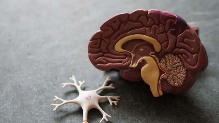
The Adrenergic System

The Adrenergic System: An Overview

The adrenergic system stands as a pivotal regulatory network within the human body, intricately orchestrating a vast array of physiological functions, most notably in neurotransmission and crucial metabolic processes. It serves as a rapid response mechanism, allowing the body to adapt to various internal and external stimuli, especially those related to stress or physical exertion. At its core, this system comprises specific chemical messengers, known as neurotransmitters, and specialized cellular components called receptors, which together facilitate communication across different tissues and organs.

This sophisticated system is primarily activated by two potent hormones and neurotransmitters: epinephrine (also known as adrenaline) and norepinephrine (noradrenaline). These vital compounds are predominantly secreted by the adrenal glands, small endocrine glands situated atop the kidneys, and also released by nerve endings throughout the body. Their release initiates a complex cascade of biochemical events that profoundly influence numerous bodily systems, from the cardiovascular and respiratory systems to metabolic pathways and endocrine regulation. The delicate balance maintained by the adrenergic system is fundamental for sustaining homeostasis and enabling the organism to respond dynamically to its environment.

The widespread impact of the adrenergic system underscores its importance in everyday functioning, from the subtlest adjustments in heart rate during a change in posture to the dramatic mobilization of resources during a perceived threat. Its influence extends to regulating blood flow distribution, modulating gastrointestinal activity, and even impacting cognitive functions like alertness and attention. Understanding the intricacies of this system is therefore crucial not only for grasping fundamental human physiology but also for comprehending the basis of many common medical conditions and the mechanisms of numerous therapeutic interventions.

Key Components: Neurotransmitters and Receptors

Central to the function of the adrenergic system are the catecholamines, epinephrine and norepinephrine, which act as its primary chemical messengers. These neurotransmitters are synthesized from the amino acid tyrosine and are stored in vesicles within nerve terminals and chromaffin cells of the adrenal medulla. Upon appropriate stimulation, such as stress or physical activity, these substances are released into the synaptic cleft or the bloodstream, traveling to target cells throughout the body. Epinephrine is widely recognized as the primary mediator of the systemic “fight-or-flight” response, while norepinephrine plays a more prominent role in localized neuronal signaling and maintaining basal vascular tone.

The cellular machinery that detects and responds to these catecholamines consists of a family of specialized proteins embedded in cell membranes, known as adrenergic receptors. These receptors are classified into distinct subtypes based on their structural characteristics, their affinity for specific neurotransmitters, and the downstream signaling pathways they activate. The classic classification includes alpha (α), beta (β), and, as described in some contexts, gamma (γ) receptors. Each subtype is strategically distributed across various tissues, ensuring that the diverse effects of epinephrine and norepinephrine are precisely targeted and regulated.

Specifically, the alpha (α) receptors are primarily activated by epinephrine and are found abundantly in the smooth muscles of blood vessels and the heart. Their activation typically leads to vasoconstriction, an increase in heart rate, and an elevation in blood pressure. Beta (β) receptors, on the other hand, respond to both epinephrine and norepinephrine and are predominantly located in the bronchi of the lungs, skeletal muscles, and the heart. Activation of beta receptors generally results in bronchodilation (relaxation of the airways), increased force of cardiac contraction, and enhanced metabolism. The gamma (γ) receptors, as described in some classifications, are found mainly in the brain and are activated by norepinephrine, playing a role in the release of certain hormones, such as cortisol and glucagon, influencing stress responses and metabolic regulation.

Historical Discovery and Elucidation

The origins of our understanding of the adrenergic system trace back to the early 20th century, with pioneering observations that laid the groundwork for modern neuroscience and pharmacology. In 1904, Thomas Renton Elliott, a British physician and physiologist, made a seminal observation that injecting adrenaline mimicked the effects of stimulating sympathetic nerves. This led him to propose that nerves release a chemical substance, rather than electrical impulses directly, to communicate with target organs, a revolutionary idea at the time. His work provided an early conceptualization of chemical neurotransmission, suggesting that adrenaline (epinephrine) might be the chemical mediator of the sympathetic nervous system.

Further pivotal insights emerged from the groundbreaking work of Otto Loewi in the 1920s, who definitively demonstrated chemical neurotransmission through his famous experiments with frog hearts. While his initial focus was on the vagus nerve and acetylcholine, his findings provided a robust experimental framework for studying chemical signaling in the nervous system. This paved the way for more detailed investigations into the substances released by sympathetic nerves. Over the subsequent decades, researchers meticulously worked to isolate and identify norepinephrine as the primary neurotransmitter released by most postganglionic sympathetic nerve terminals, solidifying the chemical basis of adrenergic signaling.

A monumental leap in understanding the adrenergic system came in 1948 with the work of Raymond P. Ahlquist, an American pharmacologist. He conducted systematic studies using various sympathomimetic drugs and observed two distinct patterns of response in different tissues. This led him to propose the existence of two fundamentally different types of adrenergic receptors, which he termed alpha (α) and beta (β). Ahlquist’s receptor classification was initially met with skepticism but was eventually confirmed through subsequent biochemical and pharmacological research, revolutionizing the field. His conceptualization of distinct receptor subtypes was critical for developing selective drugs that could target specific physiological responses, profoundly impacting the treatment of conditions ranging from heart disease to asthma. While the alpha and beta classifications became widely accepted, the concept of gamma receptors has also been explored in certain physiological contexts, contributing to the nuanced understanding of this complex system.

Physiological Mechanisms and “Fight-or-Flight”

The activation of the adrenergic system is most dramatically exemplified by the body’s rapid and coordinated “fight-or-flight” response, a fundamental survival mechanism that prepares an organism to confront or flee from a perceived threat. This intricate response involves the swift release of epinephrine and norepinephrine from the adrenal medulla and sympathetic nerve terminals, triggering a cascade of physiological changes designed to optimize immediate physical performance. The precise effects observed depend on the specific type and location of the adrenergic receptors activated, leading to a highly adaptive and integrated bodily reaction.

Upon activation, alpha receptors play a crucial role in redistributing blood flow and elevating cardiovascular parameters. When epinephrine binds to alpha receptors, particularly those in the smooth muscle of blood vessels supplying the skin, digestive tract, and kidneys, it causes these vessels to constrict. This vasoconstriction diverts blood away from non-essential organs and towards vital organs such such as the skeletal muscles, heart, and brain, ensuring they receive maximal oxygen and nutrient supply for immediate action. Concurrently, alpha receptor activation can contribute to an increase in heart rate and systemic blood pressure, further enhancing circulatory efficiency to meet the demands of a stressful situation.

Complementing the alpha receptor effects, beta receptors mediate a broader set of responses crucial for energy mobilization and enhancing respiratory and cardiac output. Activation of beta receptors in the heart significantly increases both the rate and force of myocardial contraction, augmenting the heart’s pumping capacity. In the lungs, beta receptor stimulation causes the relaxation of bronchial smooth muscles, leading to bronchodilation, which improves air intake and oxygen delivery to the bloodstream. Furthermore, beta receptors in the liver and skeletal muscles promote glycogenolysis and lipolysis, releasing glucose and fatty acids into the circulation to provide immediate energy for muscular activity, thereby boosting overall metabolism.

Beyond these well-established alpha and beta effects, the activation of gamma (γ) receptors, particularly those described as being in the brain, contributes another layer of regulatory complexity. When norepinephrine engages these receptors, it can influence the central nervous system’s response to stress, notably by modulating the release of key stress hormones. This includes triggering the secretion of cortisol from the adrenal cortex and glucagon from the pancreas. Cortisol helps manage stress by altering immune responses and energy usage, while glucagon further elevates blood glucose levels, ensuring sustained energy availability for prolonged periods of stress or exertion. Thus, the combined actions of all adrenergic receptor subtypes orchestrate a comprehensive and powerful adaptive response.

Adrenergic System in Everyday Life: A Practical Example

To fully grasp the profound impact of the adrenergic system, consider a common real-world scenario: imagine an individual, Sarah, walking alone at night when she suddenly hears a loud, unexpected noise behind her – perhaps a trash can falling or a car backfiring. This sudden sensory input triggers an immediate, unconscious alarm system within her brain, indicating a potential threat. This initial perception of danger is the crucial starting point for the cascade of physiological events mediated by her adrenergic system, preparing her for immediate protective action.

The “how-to” of the adrenergic system’s application unfolds rapidly in this scenario. First, Sarah’s brain instantly recognizes the noise as a potential threat, activating her sympathetic nervous system, the branch of the autonomic nervous system responsible for “fight-or-flight” responses. Within milliseconds, this activation signals her adrenal glands to release a surge of epinephrine and norepinephrine into her bloodstream. These potent hormones then travel throughout her body, binding to adrenergic receptors on various target cells.

The binding of epinephrine and norepinephrine to these receptors triggers a series of profound physiological changes. Alpha receptor activation causes the smooth muscles around her blood vessels to constrict, particularly those supplying her skin and digestive organs. This shunts blood away from these less critical areas and redirects it towards her skeletal muscles, heart, and brain, preparing them for rapid action. Simultaneously, beta receptor activation in her heart causes her heart rate to soar and her heart to pump more forcefully, increasing cardiac output and ensuring that her muscles and brain receive an ample supply of oxygen and nutrients. In her lungs, beta receptors cause her bronchi to dilate, allowing her to take in more air with each breath, further enhancing oxygenation. Additionally, beta receptor activation in her liver triggers the release of stored glucose, providing an immediate energy boost. If gamma receptors in her brain are also significantly activated, they might enhance the release of stress hormones like cortisol, further sharpening her focus and preparing her body for sustained vigilance or exertion. The cumulative effect of these synchronized responses is a sudden surge of strength, heightened awareness, and increased physical readiness, all designed to enable Sarah to either flee the perceived danger or confront it, even if the actual threat turns out to be benign.

Clinical Significance and Therapeutic Applications

The profound and pervasive influence of the adrenergic system makes it a cornerstone of understanding human physiology and a critical target in modern pharmacology and medicine. Its fundamental role in cardiovascular regulation, stress response, and metabolic control means that dysregulation can contribute to a wide array of pathological conditions. Consequently, drugs that modulate the adrenergic system represent some of the most widely prescribed and effective medications across various medical disciplines, highlighting its immense clinical significance.

The therapeutic applications stemming from our knowledge of adrenergic receptors are extensive and diverse. For instance, drugs known as beta-blockers, which inhibit the action of epinephrine and norepinephrine at beta receptors, are commonly prescribed for conditions such as hypertension (high blood pressure), angina (chest pain), cardiac arrhythmias, and even anxiety disorders. By reducing heart rate and force of contraction, beta-blockers alleviate strain on the heart. Conversely, beta-agonists, which stimulate beta receptors, are vital in treating respiratory conditions like asthma and chronic obstructive pulmonary disease (COPD) by inducing bronchodilation and opening up airways. In acute emergencies, a direct injection of epinephrine can be life-saving for severe allergic reactions (anaphylaxis) or cardiac arrest, rapidly restoring cardiovascular function.

Beyond the cardiovascular and respiratory systems, adrenergic modulating drugs are used in other areas. Alpha-agonists, for example, can be used as nasal decongestants by causing vasoconstriction in the nasal passages, reducing swelling. Alpha-blockers are used to treat hypertension by relaxing blood vessels and in benign prostatic hyperplasia to relax smooth muscles in the prostate and bladder neck. The continuous research into the precise mechanisms of adrenergic receptors and their subtypes allows for the development of increasingly selective drugs, minimizing side effects and maximizing therapeutic efficacy, thus improving the quality of life for countless patients globally and underscoring the system’s ongoing relevance in medical innovation.

Interconnections with Other Biological Systems

The adrenergic system does not operate in isolation; rather, it is deeply integrated and intricately interconnected with virtually every other biological system in the body, reflecting its overarching role in maintaining homeostasis and coordinating systemic responses. Its most direct and fundamental relationship is with the broader autonomic nervous system, of which it constitutes the primary effector pathway of the sympathetic nervous system. While the sympathetic branch mobilizes the body for action, its counterpart, the parasympathetic nervous system, promotes rest and digestion, creating a dynamic balance that regulates involuntary bodily functions.

Beyond the nervous system, the adrenergic system maintains crucial ties with the endocrine system. The adrenal medulla, a key component of the adrenergic system, is itself an endocrine gland that releases epinephrine and norepinephrine directly into the bloodstream, allowing for widespread hormonal effects. Furthermore, as noted with gamma receptor activation, the adrenergic system can influence the release of other hormones, such as cortisol and glucagon, which are central to stress response and metabolic regulation, respectively. This close interplay between neural and hormonal signaling ensures a robust and multifaceted response to physiological demands.

The broader category of disciplines encompassing the study of the adrenergic system includes neuroscience, physiology, and pharmacology, given its fundamental role in nerve signaling, body function, and drug action. However, its influence extends into many other fields. In immunology, adrenergic signals can modulate immune cell function, influencing inflammatory responses. In psychology, understanding adrenergic activation is critical for studying stress, anxiety, and emotional regulation. Even in areas like gastroenterology, the adrenergic system impacts gut motility and secretion. This widespread influence underscores its position as a central integrator of bodily functions, crucial for survival and adaptation across all levels of biological organization.

Pathophysiological Implications

While the adrenergic system is essential for maintaining health and coordinating adaptive responses, its dysregulation can lead to a wide spectrum of pathophysiological conditions, illustrating its delicate balance. An overactive or underactive adrenergic response, or issues with receptor sensitivity, can contribute significantly to the development and progression of various diseases, highlighting the system’s role not just in normal function but also in pathology.

One of the most common and significant pathophysiological implications of adrenergic system dysfunction involves the cardiovascular system. Overactivity of alpha receptors, for instance, can lead to persistent vasoconstriction and an increase in peripheral vascular resistance, culminating in chronic hypertension (high blood pressure). Similarly, an excessive or inappropriate activation of beta receptors in the heart can result in sustained increases in heart rate, manifesting as tachycardia, and contribute to various cardiac arrhythmias, placing undue stress on the heart and increasing the risk of cardiovascular events.

Beyond cardiovascular issues, imbalances in adrenergic signaling contribute to other systemic disorders. For example, inappropriate activation of beta receptors in the respiratory tract can lead to bronchoconstriction, narrowing the airways and exacerbating conditions like asthma. Metabolically, an overactive adrenergic system can lead to increased glucose production and reduced insulin sensitivity, contributing to hyperglycemia and potentially worsening glycemic control in individuals with diabetes. Furthermore, the persistent over-release of cortisol and glucagon, potentially influenced by gamma receptor activity as described, can lead to chronic stress responses that have detrimental effects on immune function, bone density, and overall metabolic health.

Understanding these pathophysiological mechanisms is crucial for developing targeted therapeutic strategies. Many medications aim to restore balance to the adrenergic system by either blocking overactive receptors or stimulating underactive ones. However, the complexity of the system, with its multiple receptor subtypes and overlapping functions, means that interventions must be carefully considered to achieve desired therapeutic effects while minimizing undesirable side effects, underscoring the continuous challenge and importance of research in this field.

Conclusion

In summation, the adrenergic system represents a profoundly intricate and indispensable network within the human body, serving as a master regulator of numerous physiological and metabolic processes. Its orchestrated activity, driven primarily by the release of epinephrine and norepinephrine, and mediated through a diverse family of adrenergic receptors (alpha, beta, and gamma subtypes), ensures the body’s capacity for rapid adaptation to internal and external stressors. From the immediate mobilization of resources during a “fight-or-flight” scenario to the subtle, continuous adjustments that maintain daily homeostasis, the system’s influence is both pervasive and critical.

The historical elucidation of the adrenergic system, from early observations of chemical neurotransmission to the precise classification of its receptors, underscores decades of scientific endeavor that have transformed our understanding of physiology. This knowledge has not only illuminated fundamental biological mechanisms but has also paved the way for groundbreaking therapeutic interventions. The system’s involvement in regulating vital functions such as heart rate, blood pressure, respiration, and energy metabolism highlights its central role in both health and disease, making it a key focus across fields ranging from neuroscience and pharmacology to clinical medicine.

Ultimately, the adrenergic system’s complex interconnections with other biological networks, including the autonomic nervous system and the endocrine system, emphasize its role as a crucial integrator of bodily functions. Its capacity to influence everything from cardiovascular health to stress responses and metabolic balance ensures its continuous relevance in ongoing research. Continued exploration of its intricacies promises further advancements in our ability to diagnose, treat, and prevent a wide array of human conditions, solidifying its status as one of the most vital regulatory systems in human biology.

Scroll to Top