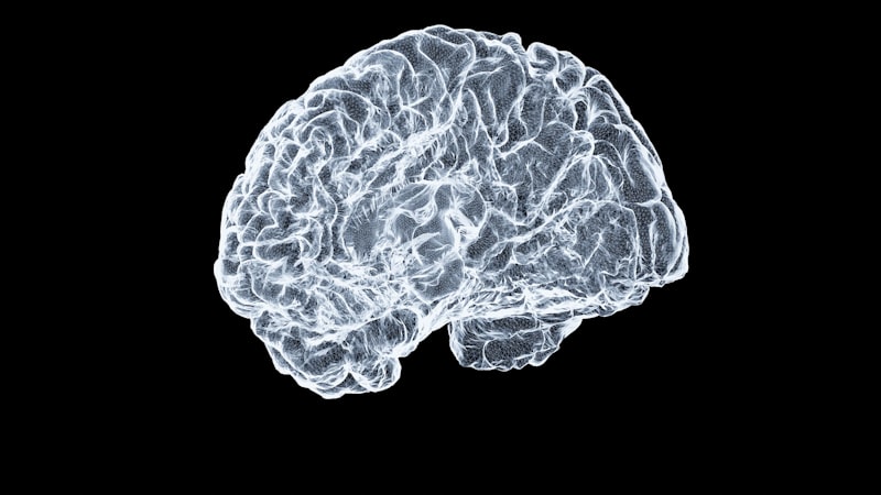
Brain Topography: Mapping Your Mind’s Hidden Architecture
- TOPOGRAPHIC MAPPING OF THE BRAIN
- The Historical Foundations of Cortical Organization
- Principles of Somatotopic Organization: The Homunculus
- Retinotopic and Tonotopic Organization
- Neurobiological Mechanisms of Map Formation
- Modern Techniques for Mapping Brain Topography
- Cortical Plasticity and Map Reorganization
- Clinical Relevance and Diagnostic Applications
- Challenges and Future Directions in Brain Mapping
TOPOGRAPHIC MAPPING OF THE BRAIN
The concept of topographic mapping of the brain refers fundamentally to the systematic organization, depiction, and classification of various functional areas within the central nervous system with explicit regard to their precise physical and spatial relations to one another. This principle dictates that neighboring points in the sensory environment or in the motor periphery are represented by neighboring neurons within the cortical or subcortical structures responsible for processing that information. Such organization is not merely a random arrangement but an ordered projection that allows for efficient processing and integration of complex stimuli, serving as a critical foundation for understanding neuroanatomy, function, and development. The study of these maps is central to contemporary neuroscience, as the fidelity and integrity of these spatial representations directly correlate with behavioral and cognitive capabilities. The topographic arrangement ensures that the brain maintains a structured, though often distorted, internal representation of the external world and the body itself, facilitating rapid comparison and integration necessary for adaptive behavior.
The fidelity and persistence of these maps underscore their profound importance in the study of neurological function and dysfunction. As early researchers noted, the robust study of the brain’s topographic organization serves a great purpose in understanding how sensory input is translated into perception and how motor commands are executed with precision. This mapping provides an essential framework for localization, allowing neuroscientists and clinicians to correlate specific behaviors or deficits with discrete anatomical regions. Furthermore, the inherent organizational structure provides crucial clues regarding the developmental mechanisms that wire the brain during embryogenesis and early life. It establishes the baseline against which the remarkable phenomenon of cortical plasticity—the capacity of the brain to reorganize its maps following injury or extensive training—is measured, providing insight into recovery potential after trauma.
Understanding the mechanisms underlying topographic mapping requires an appreciation of the complex interplay between genetic programming and activity-dependent refinement. While initial projections are often guided by molecular cues, the final fine-tuning of these maps relies heavily on neural activity patterns, ensuring that the internal representation is optimally tuned to the individual’s environment and experience. This structured approach to neural organization contrasts sharply with more distributed processing models, though both organization principles often coexist within large-scale neural networks. The study of topography thus bridges the gap between micro-level cellular interactions and macro-level cognitive functions, serving as a cornerstone for modern psychological and neurological investigations.
The Historical Foundations of Cortical Organization
The recognition that the brain possessed functionally segregated areas arranged in a systematic manner evolved significantly during the nineteenth and twentieth centuries. Early efforts focused primarily on functional localization, often driven by clinical observations of patients suffering from specific brain injuries. Key figures such as Paul Broca and Carl Wernicke established that language functions were localized to specific cortical regions, setting the precedent that the brain was not a homogenous mass but an organ segmented by function. However, the precise spatial ordering—the topographic element—was solidified through meticulous experimental work, particularly involving electrophysiological stimulation. This pioneering work moved beyond simple localization to demonstrate that functions were not only localized but arranged in an orderly, continuous fashion across the cortical surface.
One of the most profound early contributions to mapping came from the work of Korbinian Brodmann, who, in the early 1900s, generated his famous cytoarchitectural maps. Brodmann systematically analyzed the cellular composition (cytoarchitecture) of the cortex, dividing it into distinct areas (Brodmann Areas or BAs) based on variations in the layering, density, and morphology of neurons. While Brodmann’s initial classification was purely anatomical, subsequent physiological studies revealed a striking correspondence between these anatomical boundaries and distinct functional territories, reinforcing the concept that structure dictates function in a highly organized manner. These areas, such as BA 17 (the primary visual cortex), demonstrated an intrinsic structural order that mirrored their functional specialization, laying the groundwork for later, more detailed topographic studies.
Perhaps the most iconic historical demonstration of topographic mapping came from the neurosurgeon Wilder Penfield and his colleagues in the mid-twentieth century. Using gentle electrical stimulation on the exposed cortex of conscious patients undergoing epilepsy surgery, Penfield mapped the primary motor and somatosensory cortices. His findings demonstrated a consistent, continuous representation of the body surface across these strips of cortex. This empirical evidence confirmed the systematic, ordered nature of neural representation, moving the field past purely theoretical models. Penfield’s work resulted in the creation of the famous cortical homunculus, a visually striking, though highly distorted, representation of the human body mapped onto the brain, providing irrefutable proof of somatotopic organization.
Principles of Somatotopic Organization: The Homunculus
Somatotopic organization is arguably the most recognizable and studied form of brain topography. It refers specifically to the systematic mapping of the body surface onto the primary somatosensory cortex (S1) and the primary motor cortex (M1). In both regions, adjacent areas of the body—such as the thumb and index finger, or the elbow and shoulder—are represented by adjacent populations of neurons on the cortical surface. This mapping is highly continuous, yet it is neither proportional nor faithful to the actual physical size of the body parts. Instead, the size of the cortical area dedicated to a specific body part is directly correlated with the density of sensory receptors or the precision of motor control required for that area.
The resulting representation, known as the somatosensory homunculus, appears grotesquely distorted. Areas critical for fine discrimination and interaction with the environment, such as the hands, lips, and tongue, occupy disproportionately large areas of the cortex, reflecting the high resolution of sensory input received from these regions. Conversely, large body segments like the trunk or the back, which require less precise sensory feedback, are represented in relatively small areas. This differential scaling highlights a fundamental principle of topographic mapping: neural resources are allocated based on functional demands and information density, not anatomical size. The motor homunculus follows a similar, though distinct, pattern, emphasizing areas requiring complex and fine-tuned muscle control, such as the fingers and facial musculature.
The organization within the somatotopic maps is further refined by the concept of columnar organization. Within the larger topographic map, neurons are arranged in vertical columns that traverse the cortical layers, often exhibiting similar functional properties. For instance, in the somatosensory cortex, adjacent columns might respond preferentially to different types of stimuli—such as light touch, deep pressure, or joint position—or might receive input from rapidly adapting versus slowly adapting receptors. This intricate, multi-layered organization ensures that the processing of body-derived information is both spatially ordered across the surface and functionally segregated in depth, optimizing the brain’s ability to interpret and respond to complex somatosensory inputs.
Retinotopic and Tonotopic Organization
Topographic mapping is not limited to somatosensation; it is a ubiquitous principle governing most major sensory modalities, including vision (retinotopy) and audition (tonotopy). Retinotopic mapping involves the systematic projection of the visual field onto the primary visual cortex (V1), situated in the occipital lobe. The organization is such that adjacent points in the retina—and therefore adjacent points in the visual field—are mapped onto adjacent neurons in V1. This organized projection is maintained through the lateral geniculate nucleus (LGN) of the thalamus. However, similar to the somatosensory map, the retinotopic map is not a literal, scaled representation.
A key characteristic of retinotopy is the phenomenon of cortical magnification. The central part of the retina, the fovea, which is responsible for high-acuity central vision, receives a vastly disproportionate amount of cortical territory compared to the peripheral retina. This magnification mirrors the functional importance of high-resolution central vision, allocating extensive neural resources to the most information-rich part of the visual input. Furthermore, the visual map is structured according to a complex arrangement that respects the boundaries and characteristics of the visual field, often involving an inversion and reversal of the visual input as it traverses the optic pathways to the cortex, resulting in a complex yet systematic organization that is crucial for subsequent visual processing.
Tonotopic mapping describes the systematic organization of auditory frequencies along the primary auditory cortex (A1) and related subcortical structures, such as the cochlear nucleus and the inferior colliculus. In tonotopic maps, adjacent areas of the cortex respond preferentially to adjacent sound frequencies. Typically, lower frequencies are mapped to one end of the cortical area, and gradually increasing frequencies are mapped progressively toward the opposite end, creating a continuous frequency gradient. This organization is established early in the auditory pathway, originating in the mechanical structure of the cochlea, which physically separates frequencies before they are relayed to the central nervous system. This frequency-based topographic organization is essential for tasks requiring pitch discrimination and complex sound processing, such as speech and music perception.
Neurobiological Mechanisms of Map Formation
The formation of highly ordered topographic maps during neural development is one of the most remarkable achievements of the nervous system, relying on an intricate balance between precise molecular guidance and subsequent activity-dependent refinement. Initially, developing axons navigate vast distances to find their target structures through guidance cues, which are often chemical gradients released by target tissues. These cues, such as members of the Ephrin and Eph receptor families, establish a rough, initial gradient of connectivity. For example, in the retinotectal system (mapping the retina onto the superior colliculus/tectum), a high concentration of Ephrin A molecules in the posterior tectum attracts axons expressing lower levels of Eph receptors, thus enforcing a molecularly guided topographic order.
While molecular cues establish the general layout of the map, they are generally not precise enough to account for the fine-grained, high-resolution organization observed in mature maps. The subsequent refinement of connectivity is heavily dependent on electrical activity, often termed activity-dependent sharpening. During critical periods of development, spontaneous or evoked activity patterns—such as retinal waves in the visual system or correlated activity in the periphery—are crucial for strengthening appropriate synaptic connections and eliminating erroneous ones. Neurons that fire together tend to wire together, and this synchronization mechanism ensures that spatially adjacent sensory inputs activate adjacent target neurons, thereby perfecting the topographic continuity.
The stability and maintenance of these maps in the mature nervous system are maintained through ongoing plasticity mechanisms, though the degree of plasticity varies across brain regions and across the lifespan. Maintenance involves continuous molecular signaling and activity monitoring to ensure that the spatial representations remain accurate and functional. Disruptions to this balance, whether through genetic abnormalities or environmental factors (such as sensory deprivation), can lead to disorganized or poorly formed maps, resulting in significant functional deficits. This robust mechanism of formation, therefore, requires both an initial blueprint provided by molecular biology and an adaptive refinement mechanism driven by neuronal activity.
Modern Techniques for Mapping Brain Topography
The visualization and analysis of brain topography have advanced exponentially with the development of sophisticated neuroimaging and electrophysiological techniques. Earlier methods relied heavily on invasive approaches, such as microelectrode recording and direct cortical stimulation. While these methods provided high-resolution data, their applicability was limited. Modern techniques allow for non-invasive or minimally invasive mapping, enabling researchers to study topographic organization in both healthy and clinical populations with unprecedented detail.
One of the primary tools utilized today is functional Magnetic Resonance Imaging (fMRI). Specifically, techniques like phase-encoded retinotopic mapping use fMRI to measure changes in blood oxygenation (BOLD signal) in response to systematically moving visual stimuli (e.g., rotating wedges or expanding rings). By analyzing the spatial location and timing of the BOLD response across the visual cortex, researchers can precisely delineate the boundaries and fine organization of visual maps (V1, V2, V3, etc.). Similar approaches are adapted for somatosensory and motor mapping, using controlled movements or tactile stimulation to identify corresponding cortical areas, providing a robust, macro-level view of topographic organization in vivo.
Other crucial non-invasive methods include Electroencephalography (EEG) and Magnetoencephalography (MEG), which offer superior temporal resolution, capturing the dynamic nature of topographic processing. By measuring electrical or magnetic fields generated by neuronal activity, researchers can track the flow of sensory information across cortical maps in milliseconds. While their spatial resolution is generally lower than fMRI, advanced source localization techniques allow for the mapping of sensory evoked potentials onto anatomical structures, revealing the precise timing with which different parts of a topographic map are activated during perception.
At the micro-level, especially in animal models, techniques such as optical intrinsic signal imaging and multi-electrode array recordings provide high-resolution mapping data. Optical imaging measures subtle changes in light reflectance related to neural activity and blood flow, allowing for the visualization of functional maps across a large expanse of cortex with very high spatial detail. These combined methodological approaches—ranging from the high spatial resolution of fMRI to the high temporal resolution of MEG and the cellular resolution of optical imaging—provide a comprehensive toolkit for characterizing the intricate topography of the brain.
Cortical Plasticity and Map Reorganization
Contrary to the historical view of a fixed, hardwired brain, modern neuroscience emphasizes that topographic maps are highly dynamic and subject to continuous modification—a characteristic known as cortical plasticity. This reorganization capacity allows the brain to adapt to changes in sensory input, motor demands, learning, and injury. Plasticity can manifest in various ways, often resulting in the expansion or shrinkage of cortical representation fields.
One of the most dramatic forms of topographic reorganization occurs following peripheral injury, such as amputation. When a limb is lost, the cortical area previously dedicated to processing input from that limb does not remain silent. Instead, the adjacent cortical representations—often those corresponding to the face or the remaining adjacent digits—gradually invade and colonize the deprived cortical territory. This phenomenon of functional takeover demonstrates the competitive nature of map maintenance and the brain’s intrinsic capacity to reutilize available neural real estate, often contributing to phenomena like phantom limb sensation, where the stimulation of the invading area is mistakenly perceived as originating from the missing limb.
Plasticity is also intensely engaged during skill acquisition and learning. Extensive practice of a fine motor skill, such as playing a musical instrument or reading Braille, leads to a measurable expansion of the cortical territory dedicated to the body parts involved in that skill (e.g., the hand and finger representation in M1). This experience-dependent expansion reflects the strengthening of synaptic connections and the recruitment of adjacent neural populations to enhance the resolution and precision of the map for the newly mastered skill. This adaptive reorganization is critical for rehabilitation after stroke or injury, where targeted training aims to leverage plasticity to re-establish functional motor and sensory maps in damaged or surrounding healthy tissue.
Clinical Relevance and Diagnostic Applications
The understanding of topographic mapping is profoundly relevant in clinical neuroscience, forming the basis for numerous diagnostic, surgical, and therapeutic procedures. Precisely knowing the spatial arrangement of functional areas allows clinicians to predict the functional consequences of focal brain lesions, whether caused by stroke, tumor, or trauma. For example, damage localized to a specific area of the somatosensory homunculus allows for highly accurate predictions regarding which part of the body will experience sensory loss. This predictive capability is essential for accurate diagnosis and prognosis.
In neurosurgery, pre-operative mapping is a critical procedure, especially when tumors or epileptic foci are located near eloquent cortex—areas responsible for crucial functions like movement, sensation, or language. Techniques like fMRI mapping or direct cortical stimulation during surgery are used to precisely locate functional boundaries, such as the central sulcus separating M1 and S1. This meticulous mapping allows surgeons to plan the resection path to maximize tumor removal while minimizing damage to essential topographic areas, thereby preserving the patient’s motor, sensory, and cognitive functions.
Furthermore, the study of map integrity is central to understanding neurodevelopmental disorders and chronic pain conditions. Disrupted or abnormal topographic organization has been implicated in conditions such as developmental dyslexia, where auditory processing maps may be poorly formed, and in chronic pain states, where the central representation of the affected body part may be excessively enlarged or blurred. Therapeutic interventions, including focused rehabilitation and neuromodulation techniques, are increasingly being designed to normalize these pathological map changes, utilizing the principles of plasticity to restore organized functional representation.
Challenges and Future Directions in Brain Mapping
Despite significant progress, the full characterization of brain topography presents several formidable challenges. One major difficulty lies in accurately mapping higher-order cognitive functions. While primary sensory and motor areas exhibit clear, predictable topography (somatotopy, retinotopy), the organizational principles governing complex association cortices—areas involved in memory, decision-making, and executive function—are often less strictly spatial and more distributed, making traditional topographic analysis challenging. Future work must integrate spatial mapping with network analysis (connectomics) to understand how spatially ordered maps interact within larger, distributed functional networks.
Another significant challenge is achieving sufficient resolution to fully capture the complexity of the maps. Even high-resolution fMRI operates at a millimeter scale, while the fundamental processing units of topography—the cortical columns and microcircuits—are organized at the micron scale. Developing non-invasive techniques capable of resolving fine-scale topography in humans remains a key priority. Advanced magnetic resonance technologies and high-density EEG/MEG arrays are continually pushing these resolution limits, aiming to bridge the gap between macroscopic imaging and microscopic neural architecture.
Finally, future research will focus heavily on computational modeling to synthesize the vast amounts of topographical data being generated. Developing sophisticated computational models that can simulate map formation, maintenance, and reorganization will allow neuroscientists to test hypotheses about the underlying molecular and activity-dependent mechanisms that govern these fundamental organizational principles. By integrating high-resolution experimental data with robust theoretical frameworks, the field aims to move toward a complete, dynamic understanding of how the brain constructs and utilizes its internal spatial maps to mediate perception and action.