Neural Tracts: Mapping the Hidden Pathways of the Mind
The Core Definition of a Tract
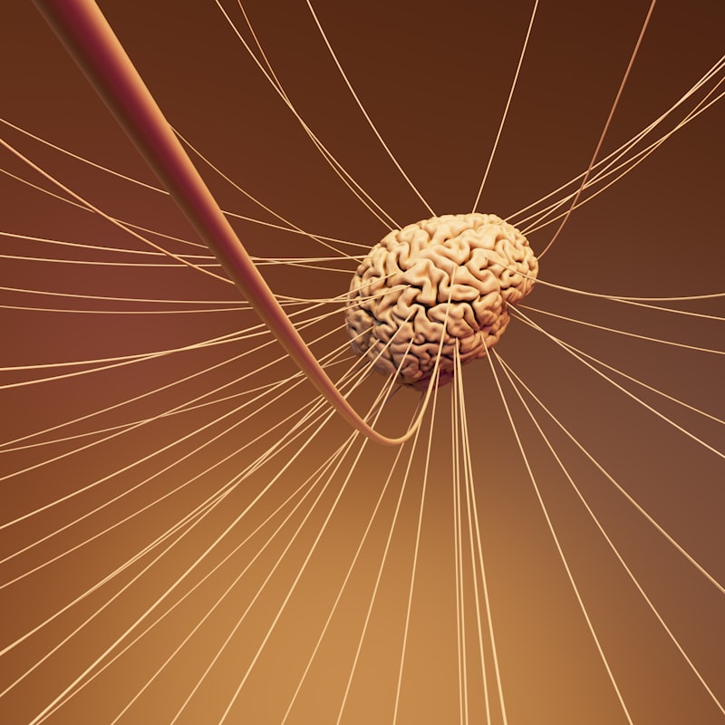
A tract, in the context of the neurosciences which heavily informs modern psychology, is defined as a bundle or set of nerve fibers, specifically axons, that are situated within the Central Nervous System (CNS). This definition distinguishes tracts from nerves, which carry similar information but are located outside the brain and spinal cord in the Peripheral Nervous System. These structured bundles of fibers act as the essential communication highways of the nervous system, transmitting action potentials rapidly and reliably between different nuclei, ganglia, and cortical regions. The consistent organization and myelination of these tracts ensure synchronous signaling, allowing for the incredibly complex and rapid processing necessary for all cognitive, emotional, and behavioral outputs. Furthermore, the terminology used to name a specific tract often implies its starting point followed by its ending point, such as the spinothalamic tract, which projects from the spinal cord to the thalamus, thereby immediately indicating its functional role in relaying sensory information.
The fundamental mechanism underlying the function of a neural tract is the principle of segregated information flow. Each tract is specialized to carry a specific type of information, whether motor commands, sensory input, or integrative signals, ensuring that data relevant to a particular psychological process arrives at the correct destination without interference. This anatomical specificity is crucial for understanding localization of function within the brain, a cornerstone of modern cognitive psychology. Without these highly organized pathways, the brain would be incapable of the coordinated activity required for tasks ranging from simple reflexes to abstract reasoning and language production. The sheer volume and density of fibers within major tracts, coupled with the insulating properties of the surrounding glia, allow for high-fidelity communication over significant anatomical distances, linking the forebrain, cerebellum, brainstem, and spinal cord into a cohesive functional unit.
Anatomical Classification and Function
Neural tracts are typically categorized into three main functional groups based on the regions they connect: association fibers, commissural fibers, and projection fibers. Association fibers connect different cortical areas within the same hemisphere, facilitating complex intra-hemispheric processing, such as linking Wernicke’s area (language comprehension) to Broca’s area (speech production). Commissural fibers, the most famous example being the corpus callosum, connect the corresponding areas of the two cerebral hemispheres, enabling bilateral coordination and information sharing essential for integrated perception and consciousness. The third category, Projection Fibers, are perhaps the most vital for connecting the cerebral cortex to subcortical structures, the brainstem, and the spinal cord, controlling both ascending (sensory) and descending (motor) information streams.
The role of projection fibers is paramount in translating psychological intent into physical action and conversely, bringing the external world into conscious experience. These descending pathways include the motor tracts that govern voluntary movement, while the ascending tracts relay somatosensory information concerning touch, temperature, pain, and proprioception. Understanding the integrity and efficiency of these projection tracts is crucial in clinical psychology and neurology, as damage to these bundles can lead to predictable deficits in sensation, motor control, and higher-order cognitive functions. For instance, the degradation of specific projection fibers is implicated in various neurodegenerative disorders, directly impacting a person’s ability to interact with their environment and maintain cognitive autonomy.
Historical Discovery of Neural Pathways
The systematic study of neural tracts gained significant momentum during the 19th century, transitioning from purely descriptive anatomy to functional neuroanatomy. Early pioneers relied heavily on post-mortem analysis of individuals who had suffered specific neurological injuries or diseases. By observing the patterns of degeneration—a process known as Wallerian degeneration—researchers could infer the trajectory and termination points of damaged fibers, thereby mapping out major pathways. Key figures like Paul Flechsig utilized myelin staining techniques to trace the maturation sequence of tracts, noting that psychological functions appeared only after their corresponding neural tracts had become fully myelinated, suggesting a developmental timeline for cognitive capabilities.
The work of Santiago Ramón y Cajal, who championed the Neuron Doctrine in the late 19th and early 20th centuries, provided the cellular foundation for understanding tracts. While Cajal focused on the individual neuron, his anatomical descriptions solidified the understanding that tracts were composed of the organized axons of these discrete cells, rather than a continuous network. Later in the 20th century, the development of sophisticated tracing methods, first using chemical markers and later advancing to non-invasive imaging techniques like Diffusion Tensor Imaging (DTI), revolutionized the ability to map these tracts in living human subjects. DTI allows clinicians and researchers to visualize the orientation and integrity of white matter tracts by measuring the diffusion of water molecules along the fiber bundles, providing unprecedented insight into the structural connectivity underlying psychological health and disorder.
Tracts in Action: A Practical Example (The Motor Command)
To illustrate the function of a neural tract in a practical, psychological context, consider the conscious decision to perform a complex voluntary movement, such as picking up a glass of water. This action requires the integration of cognitive intent with precise motor execution, primarily mediated by the Corticospinal Tract, often referred to as the pyramidal tract. The process begins with the cognitive intention formulated in the prefrontal cortex, which signals the primary motor cortex to initiate the movement plan. Neurons within the motor cortex then fire, sending their long axons downwards, forming the beginning of the corticospinal tract.
This mighty bundle of projection fibers descends through the brainstem, specifically crossing over (decussating) in the pyramids of the medulla oblongata before continuing down the spinal cord. This decussation explains why the right motor cortex controls movement on the left side of the body, and vice versa—a critical principle in clinical psychology when localizing brain injury. As the tract descends, specific fibers peel off at the appropriate spinal cord level, synapsing onto interneurons and ultimately onto the motor neurons that innervate the specific muscles required to contract the hand and arm, resulting in the successful grasp of the glass. The efficiency and structural integrity of this tract are directly responsible for the speed, smoothness, and precision of the psychological command being translated into observable behavior. If this tract is damaged, such as by a stroke in the motor cortex or a spinal cord injury, the command signal cannot reach the muscles, resulting in paralysis or paresis, fundamentally altering the individual’s psychological interaction with the physical world.
Significance in Neuropsychology and Behavior
The study of neural tracts holds profound significance for the field of Neuropsychology, as they provide the structural framework upon which all psychological phenomena are built. By understanding the specific pathways that connect sensory input to emotional centers, or cognitive planning regions to motor execution areas, researchers can accurately model the flow of information during complex tasks like decision-making, memory retrieval, and emotional regulation. The efficiency of these pathways is often correlated with measures of intelligence and cognitive reserve; thicker, more highly myelinated tracts typically support faster, more robust information processing.
Furthermore, the concept of neural tracts is critical for establishing the biological underpinnings of many psychological disorders. Conditions such as schizophrenia, autism spectrum disorder, and major depressive disorder are increasingly understood as disorders of connectivity, where the structural or functional integrity of specific white matter tracts is compromised. For example, disruptions in tracts connecting the frontal lobe (executive function) to the limbic system (emotion) are hypothesized to contribute to difficulties in emotional control and impulse regulation observed in various psychiatric conditions. Thus, mapping and assessing the health of these tracts provides objective biomarkers for diagnosis and helps guide therapeutic interventions aimed at improving functional connectivity.
Clinical Relevance and Pathologies
The clinical application of tract knowledge is pervasive across neurology and clinical psychology, particularly in diagnosing conditions where movement, sensation, or higher cognition are impaired. A classic example is Multiple Sclerosis (MS), an autoimmune disorder characterized by the destruction of the myelin sheath surrounding axons in the CNS. Since tracts rely on the myelin insulation to maintain rapid signal transmission, demyelination severely slows or blocks nerve impulses, leading to the wide range of neurological and psychological symptoms associated with MS, including fatigue, cognitive fog, and motor incoordination.
In the context of localized brain injury, such as a severe traumatic brain injury (TBI) or stroke, the specific psychological deficits observed are often predictable based on which major tracts have been severed or compressed. Damage to the arcuate fasciculus, an association tract connecting language areas, results in specific types of aphasia, hindering the ability to repeat spoken words despite intact comprehension. Therefore, the ability of clinicians to accurately diagnose and prognose patient outcomes is deeply reliant on a detailed understanding of the anatomical organization and functional projections of the various neural tracts within the Central Nervous System.
Interconnections with Cognitive Theories
The anatomical reality of neural tracts strongly reinforces psychological theories based on connectionism and network modeling. Connectionist theories posit that cognitive processes emerge from the interactions between interconnected neural units, and the tracts provide the physical wiring diagram for these interactions. The brain is not a monolithic processing unit; rather, it is a collection of specialized modules linked by specific pathways. Modern computational models of cognition, such as those used in artificial intelligence and computational neuroscience, often attempt to mirror the hierarchical and distributed nature of the brain’s white matter tracts to achieve realistic simulations of human learning and memory.
The broader category of psychology to which the study of neural tracts belongs is primarily **Biological Psychology** (or Biopsychology) and **Cognitive Neuroscience**. These fields seek to explain psychological processes—such as perception, memory, and consciousness—by examining their underlying physiological mechanisms. Related concepts that intersect with the study of tracts include the concept of **Plasticity**, which describes the brain’s ability to reorganize its tracts and functional connections following injury or learning; **White Matter Integrity**, a measure of the health and organization of these fiber bundles; and the principles of **Neural Coding**, which describe how information is actually encoded and transmitted along these anatomical pathways. The efficiency and structure of these tracts thus serve as a fundamental constraint and enabler for all higher-level psychological phenomena.