Developmental Biology: The Blueprint of Human Identity
- Introduction: The Mesonephric (Wolffian) Duct
- Embryological Origin and Early Development
- Detailed Anatomy and Histology
- Differential Fate: Sexual Dimorphism
- Hormonal Regulation of Wolffian Duct Fate
- Derivatives in the Adult Male Reproductive Tract
- Clinical Significance and Associated Anomalies
- Conclusion
- References
Introduction: The Mesonephric (Wolffian) Duct
The Wolffian duct, formally known as the mesonephric duct, represents a crucial paired structure in the early embryogenesis of all mammals, playing a decisive role in the establishment of the urogenital system. This duct system arises in close association with the mesonephros, or “middle kidney,” and initially serves as the primary conduit for excretory products during the transient mesonephric stage of renal development. Its presence is universal in early embryos, regardless of genetic sex, highlighting its fundamental position as a bipotential precursor structure. The ultimate fate of the Wolffian duct is highly dependent on the hormonal milieu established during the critical period of sexual differentiation, serving as the foundation for the complex male reproductive plumbing or undergoing nearly complete regression in the female phenotype.
Understanding the Wolffian duct is central to comprehending human reproductive anatomy and pathology. It stands in dynamic balance with the Müllerian duct (or paramesonephric duct), which serves as the precursor for the female reproductive tract. The simultaneous presence of both duct systems defines the sexually indifferent stage of development. The subsequent preservation or involution of the Wolffian duct dictates whether the embryo develops the essential internal genitalia characteristic of the male sex, including structures vital for sperm transport and fluid contribution to semen. This developmental process underscores the principle that reproductive structure is not merely built but actively maintained and sculpted by potent endocrine signals.
Beyond its direct contribution to the reproductive tract, the influence of the Wolffian duct extends indirectly to the formation of other adjacent organs, including aspects of the urinary bladder and kidneys. The precise molecular signaling pathways controlling its integration into the developing urogenital sinus and metanephric blastema are subjects of intense biological study. This comprehensive overview will detail the structural evolution, functional outcomes, hormonal control, and significant clinical manifestations associated with the proper or anomalous development of this essential embryological structure, positioning it within the broader context of developmental biology and clinical psychology related to sexual identity formation.
Embryological Origin and Early Development
The genesis of the Wolffian duct begins relatively early in human gestation, typically around the fourth week of development. It originates as a longitudinal condensation of intermediate mesoderm, running parallel to the developing pronephros and subsequently the mesonephros. This initial formation is often described as a solid cord of cells that subsequently canalizes to form a distinct tubular structure. The duct extends caudally, eventually reaching and fusing with the wall of the cloaca, which is the common terminus for the digestive, urinary, and reproductive systems in the early embryo. This critical connection establishes the drainage pathway necessary for the mesonephric kidney’s temporary function and is foundational for the future connection between the reproductive tract and the urethra.
The anatomical placement of the Wolffian duct is intrinsically linked to the mesonephric ridge, a prominent bulge on the posterior abdominal wall. The duct runs along the lateral aspect of this ridge, while the mesonephric tubules—the excretory units of the mesonephros—sprout laterally from the duct. These tubules initially connect to the developing gonad, which also originates from the mesonephric ridge. Crucially, in both sexes, some of the cranial mesonephric tubules persist and become the efferent ductules, connecting the future testis or ovary to the Wolffian duct remnant, a commonality often overlooked in simplified descriptions of reproductive system development.
The continuity and integrity of the Wolffian duct are essential precursors for its future differentiation. During this indifferent stage (prior to 6-7 weeks), the duct consists of a simple, uniform epithelial lining supported by a mesenchymal sheath. It is during this narrow window that the duct’s fate is determined. If the embryo is genetically male (XY), the SRY gene initiates testicular development, which subsequently produces the hormones necessary to stabilize and differentiate the duct. If the embryo is genetically female (XX) or lacks effective hormonal signaling, the duct rapidly begins programmed regression, illustrating the default pathway of female development versus the hormonally-driven maintenance of the male system.
Detailed Anatomy and Histology
At the peak of its development during the sexually indifferent stage, the Wolffian duct is characterized histologically by a distinct structure optimized for transport and future secretory function. The lumen is lined by a simple columnar or cuboidal epithelium. This epithelium is supported by a basement membrane and surrounded by a thick layer of mesenchymal cells, which will later differentiate into the smooth muscle and connective tissue layers required for robust peristaltic movement in the adult structures like the vas deferens. The cellular architecture of the duct indicates its fundamental role as a high-capacity conduit, demanding significant structural integrity and responsiveness to growth factors.
The epithelial cells of the Wolffian duct are highly responsive to steroid hormones, which explains their dramatic transformation under the influence of testosterone. In the absence of hormonal stimulation, these cells undergo apoptosis and cellular breakdown. However, when stabilized by androgens, the cells proliferate and differentiate into diverse specialized cell types. For example, the epithelium of the future epididymis segment develops stereocilia, which are crucial for the absorption of fluid and the subsequent maturation of sperm, while the epithelium of the vas deferens develops specialized secretory capabilities tailored to maintaining sperm viability during transit.
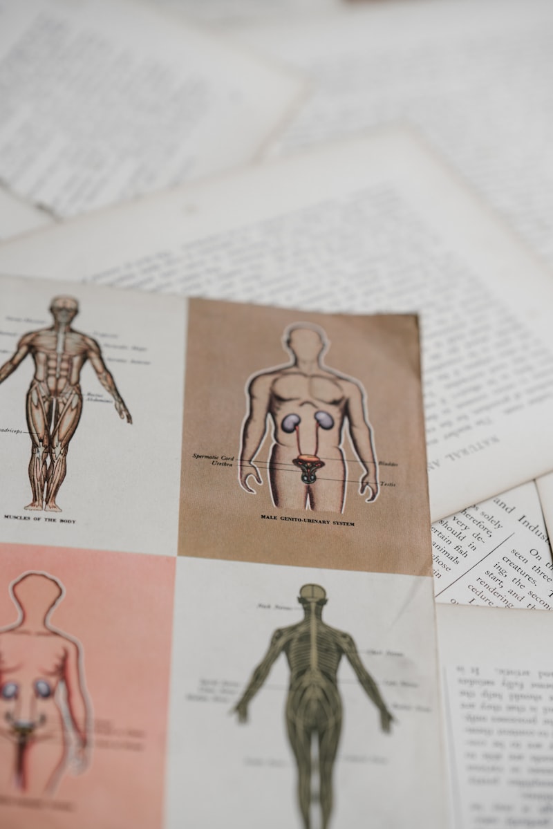
The regional specialization along the length of the duct is highly significant, reflecting the diverse functions of its adult derivatives. The cranial portion, adjacent to the gonad, differentiates into the extensive, coiled structure of the epididymis (head, body, and tail), responsible for sperm storage and functional maturation. The central portion elongates dramatically to form the vas deferens, the long, thick-walled muscular tube responsible for rapid sperm transport during ejaculation. The caudal-most segment, near the urethra, develops specialized outgrowths, forming the glandular structures known as the seminal vesicles and contributing directly to the formation of the ejaculatory ducts. This coordinated regional differentiation is a hallmark of urogenital development and necessitates precise spatial and temporal control of growth factors and steroid hormone receptors.
Differential Fate: Sexual Dimorphism
The process of internal sexual differentiation hinges entirely on the differential fates of the Wolffian and Müllerian duct systems, a paradigm governed primarily by the presence or absence of the functional testis. In the male (XY) embryo, the newly formed testis secretes two critical hormonal agents starting around week seven: Anti-Müllerian Hormone (AMH), also known as Müllerian Inhibiting Substance (MIS), and testosterone. AMH acts locally and systemically to induce the regression and subsequent disappearance of the Müllerian ducts, thus preventing the formation of the uterus and fallopian tubes. Simultaneously, testosterone, a potent androgen, is essential for the preservation, stabilization, and subsequent differentiation of the Wolffian ducts into the male internal accessory organs.
The mechanism of testosterone action is distinct from that of AMH. While testosterone itself stabilizes the Wolffian duct, promoting the survival and growth of the epididymis and vas deferens, the subsequent differentiation of some accessory glands, such as the prostate and seminal vesicles, often requires the conversion of testosterone into the even more potent androgen, dihydrotestosterone (DHT). This conversion is mediated by the intracellular enzyme 5-alpha reductase. Therefore, the internal duct derivatives (epididymis and vas deferens) are preserved directly by testosterone, while the more distal accessory glands rely on DHT, illustrating a complex, two-tiered regulatory system for complete male internal differentiation.
Conversely, in the female (XX) embryo, the default developmental pathway prevails. The absence of the SRY gene leads to ovarian development, which produces minimal testosterone during the critical period of duct differentiation. Furthermore, no AMH is produced in sufficient quantities to inhibit the Müllerian ducts, which subsequently differentiate into the uterus, fallopian tubes, and the upper third of the vagina. Consequently, the lack of androgenic support causes the Wolffian ducts to undergo programmed cell death (apoptosis) and atrophy beginning around the eighth week of gestation. While the bulk of the duct regresses, small, non-functional remnants often persist, known clinically as the epoophoron and paroophoron near the ovary, and Gartner’s ducts near the vagina, which can sometimes lead to pathological cyst formation.
Hormonal Regulation of Wolffian Duct Fate
The meticulous hormonal regulation of the Wolffian duct system provides one of the clearest and most studied examples of endocrine control over morphogenesis. The maintenance signal, testosterone, must be present continuously and at sufficient local concentration throughout the critical period, generally weeks 6 through 12 of gestation. Testosterone binds specifically to the high-affinity androgen receptors (AR) expressed abundantly within the mesenchymal and epithelial cells of the Wolffian duct. This binding initiates a complex cascade of gene expression changes that promote cell survival, proliferation, and differentiation, effectively overriding the default apoptotic and regression signals inherent to the duct structure.
The local delivery of testosterone is paramount for unilateral stabilization. The developing testis, situated immediately adjacent to the duct, produces high local concentrations of testosterone which ensures that the ipsilateral Wolffian duct is stabilized effectively. Defects in the synthesis of testosterone (e.g., due to enzyme deficiencies like 17-alpha-hydroxylase deficiency) or deficiencies in the androgen receptor itself (leading to Androgen Insensitivity Syndrome, AIS) result in the failure of Wolffian duct stabilization, even in genetically male individuals. In cases of complete AIS, where the receptor is non-functional, the Wolffian ducts regress entirely, mirroring the female developmental pattern internally, despite the presence of testes and effective AMH secretion.
While AMH is often discussed primarily in the context of Müllerian duct regression, its role is also indirectly vital to the functional outcome of the Wolffian duct. By eliminating the competing Müllerian structures, AMH ensures that the physical space and cellular resources are dedicated entirely to the Wolffian derivatives. The interplay between AMH signaling (which is active early in Sertoli cells) and testosterone signaling (which maintains the structure slightly later via Leydig cells) illustrates a highly coordinated hormonal timeline essential for establishing the definitive male urogenital anatomy. Any disruption in the timing or efficacy of either hormone production or receptor function can lead to ambiguous or incomplete internal sexual differentiation, a core issue in many disorders of sexual development (DSD).
Derivatives in the Adult Male Reproductive Tract
The adult male reproductive system is heavily reliant on the successful differentiation of the Wolffian duct derivatives, which collectively form the essential plumbing for sperm maturation, storage, and transport. The most cranial portion of the duct, along with persistent mesonephric tubules, forms the complex, highly coiled structure known as the epididymis. This structure, which can measure several meters in length when uncoiled, is critical not only for temporarily storing sperm but also for facilitating their functional maturation, enabling them to acquire progressive motility and the capacity to fertilize an egg. The duct’s epithelial cells in this region are specialized for fluid absorption and secretion, creating the optimal osmotic and chemical microenvironment for post-testicular sperm development.
Extending seamlessly from the tail of the epididymis is the vas deferens (or ductus deferens), derived from the middle section of the Wolffian duct. This structure is characterized by a remarkably thick muscular wall composed of three distinct layers of smooth muscle (inner longitudinal, middle circular, outer longitudinal), which facilitates the rapid propulsion of sperm during ejaculation via intense, coordinated peristaltic contractions. The vas deferens ascends through the inguinal canal as a key component of the spermatic cord, demonstrating the extensive migration and elongation this embryological structure undergoes during fetal development.
Finally, the caudal portions of the Wolffian duct contribute significantly to the glandular components and terminal transport structures. Near the base of the bladder, the terminal end dilates to form the ampulla of the vas deferens, which acts as a temporary reservoir. Outgrowths from this area develop into the seminal vesicles, paired glands responsible for producing the majority of the volume of seminal fluid, a substance rich in fructose (energy source) and various clotting factors necessary for the temporary retention of semen in the female reproductive tract. The convergence of the duct from the seminal vesicle and the terminal vas deferens forms the short ejaculatory duct, which then traverses the prostate gland to empty into the prostatic urethra, thereby completing the integrated male transport system.
Clinical Significance and Associated Anomalies
Disorders affecting the development or differentiation of the Wolffian duct structures have profound clinical significance, often leading to issues related to fertility, ambiguous genitalia, and general urological health. One major category of anomalies involves defects in stabilization due to hormonal signaling failures, as seen in various forms of Disorders of Sexual Development (DSD). For instance, patients with partial Androgen Insensitivity Syndrome (PAIS) may exhibit variable development of Wolffian derivatives, ranging from slightly hypoplastic structures to near-complete absence, directly correlating with the severity of androgen receptor dysfunction and the resulting inability to respond to circulating testosterone.
Structural anomalies of the Wolffian duct derivatives are also common causes of male infertility. Congenital absence of the vas deferens (CAVD) is a key example, often resulting in obstructive azoospermia (absence of sperm in the ejaculate). While CAVD can occur in isolation, it is frequently associated with mutations in the Cystic Fibrosis Transmembrane Conductance Regulator (CFTR) gene, suggesting a shared developmental pathway or an essential role for CFTR in the final maturation and patency of the duct system. Similarly, abnormalities in the coiled structure of the epididymis can impair sperm maturation and transport kinetics, contributing significantly to fertility challenges.
In females, although the Wolffian duct typically regresses almost entirely, persistent remnants can occasionally become clinically relevant. These remnants, particularly Gartner’s ducts, may form epithelial-lined cysts along the lateral walls of the vagina or adjacent to the uterus. While usually benign and asymptomatic, these cysts can sometimes grow large enough to cause pelvic pain, dyspareunia, or mass effects, occasionally requiring surgical excision. Furthermore, the proper development of the Wolffian duct system is sometimes implicated in the complex pathophysiology of conditions like cryptorchidism (undescended testes), suggesting that intrinsic developmental defects may contribute to the failure of testicular descent into the scrotum.
Conclusion
The Wolffian duct represents a quintessential structure in developmental biology, illustrating the elegant efficiency of bipotential embryonic precursors. Its destiny—whether to form the vital components of the male reproductive tract (epididymis, vas deferens, seminal vesicles) or to regress into vestigial remnants in the female—is entirely dictated by the precise and timely presence of testicular hormones, particularly testosterone. This process highlights the critical nature of endocrine signaling in determining internal anatomical sex, a process that is frequently disrupted in clinically relevant disorders of sexual development and intersex conditions.
The integrated nature of the urogenital system means that anomalies originating in the Wolffian duct often have far-reaching consequences, affecting fertility, urinary tract anatomy, and the overall psychological well-being associated with sexual development. Conditions ranging from congenital absence of the vas deferens to the formation of benign Gartner’s duct cysts underscore the complexity involved in the complete differentiation or regression of this structure. Research continues to explore the intricate genetic and molecular pathways that govern the stabilization, elongation, and regional specialization of the Wolffian duct epithelium and its surrounding mesenchyme, continually refining our understanding of mammalian sexual differentiation.
In summary, the Wolffian duct is far more than a transient embryological feature; it is the structural cornerstone of the internal male genitalia. Its study provides fundamental insights into hormone action, developmental genetics, and the etiology of numerous reproductive pathologies, reinforcing its position as a central topic in advanced human anatomy and developmental psychology, particularly concerning the biological foundations of sex and gender identity.
References
-
Bowen, J. L., & O’Rahilly, R. (2013). Human Embryology and Teratology (3rd ed.). Hoboken, NJ: Wiley-Blackwell.
-
Hassan, M. M., & Al-Azemi, M. (2012). Role of the Wolffian ducts in the etiology and pathogenesis of cryptorchidism. International Journal of Fertility & Sterility, 5(3), 175-182.
-
Kumar, S., & Aggarwal, D. (2015). Wolffian duct and its role in male reproductive system. International Journal of Anatomy and Research, 3(3), 1162-1164.