Acrocephaly: Understanding the Impact of Cranial Shape
Introduction and Definition of Acrocephaly
Acrocephaly, sometimes referred to interchangeably as oxycephaly or turricephaly, describes a profound developmental disorder characterized by an exceptionally high, peaked, or conical skull shape. This specific cranial deformity results from premature fusion of certain cranial sutures, a condition broadly termed craniosynostosis. Typically, acrocephaly arises from the early closure of both the coronal and sagittal sutures, forcing the growing brain to expand vertically rather than horizontally or anteroposteriorly. This restriction of normal cranial vault expansion leads to a characteristic “tower skull” appearance, which is often asymmetrical and globally restrictive to brain growth. The psychological significance of acrocephaly stems directly from the resulting compression and distortion of cerebral structures, leading frequently, though not invariably across all variants, to severe mental retardation and significant developmental challenges, establishing it as a critical area of study within neurodevelopmental psychology and medical genetics.
The structural anomaly inherent in acrocephaly is not merely cosmetic; it is a mechanical constraint on the central nervous system. The normal function of cranial sutures is to allow flexibility and continuous expansion of the skull throughout infancy and early childhood, accommodating the rapid growth trajectory of the brain. When these sutures ossify prematurely, the resulting reduction in intracranial volume creates a cascade of pathological events. The definition of acrocephaly centers on this failure of geometric expansion, necessitating compensation through vertical growth at patent sutures, resulting in the distinctive high cranial vertex. Clinically, the condition must be differentiated from other forms of craniosynostosis, such as scaphocephaly (sagittal fusion) or brachycephaly (bilateral coronal fusion without the extreme vertical growth), although acrocephalic syndromes often incorporate elements of brachycephaly, particularly severe midfacial retrusion.
Early identification of acrocephaly is crucial because the severity of the intellectual impairment is often correlated with the degree of intracranial pressure (ICP) and the timing of surgical intervention. The original description of the disorder emphasized its association with severe mental retardation, highlighting the fact that the restricted growth environment severely compromises neural development and function. While modern medical management, including aggressive surgical decompression, has improved outcomes for some individuals, the underlying genetic and morphological defects often predispose patients to complex cognitive and motor deficits. Thus, acrocephaly serves as a powerful example of how early physical constraints directly dictate later psychological and intellectual capacity, demanding a comprehensive, multidisciplinary approach involving neurosurgeons, geneticists, and developmental psychologists.
Etiology and Pathophysiology of Cranial Restriction
The core pathophysiology of acrocephaly involves aberrant signaling pathways that govern the timing of osteoblast differentiation and subsequent mineralization at the synchondroses and sutures. The most common underlying cause is a mutation in genes encoding Fibroblast Growth Factor Receptors (FGFRs), particularly FGFR2 and FGFR3, which regulate bone growth and development. These mutations typically result in a gain-of-function mechanism, meaning the receptor is constitutively active or overly sensitive to ligand binding, triggering premature and excessive fusion of the calvarial sutures, specifically the coronal sutures, often combined with other fusions. This premature fusion halts growth across the affected vector, forcing compensatory expansion in the only directions remaining, which is typically superiorly (upward), resulting in the characteristic conical or pointed skull shape.
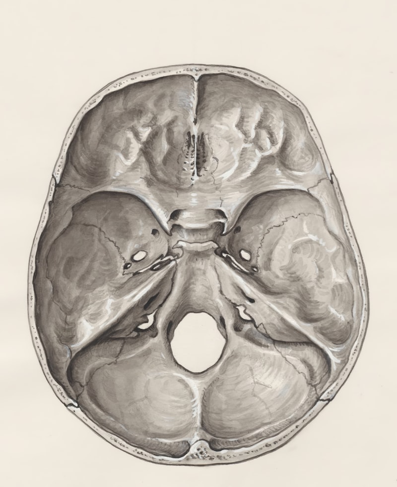
The mechanical consequences of this early fusion are dire. As the brain continues its obligatory growth, the fixed cranial volume rapidly leads to significantly increased intracranial pressure (ICP). Chronic elevation of ICP is the primary driver of the neurological symptoms associated with acrocephaly. Elevated ICP can impede normal cerebrospinal fluid circulation, potentially leading to hydrocephalus, and, more critically, it compresses the delicate cerebral cortex and underlying white matter. This chronic compression is directly responsible for optic nerve atrophy, visual impairment, and the progressive destruction of neuronal networks necessary for higher cognitive function. The magnitude of ICP increase is often proportional to the number of sutures involved and the age at which fusion occurred, highlighting the urgency of addressing the underlying restriction before irreversible neurological damage occurs.
Furthermore, the restricted growth extends beyond the cranial vault to the cranial base and midface. In many acrocephalic syndromes, the midfacial bones, including the maxilla and zygoma, are also underdeveloped (midfacial hypoplasia). This structural deficit severely compromises the orbital cavities, leading to shallow orbits (orbital hypoplasia) and subsequent ocular manifestations such as exophthalmos (protruding eyes), a hallmark symptom in several specific acrocephalic syndromes. The combination of restricted cranial growth, compromised CSF dynamics, and severe facial skeletal defects necessitates complex surgical interventions aimed not only at decompressing the brain but also at reconstructing the orbitofacial skeleton to protect vision and establish functional airways.
Clinical Manifestations and Neurological Impact
The clinical presentation of acrocephaly is dominated by distinct craniofacial features and severe neurological sequelae resulting from chronic intracranial hypertension. Physically, patients exhibit a skull that is disproportionately high and narrow, often described as tower-shaped (turricephaly). Associated features frequently include a high, sometimes bossed, forehead, and severe midfacial retrusion, giving the face a concave or ‘dished-out’ appearance. Ocular symptoms are pervasive and critical, stemming from the shallow orbital cavities; these include severe proptosis (exophthalmos), which renders the eyes vulnerable to exposure keratitis, and strabismus. More seriously, sustained elevated ICP manifests clinically as persistent headaches, vomiting, lethargy, and papilledema, which, if unmanaged, progresses rapidly to secondary optic atrophy and permanent blindness.
The neurological and developmental consequences are perhaps the most devastating aspect of acrocephaly, leading to the classification of many severe cases alongside severe mental retardation in the original entry. The combination of chronic cerebral compression and potentially associated brain anomalies (such as corpus callosum agenesis or specific cortical malformations common in some related syndromes) severely compromises cognitive development. While the spectrum of intellectual disability varies widely depending on the specific genetic etiology and the efficacy of early decompression, many patients experience global developmental delay, speech and language deficits, and significant challenges in adaptive functioning.
In addition to cognitive deficits, behavioral and psychological issues are frequently observed. These may include features of Autism Spectrum Disorder (ASD), hyperactivity, impulsivity, and difficulties with social interaction. The chronic physical discomfort associated with headaches and visual impairment, coupled with the cosmetic challenges of severe facial dysmorphology, also contributes significantly to poor quality of life and psychological distress. Therefore, the management plan for acrocephaly must extend far beyond neurosurgery, requiring extensive psychological assessment, speech therapy, occupational therapy, and ongoing educational support tailored to the specific cognitive profile resulting from the early developmental insult.
The Crouzon Syndrome Variant
Crouzon syndrome, often referred to as craniofacial dysostosis, represents a classic and well-defined variant of acrocephaly, directly referenced in the initial description. This disorder is characterized by the triad of acrocephaly (or generalized craniosynostosis), exophthalmos (protruding eyes), and midfacial hypoplasia. It is typically inherited in an autosomal dominant pattern and is most often caused by specific mutations in the FGFR2 gene. Unlike some other acrocephalic syndromes, Crouzon syndrome typically does not involve limb abnormalities. However, the severity of the craniofacial involvement is profound, often involving the bilateral coronal, sagittal, and lambdoid sutures, leading to a complex restriction pattern that significantly impacts the face and orbits.
The defining characteristic of Crouzon syndrome, beyond the high skull, is the severe facial involvement. Patients exhibit maxillary hypoplasia (underdeveloped upper jaw), resulting in a characteristic Class III malocclusion (underbite) and respiratory difficulties due to reduced nasopharyngeal space. The orbits are notoriously shallow and divergent, leading to significant exophthalmos and ocular complications. Furthermore, the original content noted that these symptoms are associated with small orbital cavities and increased intracranial pressure, which is a constant threat throughout childhood. The ICP often arises not just from the cranial restriction but also from venous outflow obstruction due to the malformed cranial base. This sustained pressure is the primary factor dictating the need for early and serial cranial vault expansion and decompression procedures.
The genetic basis of Crouzon syndrome is highly relevant to the concept of varying penetrance, as mentioned in the original source text. While it is caused by a single dominant gene, the clinical expression among carriers can be remarkably diverse. Some individuals may present with relatively mild skull deformity and moderate exophthalmos, experiencing only minimal cognitive impact, while others may suffer from severe, multi-sutural synostosis requiring complex, life-saving surgery in infancy, along with profound visual and cognitive impairment. This wide variation in expressivity means that not all individuals who possess the dominant gene are equally affected, posing challenges for predictive counseling and requiring individualized treatment plans based on the severity of the structural and functional deficits.
The Apert Syndrome Variant
Apert syndrome, or acrocephalosyndactyly, is another major form of acrocephaly and is distinguished by a unique combination of severe craniofacial malformations and complex bilateral limb defects. The syndrome is caused almost exclusively by one of two specific mutations (Ser252Trp or Pro253Arg) in the FGFR2 gene, inherited in an autosomal dominant fashion, although most cases arise from spontaneous mutations. The cranial presentation involves severe acrocephaly, often manifesting as turribrachycephaly, characterized by a high, steeply sloping forehead and a flat occiput. Cranial base synostosis is typically extensive, leading to severe midfacial hypoplasia and a high risk of chronically elevated ICP and associated severe mental retardation.
The features that uniquely define Apert syndrome, setting it apart from Crouzon syndrome and others, are the characteristic hand and foot anomalies, referred to in the original text as syndactylia (fingers grown together). This syndactyly is typically complex and symmetrical, involving bony and soft tissue fusion of the second, third, and fourth digits, often resulting in a severe “mitten” or “spoon” hand deformity. The degree of fusion significantly impairs fine motor skills and requires extensive, often staged, reconstructive hand surgery beginning in infancy. The combination of severe intellectual disability and major limb impairment profoundly affects the patient’s functional independence and quality of life.
Furthermore, Apert syndrome is consistently associated with hypertelorism (eyes wide apart), severe proptosis, and a downward-slanting palpebral fissure. The midface is severely underdeveloped, resulting in a concave facial profile and chronic upper airway obstruction, often necessitating tracheostomy or aggressive advancement of the midface skeleton later in childhood. Beyond the skeletal defects, patients with Apert syndrome frequently exhibit visceral anomalies, including cardiac, genitourinary, and gastrointestinal defects, and are at a significantly higher risk for structural brain malformations, such as hydrocephalus, which contribute substantially to the cognitive impairment observed in this population. The complexity of these manifestations mandates highly coordinated care across multiple pediatric subspecialties.
The Grieg Syndrome and Differential Diagnosis
The Grieg syndrome, correctly identified in the original text as Grieg cephalopolysyndactyly (GCPS), constitutes another genetically distinct disorder that combines features of acrocephaly with specific digital anomalies and is characterized by a high degree of hypertelorism. Unlike Apert syndrome, where the primary digital defect is complex syndactyly, Grieg syndrome involves a combination of hypertelorism and polydactyly (extra digits) or syndactyly, often less severe than the complex fusion seen in Apert’s. The original text accurately noted the core distinction: in the Grieg syndrome, the high skull is combined with a greater degree of telorism but without the defect in the fingers described in Apert syndrome, although subtle or minor hand/foot anomalies often exist.
GCPS is caused by mutations or deletions involving the GLI3 gene, a transcription factor crucial for patterning during embryonic development, particularly in the midline and limbs. The cranial manifestation can range from mild macrocephaly to full-blown acrocephaly, but the most consistent craniofacial feature is pronounced orbital separation, or hypertelorism, which can be quite severe, alongside a prominent forehead (frontal bossing). Crucially, the neurological prognosis in GCPS is generally more favorable than in Crouzon or Apert syndromes; while developmental delays can occur, severe mental retardation is less common, depending on the specific mutation and the presence of associated central nervous system malformations.
The complexity and overlapping features among acrocephalic syndromes necessitate rigorous differential diagnosis. Clinicians must distinguish between these conditions because the management, prognosis, and recurrence risks are highly specific to the underlying genetic locus. Key differentiators include:
- Apert Syndrome: Defined by severe, complex syndactyly of hands/feet combined with severe craniosynostosis.
- Crouzon Syndrome: Defined by craniosynostosis and midfacial hypoplasia/exophthalmos, but lacking limb abnormalities.
- Grieg Syndrome: Defined by prominent hypertelorism and specific forms of polysyndactyly, often with a milder cranial presentation.
Accurate diagnosis, achieved through molecular genetic testing, is vital for predicting the long-term cognitive outcome and for planning staged surgical corrections for the craniofacial and limb anomalies.
Genetic Basis and Variability of Penetrance
The unifying theme across the primary acrocephalic syndromes—Crouzon, Apert, and Grieg—is their etiology as single-gene disorders with an autosomal dominant inheritance pattern. As noted in the original entry, there is strong evidence that these variations are due to a dominant gene, at least in some families. This means that a mutation in only one copy of the causative gene (e.g., FGFR2 or GLI3) is sufficient to cause the disorder. However, a significant proportion of cases, particularly Apert syndrome, arise from de novo (new) mutations in the affected individual, whose parents are unaffected. Understanding the specific gene involved is fundamental, as mutations in the FGFR family result in disorders primarily affecting the bone matrix and cell signaling, leading directly to premature suture fusion.
The concept of varying penetrance is essential to grasping the clinical diversity observed in acrocephaly. Penetrance refers to the likelihood that an individual carrying a specific pathogenic genotype will actually express the associated phenotype. In acrocephalic syndromes, particularly Crouzon syndrome, penetrance is high but the expressivity—the degree or severity of the phenotype—is highly variable. This variability explains why wide variations exist in the degree of skull deformity and exophthalmos even among family members carrying the identical mutation. Factors influencing this variability are complex and include subtle differences in genetic background, epigenetic influences, and possible interactions with modifying genes that affect the downstream signaling pathways regulated by the primary mutated gene.
This genetic heterogeneity and variable expressivity have profound implications for genetic counseling. Families require clear communication regarding the recurrence risk and the uncertainty surrounding the potential severity of the condition in an affected offspring. For dominant disorders like these, the recurrence risk in subsequent children, if one parent is affected, is 50 percent. However, if the case is the result of a de novo mutation (as often seen in Apert syndrome), the parental risk is extremely low, though there is a slight, increased risk due to the possibility of parental germline mosaicism. Molecular confirmation of the specific mutation is therefore mandatory not only for accurate diagnosis and prognosis but also for providing informed reproductive counseling, ensuring that families understand the full spectrum of potential outcomes dictated by the complex interplay of dominant inheritance and varying penetrance.