Alpha Motor Neurons: The Engines of Human Action
- The Core Definition of Alpha Motor Neurons
- Historical Context and Discovery
- Detailed Structure and Function
- A Practical Example: Lifting a Cup of Coffee
- Significance and Impact in Psychology and Medicine
- Connections and Relations to Other Concepts
- Broader Category: Motor Control and Neurophysiology
- Clinical Relevance and Damage
The Core Definition of Alpha Motor Neurons
An alpha motor neuron (AMN) represents a pivotal type of efferent neuron found in the central nervous system, specifically within the spinal cord and brainstem, whose primary function is to directly innervate and control the contraction of skeletal muscle fibers. These large, multipolar lower motor neurons are the final common pathway for motor commands originating from higher brain centers and local spinal reflexes, translating neural signals into physical movement. Their direct connection to extrafusal muscle fibers ensures the precise and powerful contractions necessary for virtually all voluntary and many involuntary movements, from intricate fine motor skills to robust gross motor actions.
The fundamental mechanism underpinning the function of an alpha motor neuron involves the generation and propagation of an action potential. When an appropriate excitatory signal reaches the AMN, it depolarizes, triggering an action potential that travels down its long axon to the neuromuscular junction. At this specialized synapse, the arrival of the action potential prompts the release of the neurotransmitter acetylcholine into the synaptic cleft. Acetylcholine then binds to receptors on the muscle fiber membrane, initiating a cascade of events that ultimately leads to muscle fiber depolarization and contraction. This intricate electrochemical process is the bedrock of all skeletal muscle movement, highlighting the AMN’s indispensable role in motor control.
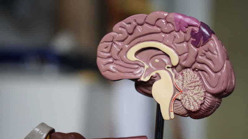
Alpha motor neurons are specifically housed within the ventral horn of the spinal cord’s gray matter and in the motor nuclei of the brainstem for cranial nerves. They are characterized by their large cell bodies and extensive dendritic trees, which allow them to integrate numerous synaptic inputs from both descending pathways (e.g., from the motor cortex) and local spinal interneurons. Each AMN, along with all the muscle fibers it innervates, constitutes a motor unit, which is the smallest functional unit of motor control. The size of a motor unit varies considerably depending on the muscle’s function, with small motor units providing fine control (e.g., eye muscles) and large motor units generating powerful forces (e.g., leg muscles).
Historical Context and Discovery
The understanding of motor neurons and their role in movement has evolved significantly over centuries, rooted in early anatomical observations and later solidified by physiological experiments. While the specific term “alpha motor neuron” is a modern construct, the concept of a nerve controlling muscle contraction can be traced back to ancient Greek physicians. However, the scientific foundation for our current knowledge began to form with the advent of microscopy and experimental physiology in the 19th century. Key figures like Charles Bell and François Magendie, through their independent work on the spinal nerves in the early 1800s, established the distinction between sensory and motor roots of spinal nerves, a fundamental discovery known as the Bell-Magendie Law. This laid the groundwork for understanding that different nerve pathways conveyed different types of information.
Towards the late 19th and early 20th centuries, pioneering neuroanatomists like Santiago Ramón y Cajal, utilizing staining techniques developed by Camillo Golgi, meticulously mapped the intricate structures of the nervous system. Cajal’s work provided definitive evidence for the neuron doctrine, postulating that individual neurons are discrete units, and importantly, he detailed the morphology of motor neurons within the spinal cord. Simultaneously, physiologists such as Charles Sherrington conducted groundbreaking research on reflexes and synaptic transmission. Sherrington introduced concepts like the “final common pathway” to describe the motor neuron’s role as the converging point for all neural influences on muscle, a concept directly applicable to the alpha motor neuron. His work, along with that of others, firmly established the motor neuron as the critical link between the nervous system and muscle.
The specific classification of “alpha” motor neurons came later as researchers distinguished them from other types, particularly gamma motor neurons, which innervate muscle spindles and regulate muscle tone and proprioception. This distinction allowed for a more nuanced understanding of motor control, recognizing that different motor neuron populations serve specialized roles in orchestrating complex movements. The ongoing study of alpha motor neurons continues to reveal deeper insights into their electrophysiological properties, their synaptic integration capabilities, and their vulnerability in neurodegenerative diseases, building upon a rich historical legacy of neurological discovery.
Detailed Structure and Function
The structure of an alpha motor neuron is exquisitely adapted to its function as the primary executor of muscle contraction. These neurons possess a large cell body, or soma, typically located in the ventral horn of the spinal cord or in the motor nuclei of the brainstem. Extending from the soma are numerous dendrites, which are tree-like processes that receive thousands of synaptic inputs from various sources, including sensory neurons, interneurons within the spinal cord, and descending pathways from the brain (such as the corticospinal tract). This extensive dendritic arborization allows the AMN to integrate a vast amount of information, processing excitatory and inhibitory signals to determine whether an action potential will be fired.
From the cell body, a single, long axon projects out of the central nervous system, often traveling considerable distances to reach its target muscle. This axon is typically myelinated, which significantly increases the speed of action potential conduction, ensuring rapid communication between the nervous system and the muscle. Upon reaching the muscle, the axon branches extensively, with each branch forming a synapse with a different muscle fiber at a specialized structure called the neuromuscular junction. The collection of an alpha motor neuron and all the muscle fibers it innervates is termed a motor unit. The number of muscle fibers per motor unit varies greatly depending on the precision required for a given movement; muscles requiring fine control, like those in the fingers or eyes, have small motor units, while powerful muscles, such as those in the quadriceps, have large motor units.
Beyond initiating voluntary movement, alpha motor neurons also play a critical role in maintaining muscle tone and regulating reflexive responses. Muscle tone refers to the continuous, passive partial contraction of the muscles, which helps maintain posture and balance and prepares muscles for immediate action. This tonic activity is modulated by a constant stream of excitatory and inhibitory inputs to the AMNs. Furthermore, AMNs are integral components of reflex arcs, such as the stretch reflex. When a muscle is suddenly stretched, sensory receptors within the muscle (muscle spindles) send signals to the spinal cord, directly exciting the AMNs that innervate that same muscle, causing it to contract reflexively. This rapid, involuntary response is crucial for protection and balance, demonstrating the AMN’s fundamental involvement in both conscious and subconscious aspects of motor function.
A Practical Example: Lifting a Cup of Coffee
To illustrate the intricate function of alpha motor neurons, consider the seemingly simple act of reaching for and lifting a cup of coffee. This everyday action requires precise coordination and varying degrees of force, all orchestrated by the nervous system with AMNs as the final executive command. When you decide to lift the cup, a complex series of neural commands originates in your brain’s motor cortex, which then descends through various pathways, including the corticospinal tract, to the spinal cord.
As these descending signals arrive at the appropriate segments of the spinal cord, they converge upon the alpha motor neurons located in the ventral horn. For instance, AMNs innervating the biceps brachii and forearm flexors will receive excitatory input. Simultaneously, AMNs controlling antagonist muscles, like the triceps, will receive inhibitory input to ensure smooth, coordinated movement without opposing forces. Each activated AMN then generates an action potential, which rapidly travels down its axon to the neuromuscular junction of its respective skeletal muscle fibers.
At the neuromuscular junction, the release of acetylcholine neurotransmitter triggers the muscle fibers to contract. The number of AMNs recruited and the frequency at which they fire action potentials determine the force of the contraction. For lifting a light coffee cup, fewer motor units are recruited and fire at lower frequencies. If the cup is unexpectedly heavy, sensory feedback from proprioceptors (like muscle spindles and Golgi tendon organs) quickly modulates the activity of the AMNs, causing more motor units to be recruited or existing ones to fire at higher frequencies, thereby increasing the contractile force to prevent dropping the cup. This continuous feedback loop, mediated by the adaptable responses of the alpha motor neurons, ensures that the movement is executed smoothly, effectively, and with appropriate force, highlighting their indispensable role in dynamic motor control.
Significance and Impact in Psychology and Medicine
The concept of the alpha motor neuron is of immense significance to the field of psychology, particularly in understanding the biological basis of behavior, motor control, and the neural underpinnings of learning and action. From a psychological perspective, AMNs represent the ultimate physiological output of complex cognitive processes, including planning, decision-making, and execution of voluntary movements. Understanding how these neurons integrate diverse inputs helps psychologists comprehend how intentions are translated into actions, how motor skills are acquired and refined, and how neurological damage impacts behavior and psychological well-being. The study of AMNs also informs theories of motor learning, demonstrating how repeated practice can optimize the recruitment and firing patterns of these neurons, leading to greater efficiency and precision in movement.
In the broader scientific and medical communities, the impact of understanding alpha motor neurons is profound. Their role as the “final common pathway” makes them a critical focus in neurology, neurorehabilitation, and the study of motor disorders. Knowledge of AMN function is directly applied in diagnosing and treating conditions such as Amyotrophic Lateral Sclerosis (ALS), Poliomyelitis, spinal cord injuries, and various peripheral neuropathies. These conditions often involve damage or degeneration of AMNs, leading to muscle weakness, paralysis, and atrophy, collectively known as lower motor neuron syndrome. Therapeutic interventions, including physical therapy, occupational therapy, and pharmacological treatments, often aim to preserve AMN function, mitigate symptoms, or facilitate neural plasticity to recover lost motor control.
Furthermore, the principles governing AMN function have found applications in fields beyond traditional medicine. In the realm of biomechatronics and neuroprosthetics, researchers are developing brain-computer interfaces (BCIs) and prosthetic limbs that attempt to bypass damaged AMNs or restore their function. By decoding signals from the brain or remaining motor neurons, these technologies aim to allow individuals with paralysis to control external devices or move prosthetic limbs, directly translating neural commands into action, much like AMNs do in a healthy nervous system. This interdisciplinary research underscores the fundamental importance of AMNs as the link between thought and movement, continuously pushing the boundaries of what is possible in restoring motor function and enhancing human capabilities.
Connections and Relations to Other Concepts
The alpha motor neuron does not operate in isolation but is intricately connected within a vast network of neural structures and processes. It forms a crucial part of the broader system of motor control. A primary distinction is often made between upper motor neurons and lower motor neurons. Upper motor neurons originate in the brain (e.g., in the motor cortex) and descend to synapse with lower motor neurons in the brainstem or spinal cord. The alpha motor neuron is a type of lower motor neuron, acting as the final common pathway for all motor commands, whether originating from conscious thought or reflexive actions. Damage to upper motor neurons typically results in spasticity and hyperreflexia, while damage to lower motor neurons (like AMNs) leads to flaccid paralysis, muscle atrophy, and diminished reflexes.
Another critical relationship exists with gamma motor neurons. While alpha motor neurons innervate the extrafusal muscle fibers (those responsible for generating force), gamma motor neurons innervate the intrafusal muscle fibers located within muscle spindles. These spindles are sensory receptors that detect changes in muscle length and the rate of length change, contributing to proprioception. The co-activation of alpha and gamma motor neurons (alpha-gamma co-activation) ensures that muscle spindles remain sensitive to changes in muscle length even during muscle contraction, maintaining accurate sensory feedback about muscle position and movement. This coordinated activity is essential for precise motor control and maintaining muscle tone.
Furthermore, AMNs are central to the functioning of the reflex arc, the neural pathway that mediates a reflex action. In the classic stretch reflex, sensory input from muscle spindles directly excites the alpha motor neuron, causing a rapid, involuntary muscle contraction. Local spinal interneurons also play a significant role, receiving input from various sources and modulating AMN activity through both excitatory and inhibitory synapses. These interneurons enable complex spinal reflexes, reciprocal inhibition (inhibiting antagonist muscles during agonist contraction), and contribute to rhythmic movements like walking. The alpha motor neuron, therefore, serves as the critical junction where complex descending commands, local spinal circuitry, and sensory feedback converge to produce coordinated and purposeful movement.
Broader Category: Motor Control and Neurophysiology
The alpha motor neuron fundamentally belongs to the broader subfield of Neuroscience, specifically within the domains of Motor Control and Neurophysiology. Motor control is a multidisciplinary field that studies how the central nervous system regulates the musculoskeletal system to produce coordinated movements. It encompasses everything from the initial planning of movement in higher brain centers to the final execution by muscle fibers, with the alpha motor neuron serving as the critical interface. Understanding AMNs is indispensable for comprehending the principles of motor learning, motor skill acquisition, and the neural mechanisms underlying balance, posture, and locomotion.
Within neurophysiology, the study of alpha motor neurons delves into their electrical and chemical properties, synaptic integration, and the ionic mechanisms that govern their excitability. Researchers in this area examine how AMNs generate action potentials, how they respond to different neurotransmitters, and how their firing patterns contribute to muscle force generation and modulation. This involves detailed investigations at the cellular and molecular levels to elucidate the precise mechanisms by which neural signals are transduced into muscular contractions, providing a foundational understanding of how the nervous system communicates with the body’s effectors.
The importance of alpha motor neurons extends into clinical neurology and rehabilitation, where their health and function are directly correlated with an individual’s motor capabilities. As the final common pathway, their integrity is paramount for normal movement. Thus, disorders affecting AMNs are central topics in clinical neuroscience, driving research into treatments and interventions for conditions that impair motor function. The study of AMNs bridges basic neuroscience with applied clinical science, making them a cornerstone concept in understanding the physiological basis of movement and its disruption.
Clinical Relevance and Damage
The integrity and proper functioning of alpha motor neurons are absolutely critical for normal motor function, making them highly significant in clinical contexts. Damage or degeneration of these neurons can lead to a range of debilitating conditions collectively known as lower motor neuron lesions. These lesions can result from various etiologies, including direct trauma to the spinal cord or peripheral nerves, viral infections, genetic disorders, and neurodegenerative diseases. The clinical presentation of alpha motor neuron damage typically includes symptoms directly attributable to the loss of innervation to skeletal muscles, profoundly impacting a person’s ability to move and perform daily activities.
One of the most devastating conditions involving progressive alpha motor neuron degeneration is Amyotrophic Lateral Sclerosis (ALS), also known as Lou Gehrig’s disease. ALS is characterized by the progressive loss of both upper and lower motor neurons, including AMNs, leading to muscle weakness, atrophy, fasciculations (visible twitching of muscle), and ultimately paralysis. Another historical example is Poliomyelitis, a viral infection that specifically targets and destroys alpha motor neurons in the spinal cord, causing flaccid paralysis. While largely eradicated in many parts of the world due to vaccination, post-polio syndrome can still affect survivors. Other conditions like Guillain-Barré syndrome, peripheral neuropathies, and nerve root compressions can also temporarily or permanently impair AMN function.
The consequences of AMN damage are distinct from those of upper motor neuron lesions. When alpha motor neurons are damaged, the muscles they innervate lose their direct excitatory input, leading to symptoms such as muscle weakness, which can progress to complete paralysis (flaccid paralysis). Over time, the denervated muscles will undergo severe muscle atrophy due to disuse and lack of trophic factors supplied by the neurons. Additionally, areflexia or hyporeflexia (diminished or absent reflexes) is a hallmark sign, as the reflex arc is interrupted at its final efferent pathway. The presence of fasciculations is also a classic sign, representing spontaneous, irregular contractions of muscle fibers due to nerve irritation or degeneration. Understanding these clinical manifestations is crucial for accurate diagnosis, prognosis, and the development of targeted therapies aimed at improving the quality of life for individuals affected by these debilitating motor neuron disorders.